
Abstract
Polyglucosan bodies (Lafora bodies) were identified in a juvenile gray-headed flying fox (Pteropus poliocephalus) with neurological signs. The structures were only noted in the brain stem, and no associated degenerative changes were present. These structures have not been previously identified in any species in the order Chiroptera.
A captive male, juvenile gray-headed flying fox (Pteropus poliocephalus; order Chiroptera, suborder Megachiroptera family Pteropodidae) was presented to Regional Veterinary Laboratory (Camden, NSW, Australia) with a history of weakness and apparent ataxia, primarily of the hind legs. The animal was assessed as having marked ataxia of the hind limbs and was noted to struggle, maintaining its grip on the cage mesh with its wing digits. Due to the potential of Australian bat lyssavirus (ABLV) infection, the animal was euthanized immediately with intraperitoneal barbiturate.
At necropsy, the animal was in moderate body condition. Internally, there were multiple 1–2-mm pale foci scattered throughout the hepatic parenchyma. No other significant findings were noted, and the brain was removed to the level of the first cervical segment and sectioned midsagitally. No further processing was carried out until infection with the zoonotic ABLV was excluded. One half of the brain and sections of both parotid salivary glands were sent to the Australian Animal Health Laboratory (East Geelong, VIC, Australia), and both immunohistochemistry and fluorescent antibody tests were negative for ABLV. Histologically, scattered foci of hydropic vacuolation were present in the liver. The brain stem contained numerous lightly amphophilic (hematoxylin and eosin-staining), spherical inclusion bodies (Fig. 1). These homogenous globular structures ranged between 5 and 20 μm in diameter. Occasional smaller fused clusters of bodies were also noted. The peripheral margins were smooth. The structures were Von Kossa negative and periodic acid-Schiff positive, with lack of degradation after diastase treatment, indicating that the bodies were nonminer-alized and composed of complex polysaccharides. A full range of tissues were examined histologically, including the cranial spinal cord and multiple sections of skeletal muscle. No significant changes were noted.
Due to limits of sample size, sections of tissue were selected for electron microscopy from the paraffin-embedded block. Numerous 2-mm sections of tissue were excised from the block and deparaffinized in xylene for 1 hr. The specimens were transferred to acetone and then rehydrated in an ethanol series. The specimens were retrimmed and transferred to Karnovsky fixative for 6 hr, and then rinsed and transferred to 2% uranyl acetate for 1 hr. After dehydration, the tissue specimens were transferred to acetone, embedded in Spurr resin, and allowed to polymerize at 70°C for 10 hr. Ultrathin sections were mounted on copper grids, stained with uranyl acetate and lead citrate, and examined on a transmission electron microscope. a Ultrastructurally, numerous non–membrane-bound inclusions were observed composed of filamentous masses (Fig. 2). The inclusions were not able to be confidently identified within neuronal cell bodies.
Lafora bodies, or polyglucosan bodies, are inclusions composed of complex glycoprotein polymers in tissue. 3 The former term specifically refers to a condition in human medicine in which an autosomal recessive mutation leads to deposition of polyglucosan bodies, which are associated with a progressively deteriorating, eventually lethal epileptic syndrome. 9 Polyglucosan bodies are occasionally noted incidentally or in a sporadic, late-onset neurological syndrome. The inclusions are characteristically distributed in the cerebral cortex, thalamus, globus pallidus, and substantia nigra. Additionally, the retinal ganglia, peripheral nerves, hepatocytes, striated muscle, and sweat gland ductular epithelium often include inclusions. 4,10 In the current case, no inclusions were noted in the skin, liver, retina, parotid and mandibular salivary glands, skeletal muscle, or any other portion of the central nervous system, including cerebellum and cerebrum.
In nonhuman species, the incidence of polyglucosan bodies has been infrequently documented in dogs and cats and often associated with neurological signs. 5,7 Additionally, the presence of polyglucosan bodies has been demonstrated in a fox, 8 a cockatiel, 2 and cattle. 11,12 In published cases, the presence of inclusions associated with clinical neurological disease (i.e., Lafora bodies) are typically intraneuronal. The histological appearance of the inclusions in published reports varies from homogenous and concentrically lamellar to those possessing peripherally radiating patterns. In the current case, the appearance was distinctly homogenous. 14 However, the ultrastructural location of inclusions was not able to be confidently located, possibly because of artifactual changes related to formalin fixation. Corpora amylacea, more commonly noted and often incidental polyglucosan bodies, typically develop within astrocytic processes. 13 Additionally, corpora amylacea are frequently lamellar in appearance. 6 The consensus in the veterinary literature is that the presentation of polyglucosan bodies within the neuropil is more consistent with incidental histological changes in aging animals. 1,3,6,9,13 One study of aging changes in the dog consistently found low numbers of polyglucosan bodies within the neuropil of aging animals but not in young controls. 1 In contrast, the present case was a juvenile male with distinct neurological signs. The challenge of distinguishing between ataxia or weakness in the bat was not successfully determined by the authors, and the link between these polyglucosan bodies and the neurological condition in this animal remains enigmatic.
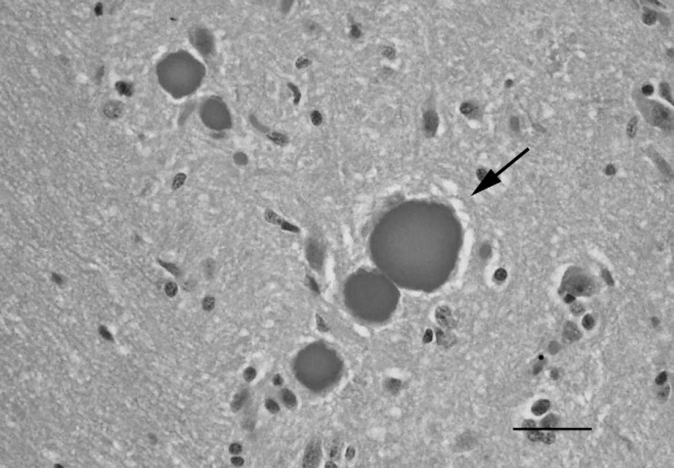
Brain stem; gray-headed flying fox (Pteropus poliocephalus). Polyglucosan bodies within the neuropil (arrow). The deposits are lightly amphophilic and spherical. Hematoxylin and eosin. Bar = 30 μm.
Gray-headed flying foxes are common fructivorous bats located along often heavily populated regions of eastern Australia. The known presence of ABLV within this population has led to increasing submissions from the general public to government laboratories. The present case highlights the challenges of dealing with a host population in which a known fatal zoonotic encephalop-athy is present, in which sample sizes are necessarily limited, and in which very little is known regarding general pathological conditions.
Acknowledgements. The authors wish to thank Mr. Lowan Turton for his assistance with photography.
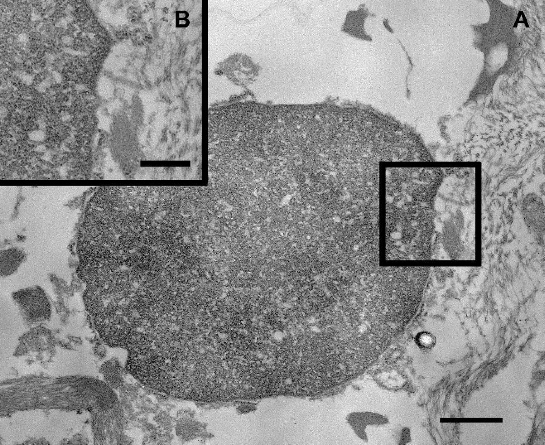
Transmission electron micrograph.
Footnotes
a.
Philips 208S TEM, FEI Co., Hillsboro, OR.