
Abstract
A 10-year-old female Egyptian fruit bat (Rousettus aegyptiacus) was evaluated for weakness and lethargy. Despite clinical improvement after supportive therapy, the bat was found dead the following day. Gross necropsy identified a mass associated with the duodenal wall and right renal cortex. Microscopically, the duodenal and gastric tunica muscularis and serosa and the right renal cortex were effaced by interlacing bundles of neoplastic spindle cells that were diffusely and strongly positive for vimentin and alpha-smooth muscle actin. The neoplastic cells also had mild to moderate cytoplasmic labeling for laminin and S100 and were negative for c-kit and desmin. On the basis of cell morphology and the immunophenotype, this tumor was diagnosed as a gastrointestinal leiomyosarcoma.
A 10-year-old female Egyptian fruit bat (Rousettus aegyptiacus) was evaluated for erratic flying and weakness. The bat was housed with a colony of Egyptian fruit bats and Jamaican fruit-eating bats (Artibeus jamaicensis). The bat was anesthetized with 5% isoflurane via facemask for a thorough physical examination and venipuncture. The bat weighed 115 g and was in good body condition. Physical exam revealed moderate dehydration. A possible hepatomegaly was noted upon abdominal palpation. The bat had decreased range of motion in the left elbow, which correlated with degenerative joint disease previously diagnosed by radiography. Supportive therapy included administration of lactated Ringer solution a (50 ml/kg subcutaneously), enrofloxacin b (4.35 mg/kg subcutaneously), dexamethasone c (0.87 mg/kg intramuscularly), and 50% dextrose d (7 ml/kg orally). Anesthetic recovery was normal, and the bat was noted to be active and eating well after the procedure. The bat was returned to the exhibit but was found dead the following day.
Immunohistochemical markers used in the characterization of a leiomyosarcoma in an Egyptian fruit bat (Rousettus aegyptiacus).*
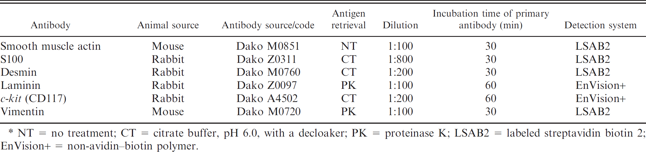
NT = no treatment; CT = citrate buffer, pH 6.0, with a decloaker; PK = proteinase K; LSAB2 = labeled streptavidin biotin 2; EnVision+ = non-avidin—biotin polymer.
Gross examination revealed a 3–4-mm, pale, tan abdominal mass associated with and infiltrating the duodenal wall and right renal cortex. The thoracic cavity contained approximately 2 ml of yellow serous fluid. The lungs were diffusely congested and had a small number of 1–2-mm white foci on the pleura of the caudal lobes (histologically consistent with pleural fibrosis). The hepatic parenchyma was diffusely friable and brown. Tissues were fixed in 10% neutral buffered formalin and submitted to the Animal Disease Diagnostic Laboratory at Purdue University (West Lafayette, IN) for histopathologic examination.
Microscopically, the small intestinal tunica muscularis and serosa were segmentally effaced by haphazardly arranged, interlacing bundles of infiltrative neoplastic spindle cells (Fig. 1). The neoplastic cells were elongate, with scant to moderate amounts of pale, eosinophilic cytoplasm and variably sized round to elongate, euchromatic nuclei that frequently contained a prominent nucleolus (Fig. 2). Twenty-six mitotic figures were observed in 5 high-power fields (400x) examined. The neoplastic cells focally infiltrated into the adjacent pancreatic parenchyma. The neoplastic cells were also observed segmentally effacing the gastric tunica muscularis and serosa and focally infiltrating the right renal cortex (corresponding to the grossly described mass associated with the right renal cortex). The subcapsular hepatic parenchyma contained a focus of acute coagulative necrosis surrounded by hepatocytes with moderate amounts of intracytoplasmic brown pigment. Bile canaliculi contained abundant bile, consistent with marked cholestasis.
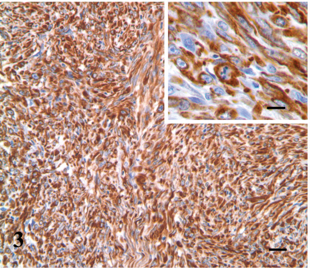
A tentative diagnosis of spindle cell sarcoma was made. Differential diagnoses included malignant schwannoma, leiomyosarcoma, and gastrointestinal stromal tumor (GIST). An immunohistochemical panel was performed with the use of previously reported standardized techniques for canine tissues, 6 which are summarized in Table 1. Immunostaining was performed with the use of an autostainer. e All antibody incubations were performed at room temperature. The immunologic reaction was detected with the chromogen substrate 3,3′-diaminobenzidine-H2O2 combination. Positive control tissues were canine tissues. Internal controls exhibiting identical immunolabeling to equivalent canine tissues were present in tissues from the fruit bat (vascular and intestinal smooth muscle: smooth muscle actin [SMA], vimentin, desmin; myenteric ganglia: S100; myenteric interstitial cells of Cajal: c-kit). The negative control tissues were treated with identical procedures, replacing the primary antibody with nonimmune immunoglobulins from the same species. More than 95% of the neoplastic spindle cells had strong, diffuse cytoplasmic labeling for alpha-SMA (α-SMA; Fig. 3) and vimentin (Fig. 4). Fewer neoplastic cells had weak to moderate cytoplasmic labeling for laminin (Fig. 5), and weak to moderate cytoplasmic, and less frequently nuclear, labeling for S100 (Fig. 6). No labeling for desmin or c-kit was observed. The morphology of the neoplastic cells and presence of strong and diffuse cytoplasmic labeling for vimentin and α-SMA supported a diagnosis of leiomyosarcoma.
Differential diagnoses for spindle cell sarcomas of the gastrointestinal tract can include leiomyosarcoma, malignant schwannoma, gastrointestinal stromal tumor, and fibrosarcoma. The positive labeling of the neoplastic cells for SMA ruled out a malignant schwannoma and fibrosarcoma and suggested a myogenic tumor. In addition, laminin was included in the immunohistochemical profile to distinguish fibrosarcoma, in which the neoplastic cells do not produce basement membrane, from leiomyosarcoma. 1 Gastrointestinal stromal tumors are neoplasms that are believed to arise from the interstitial cells of Cajal, the c-kit—positive and so-called pacemaker cells of the gastrointestinal myenteric plexus. Diagnosis of GISTs frequently relies on positive immunoreactivity to c-kit, a property of these neoplasms that is observed in more than 95% of human GISTs. 5 Gastrointestinal stromal tumors can also be concurrently positive for SMA and S100. 3 The lack of c-kit labeling in these neoplastic cells ruled out a GIST. Leiomyosarcomas traditionally have cytoplasmic labeling for SMA, vimentin, laminin, desmin, and variably, S100. 4 Desmin labeling of the described neoplastic smooth muscle cells in the current case was absent. Weak labeling for desmin was also noted in a microchip-associated leiomyosarcoma in an Egyptian fruit bat. 8 One study found that human leiomyosarcomas exhibited a much lower level of desmin immunoreactivity than leiomyomas; therefore, desmin immunoreactivity might correlate with malignancy. 7 This could, in part, explain the absence of desmin labeling in this tumor.
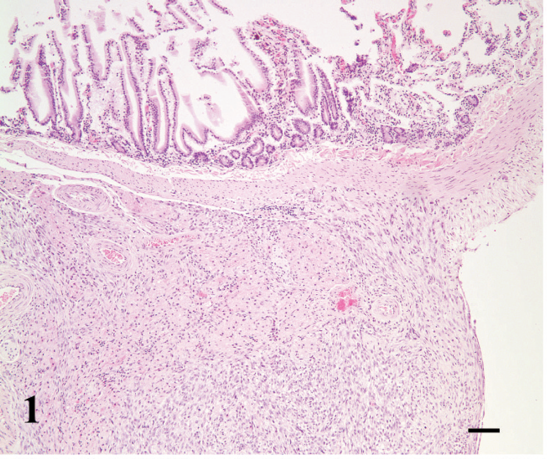
Duodenal mass, leiomyosarcoma; Egyptian fruit bat (Rousettus aegyptiacus). The neoplasm infiltrates the duodenal wall, segmentally effacing the duodenal muscularis externa and serosa. Hematoxylin and eosin. Bar = 167 μm.
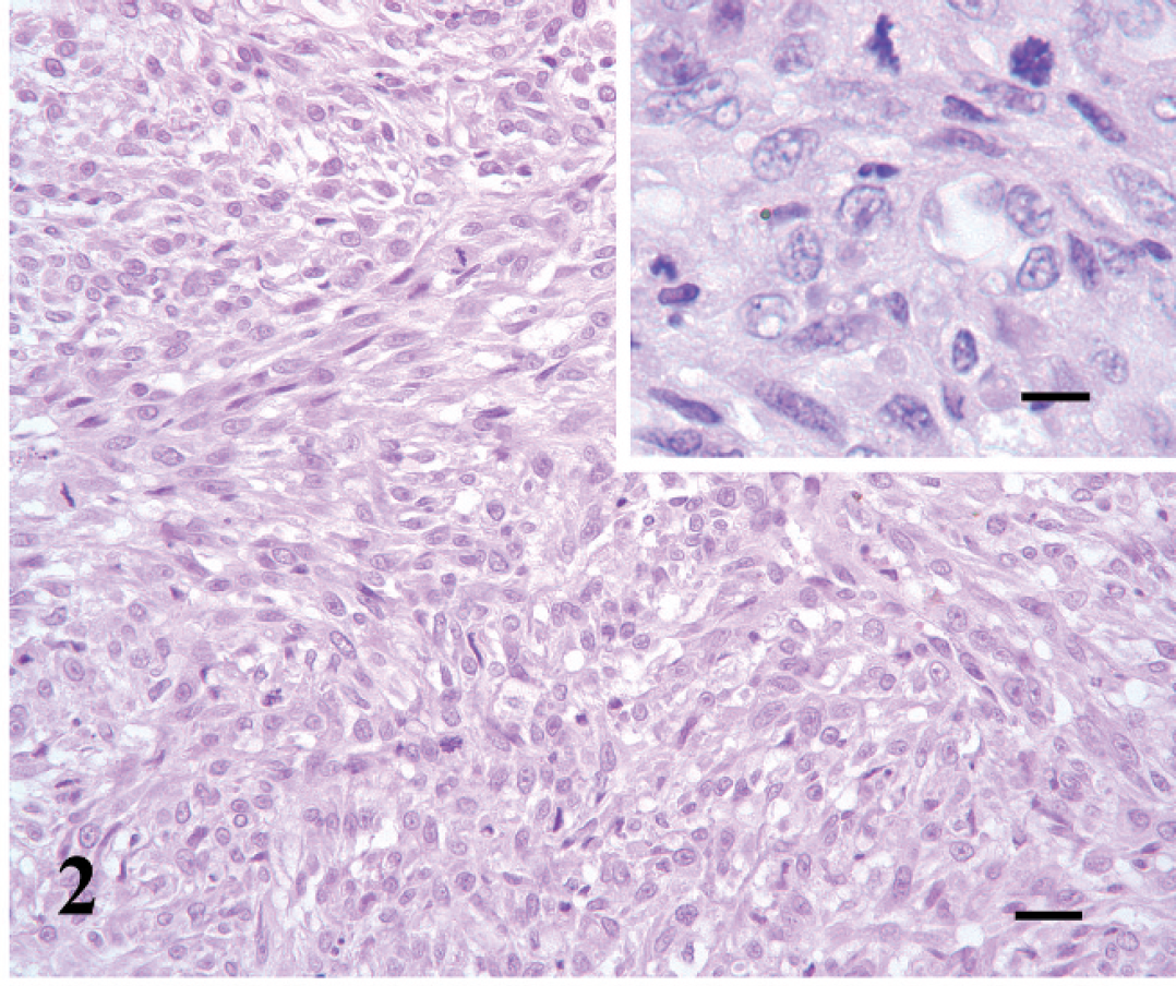
Duodenal mass, leiomyosarcoma; Egyptian fruit bat (Rousettus aegyptiacus). The neoplasm is highly cellular and composed of spindle-shaped neoplastic cells that form streaming and interwoven bundles interspersed with scant collagen. Hematoxylin and eosin. Bar = 42 μm. Inset: Bar = 16.8 μm.
Duodenal mass, leiomyosarcoma; Egyptian fruit bat (Rousettus aegyptiacus). Approximately 95% of the neoplastic cells have diffuse, strong cytoplasmic labeling for vimentin. Immunoperoxidase-3,3′-diaminobenzidine. Bar = 67 μm. Inset: Bar = 27 μm.
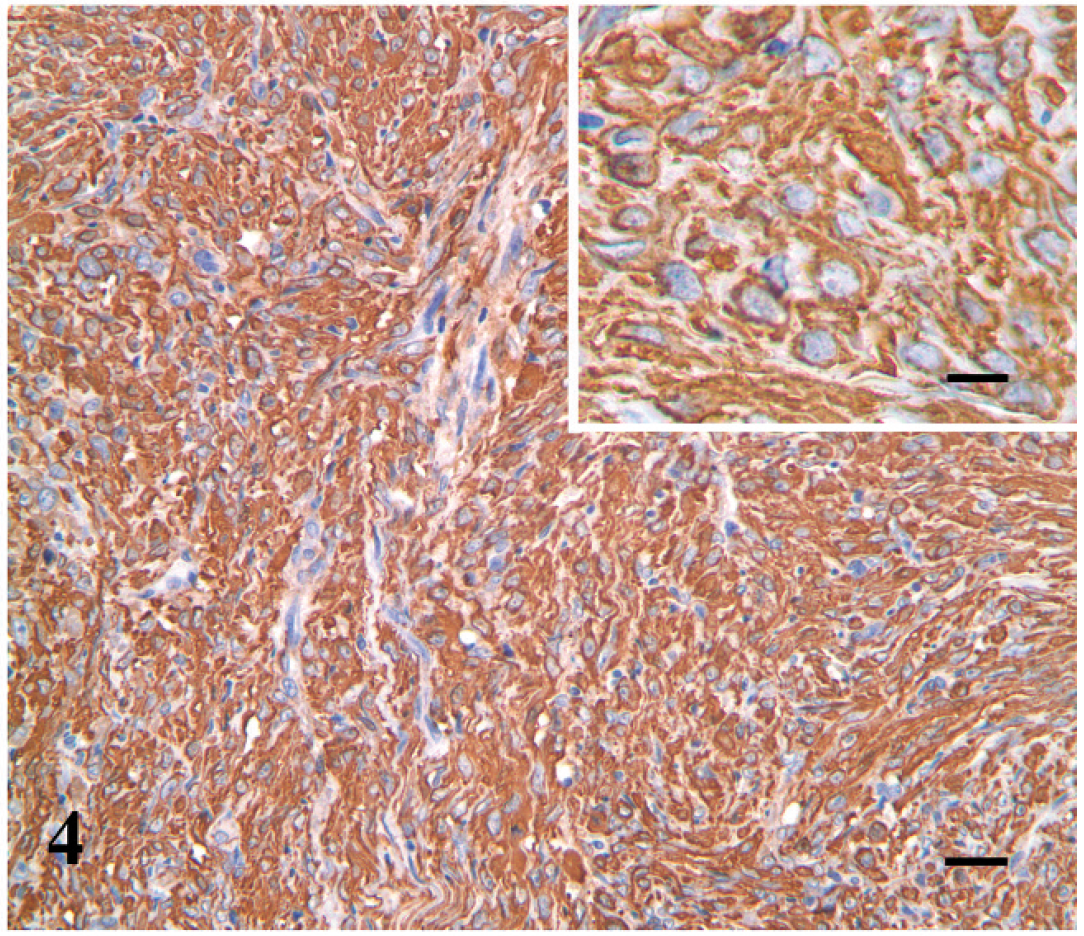
Duodenal mass, leiomyosarcoma; Egyptian fruit bat (Rousettus aegyptiacus). Approximately 95% of the neoplastic cells have diffuse, strong cytoplasmic labeling for alpha smooth muscle actin. Immunoperoxidase-3,3′-diaminobenzidine. Bar = 67 μm. Inset: Bar = 27 μm.
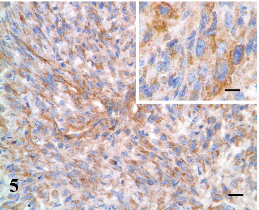
Duodenal mass, leiomyosarcoma; Egyptian fruit bat (Rousettus aegyptiacus). Some neoplastic cells have moderate cytoplasmic labeling for laminin. Immunoperoxidase-3,3′-diaminobenzidine. Bar = 67 μm. Inset: Bar = 27 μm.
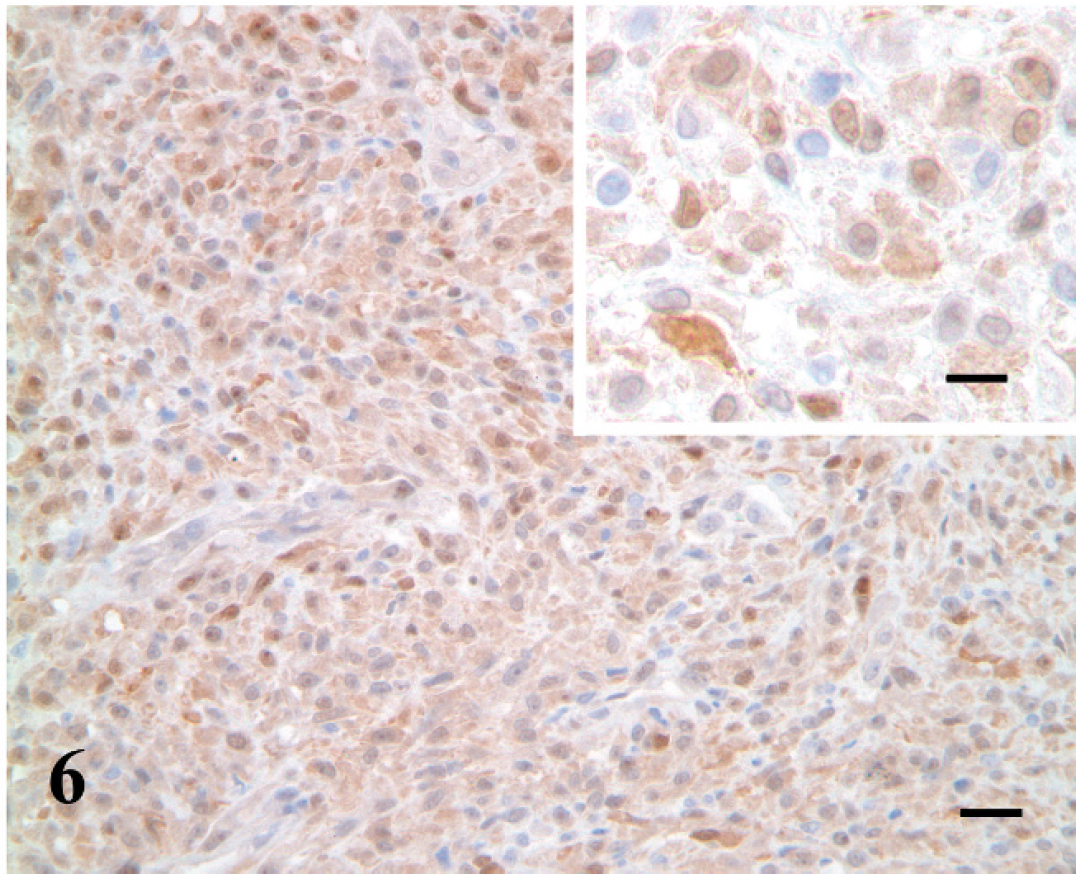
Duodenal mass, leiomyosarcoma; Egyptian fruit bat (Rousettus aegyptiacus). Few neoplastic cells have mild to moderate cytoplasmic and less frequent nuclear labeling for S100. Immunoperoxidase-3,3′-diaminobenzidine. Bar = 67 μm. Inset: Bar = 27 μm.
Postmortem examination in the aforementioned microchip case 8 also revealed metastatic foci on the diaphragm and the gastrosplenic and hepatoduodenal ligaments, in the connective tissue surrounding the adrenal glands, and throughout the hepatic parenchyma. In addition to the microchip-associated dermal leiomyosarcoma, other reports of leiomyosarcomas in chiropterans include a cutaneous leiomyosarcoma in a Townsend's big-eared bat (Corynorhinus townsendii; previously known as the long-eared bat [Plecotus townsendii virginianus]) 2 and a uterine leiomyosarcoma in a Seba's short-tailed bat (Carollia perspicillata). 8 However, it appears that information on diagnosis and treatment of malignant neoplasms in chiropterans is scant, and further research is warranted.
Acknowledgements The authors thank the animal health center staff and keeper staff at the Potawatomi Zoo for their assistance with this case and the care of the bats.
Footnotes
a.
Lactated Ringer solution, Hospira Inc., Lake Forest, IL.
b.
Baytril®, Bayer Health Care LLC, Shawnee Mission, KS.
c.
Dexaject®, IVX Animal Health Inc., St Joseph, MO.
d.
IVX Animal Health Inc., St. Joseph, MO.
e.
Dako North America Inc., Carpinteria, CA.