
Abstract
Chinchillas (Chinchilla lanigera) are known to be very sensitive to aflatoxins, and often a large number of animals die if toxicosis occurs. An outbreak of acute aflatoxicosis on a chinchilla farm in Argentina is described in the present study. A commercial feed suspected of causing the death of 200 animals was sampled. Livers from 9 dead chinchillas were analyzed for their macroscopic and microscopic characteristics via necropsy and histopathology. Aflatoxins B1, B2, G1, and G2 were determined, by thin layer chromatography, to be in the feed. Macroscopic inspection of livers revealed general enlargement, pale-yellowish coloration, hypertrophy, rounded borders, and increased friability. Size and color were remarkably different from a healthy organ. Histopathologic analyses of hepatic parenchyma showed severe, diffuse cytoplasmic vacuolation of hepatocytes. Sudan III staining confirmed the presence of lipid within the vacuoles. The feed was positive for aflatoxin B1 in quantities that exceeded the recommended levels. Histologic lesions were typical of aflatoxin intake. Monitoring feed for mycotoxins is crucial to prevent outbreaks of toxicosis, to improve management practices, and to diminish exposure risk of animals and humans to these harmful toxins.
Aflatoxins (AF) are a group of naturally occurring mycotoxins produced by Aspergillus fungi, especially Aspergillus flavus and Aspergillus parasiticus, which grow in a wide variety of improperly stored food commodities. 8 The ingestion of AF-contaminated food and feed is a matter of worldwide concern, which causes acute and chronic toxicosis in humans and domestic and laboratory animals. Aflatoxins are carcinogenic and are potent liver toxins. 4 Aflatoxins B1 (AFB1), B2 (AFB2), G1 (AFG1), and G2 (AFG2) are 4 naturally occurring forms of AFs, with AFB1 being the most potent, prevalent, and carcinogenic mycotoxin. Aflatoxin B1 has been classified as a 1B human carcinogen by the International Agency for Research on Cancer. 10,17 After ingestion, AFs are absorbed and transported to the liver via the circulatory system. They undergo biotransformation to the 8,9-epoxide derivative that form complexes with endogenous proteins and nucleic acids, which result in damage to the liver. 5,6,9,11
In all species, the liver is the primary target organ of acute injury caused by AFs. Acute aflatoxicosis is frequently associated with the ingestion of large doses of AFs, which cause typical hepatic changes, such as liver enlargement, yellowish color because of fat accumulation, and lipid vacuolation, which are confirmed by necropsy and histopathology. 4,13
Chinchillas (Chinchilla lanigera) are rabbit-sized crepuscular rodents native to the Andes Mountains in South America. Because of hunting, this species practically no longer exists in its natural habitat. Chinchillas are farm raised and are currently used by the fur industry and as pets. The growing international demand for chinchilla fur makes the breeding of these animals a highly profitable activity in Argentina. Many cases of acute aflatoxicosis have been reported worldwide in dogs, pigs, or cattle that consume naturally contaminated feed. 2,3,14 Chinchillas are known to be very sensitive to mycotoxins, and a large number of animals often die if acute aflatoxicosis occurs. Clinical signs that may indicate mycotoxicosis on a farm include low feed intake, diarrhea, weight loss, poor condition of the skin, fur discoloration, sudden death, and a predisposition to secondary infections (Labala J: 2008, Micotoxinas de chinchillas [Mycotoxins of chinchillas]. Available at: http://www.cuencarural.com/granja/chinchillas/micotoxinas-de-chinchillas. Accessed Jan. 16, 2008. In Spanish). However, data about aflatoxicosis in fur production animals has not been reported. The current study describes acute aflatoxicosis on a chinchilla farm in Argentina that caused the death of 200 animals. The aims of the present study were 1) to determine the AF concentrations present in feed, and 2) to evaluate macroscopic and histologic changes in the livers of dead chinchillas.
A 2-kg feed sample taken from a chinchilla farm located in the province of Córdoba, in the central region of Argentina, was received for laboratory analysis. Feed samples had undergone a pelleting process by an expander at 90°C for 60 min. This oat-based commercial feed was suspected to have caused the death of 200 animals on the farm.
All chinchillas were kept under the same husbandry conditions on the farm. In Argentina, a medium-sized chinchilla farm consists of approximately 170 animals. It takes approximately 3 years to reach this number of animals, with an estimated production of 360 furs a year. Chinchilla farming is a highly profitable business with major European consumers of pelts. In the present study, the hatchery had 200 animals that all received the same feed. All of these animals died naturally after the consumption of feed. The suspected etiology of disease was acute aflatoxicosis.
Feed was randomly sampled by taking small portions of material from different places of the storage bin; sampling was performed by the farm workers. In the laboratory, the feed was quartered to obtain smaller 500-g subsamples, which were milled and stored at —5°C until mycotoxin analyses. Two of the subsamples were analyzed for AFs as duplicates.
Aflatoxins B1, B2, G1 and G2 were determined in the chinchilla feed samples. These analyses were performed by thin layer chromatography (TLC), following published guidelines in the Official Methods of Analysis manual. 19 Mixed ground feed (25 g) was extracted with a solution that consisted of 125 ml of methanol:water (60:40 v/v), 80 ml hexane, and 2 g NaCl. The feed and extraction solution were shaken for 30 min in an orbital shaker. The mixture was passed through filter paper, a and 25 ml of the methanol:water phase filtrate was extracted twice with 25 and 15 ml chloroform, respectively. The chloroform phase was then vacuum dried by using a rotatory evaporator, and the extract was redissolved in 200 μl of benzene:acetonitrile (98:2 v/v). The extract was screened for AFB1, AFB2, AFG1, and AFG2 by spotting 2 μl, 5 μl, and 10 μl of each extract together with the toxins' standard solutions on silica gel 60 TLC aluminum sheets b (20 × 20 cm) and was developed with chloroform:acetone (90:10 v/v). The aluminum sheets were placed vertically into a glass chamber that contained the chloroform:acetone developing solvent mix in a volume such that only the very bottom of the sheet was covered in the liquid. The solvent mix slowly ascended the TLC plate by capillary action and developed the chromatogram. When the solvent has ascended the plate within 1–0.5 cm from top, it was removed from the incubation chamber. Chromatograms were air-dried and observed under 365-nm ultraviolet (UV) light. The relative amounts of AFs were quantitatively determined by visual comparison with standard solutions of known toxin concentration under UV light. Fluorescent spots that corresponded to AFs were identified in the chromatogram by comparing the Rf value of the spots present in the samples with the value of the standard solution of AF (Rf = distance traveled by the compound/distance traveled by the solvent front) and by their color under UV light (AFB1 and AFB2 = blue, AFG1 and AFG2 = green). Analyses of the pelletized feed for AFs by TLC revealed that the feed sample was contaminated at a mean level of 212 ppb ± 8.48 ppb of AFB1. Aflatoxins B2, G1, and G2 were not detected in feed samples.
Livers from 9 chinchillas that died naturally during the disease outbreak and livers from 9 healthy chinchillas slaughtered for commercial pelt recovery were analyzed for their macroscopic and microscopic characteristics via necropsy and histopathology. All of the dead chinchillas were from the aflatoxicosis outbreak on the same farm. Livers from healthy chinchillas were obtained from another farm that used AFB1-free feed. Macroscopic characteristics evaluated included the general size (lateral width), weight, and color of the livers. Liver tissue for histology was fixed in 10% neutral buffered formalin, trimmed, processed routinely, embedded in paraffin, sectioned at 5-μm thickness, and stained with hematoxylin and eosin (HE). Additional frozen sections of liver also were cut on a freezing microtome and stained with Sudan III to demonstrate fat. Histopathologic analysis evaluated hepatocellular characteristics such as cytoplasmic vacuolation, nodular hyperplasia, and bile-duct proliferation.
Macroscopic inspection of the livers revealed general enlargement, pale-yellowish coloration, hypertrophy, rounded hepatic borders, and increased friability (Table 1). Livers from chinchillas with aflatoxicosis were 38–71% larger than those from control animals. Livers from healthy animals taken from another farm (controls) were smaller and had sharp borders and a reddish-brown color. The color of the livers from chinchillas with acute aflatoxicosis was yellowish gray to pale yellow with gray spots (8 of 9 affected livers; Fig. 1). The average hepatic weight (mean ± standard deviation) of affected animals was 129.6 ± 23.5 g, whereas that for healthy livers was 83.6 ± 6.7.
Histopathology revealed severe, diffuse cytoplasmic vacuolation, with the appearance of many large and fewer small cytoplasmic vacuoles in hepatocytes in HE-stained tissue sections (Fig. 2). Frozen sections of liver stained with Sudan III confirmed the presence of lipid within the cytoplasmic vacuoles (Fig. 3).
Macroscopic characteristics of livers from healthy chinchillas (controls) and chinchillas with acute aflatoxicosis.
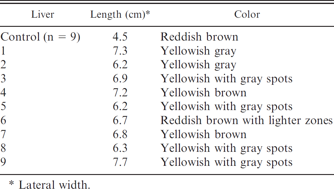
Lateral width.
Most studies of AFs mainly focus on chronic toxicosis and carcinogenic effects. In the current study, the acute hepatotoxic effects of naturally occurring AFB1 on the livers of 9 chinchillas that died from acute aflatoxicosis were investigated. Aflatoxicosis was confirmed by TLC analysis of the feed that the animals had consumed. This feed was positive for AFB1 in quantities (212 ± 8.48 ppb) that exceeded the putative safe concentrations of AF in animal feed and feed ingredients (10 ppb). 16
The diagnosis of mycotoxicosis is a common challenge for veterinarians, because the mycotoxin-induced disease syndromes can easily be confused with other diseases caused by pathogenic microorganisms. Histologic evaluation of the livers of affected animals and analysis of the feed for mycotoxin content are crucial to confirm the clinical diagnoses. Histopathology confirmed that the lesions found the 9 affected livers were typical of acute aflatoxicosis and are comparable with research findings in vivo after ingestion of AF-contaminated feed by experimental animals. In one such study, bile-duct hyperplasia, hepatocellular degeneration, fatty change of hepatocytes, and mononuclear-cell infiltration of the hepatic parenchyma were observed in broiler chickens fed 1 ppm AFs. 7,15 In a 2005 research study, broilers were fed a combination of AFs and fumonisins. The livers of affected birds were enlarged, yellowish, friable, and had rounded borders. 12 The HE-stained tissue sections were characterized by multifocal cytoplasmatic vacuolation, with a variable location within hepatic lobes. Hepatocellular damage manifested by marked cytoplasmic vacuolation and pyknotic nuclei was reported in a 2006 study of rats administered 2 mg/kg body weight of AFB1. 18
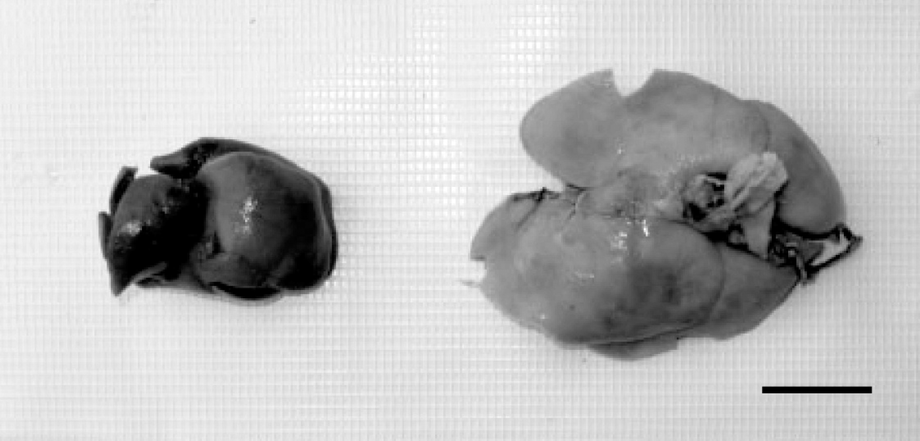
Liver of a healthy chinchilla (left) compared with the enlarged and lighter colored liver of a chinchilla that died of acute aflatoxicosis (right). Bar = 2 μm.
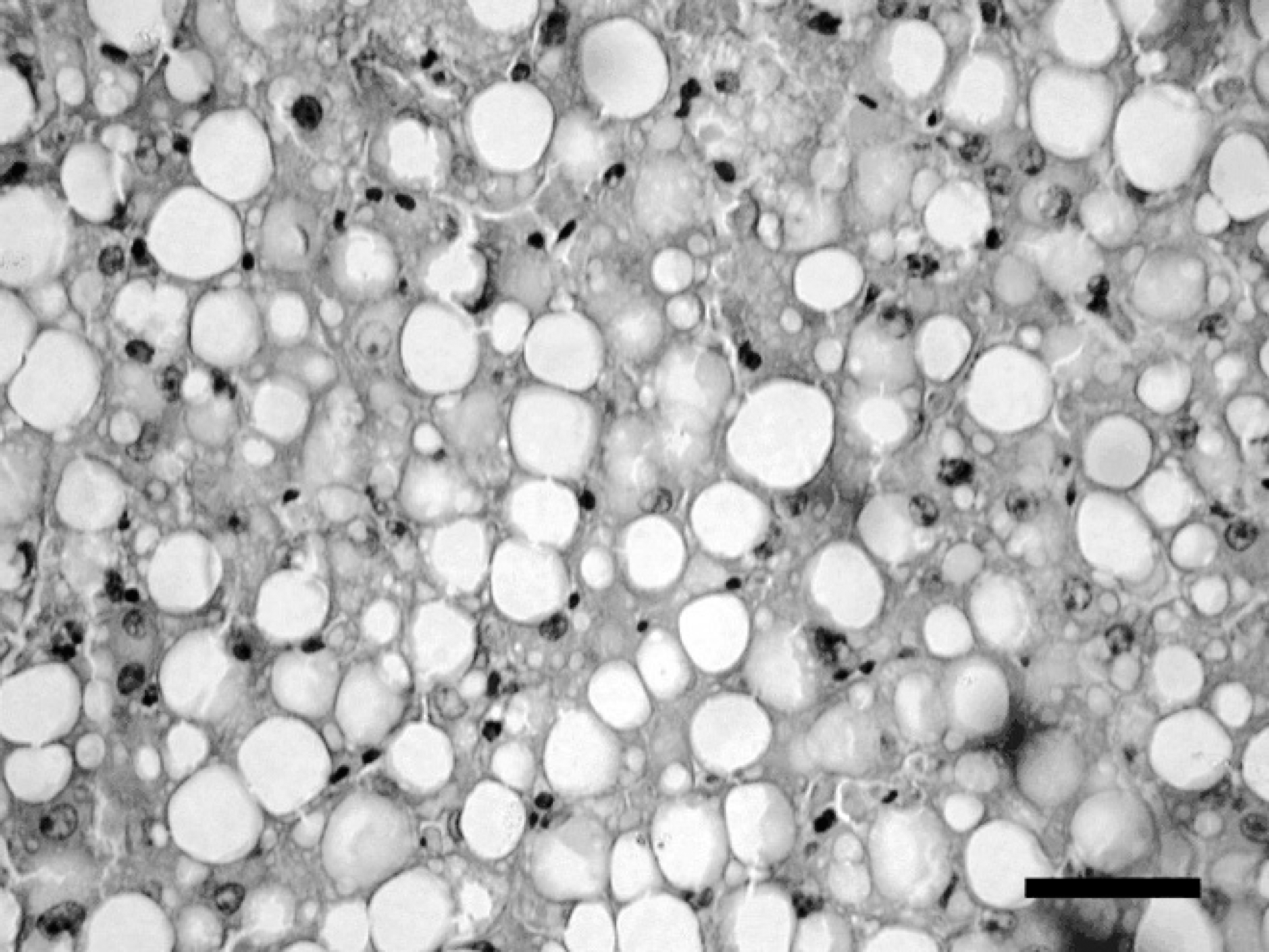
Diffuse cytoplasmic vacuolation of hepatocytes is present in a section of liver from a chinchilla with acute aflatoxicosis. Hematoxylin and eosin. Bar = 20 μm.
The liver is the primary target organ of acute injury from AF ingestion in all species. Although it is difficult to prove that a particular disease outbreak was caused by a mycotoxin, a number of reports in the veterinary literature are reasonably well documented. 4 A diagnosis of mycotoxicosis is usually made by feed analysis and histopathology because clinical signs of aflatoxicosis can be nonspecific and confusing.
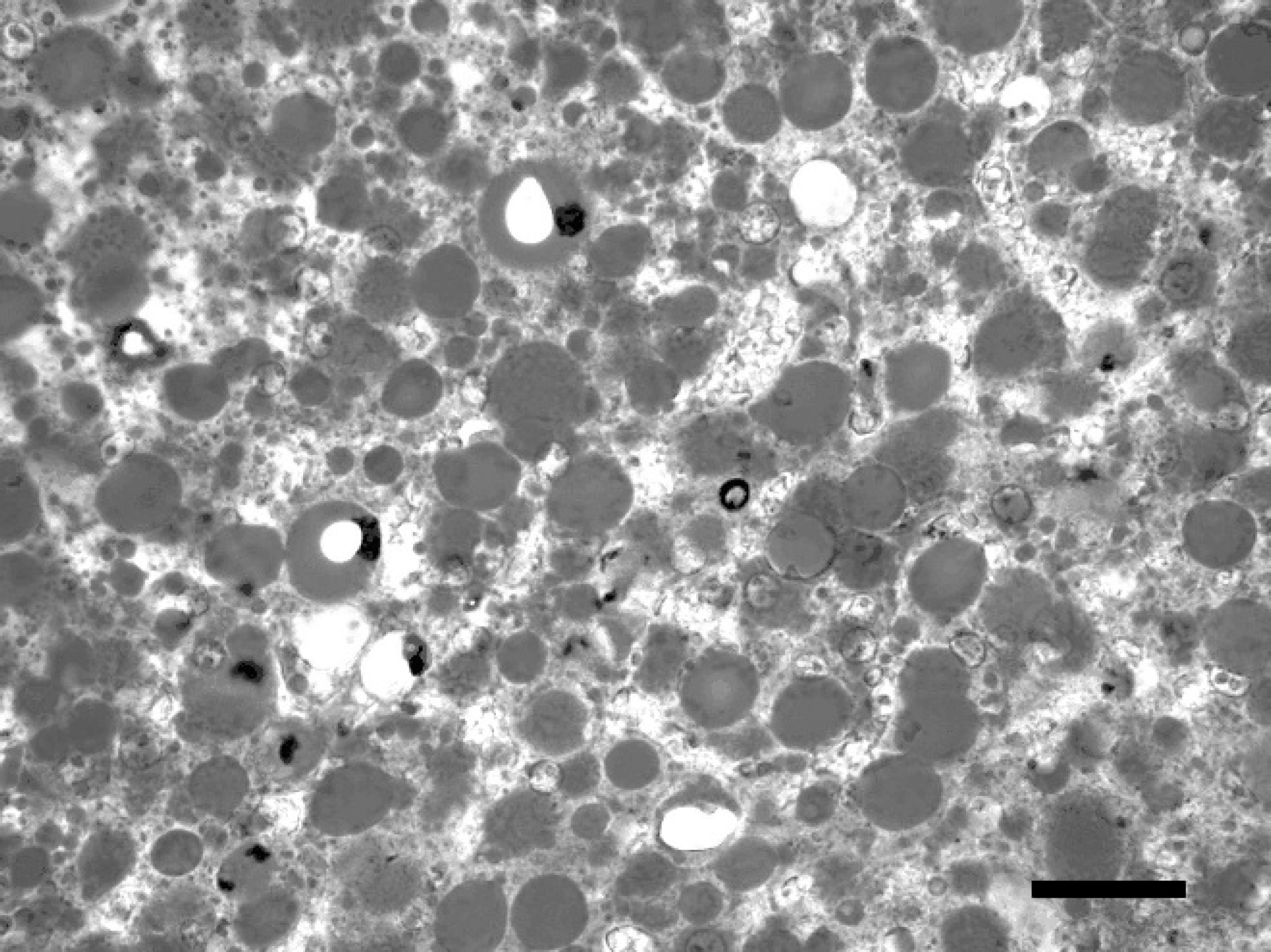
Hepatocellular cytoplasmic vacuoles stain positively for lipid in a frozen section of liver from a chinchilla with acute aflatoxicosis. Sudan III stain. Bar = 20 μm.
Acute cases of mycotoxicosis are infrequent. However, sublethal doses of mycotoxins produce a chronic toxicity that can result in cancer, primarily liver cancer, in some species. 1 Aflatoxins have elicited great public health concern because of their widespread occurrence in several dietary staples such as peanuts, tree nuts, corn, dried fruits, silage, and forages, all of which are used as animal feed ingredients. 4 Monitoring these substrates for mycotoxins, especially AFB1, is crucial to prevent outbreaks of acute mycotoxicosis, such as that presented in the current study, to improve farm management practices, and to diminish exposure risk of animals and humans to these harmful toxins.
Acknowledgements. This work was carried out, with thanks, by grants from SECYT-UNRC, FONCYTPICTO, and SPU-CAPES. The authors also thank Mr. Daniel Alaminos for his assistance in getting the feed samples and animal material.
Footnotes
a.
Whatman no. 4, Whatman Inc., Clifton, NJ.
b.
Merck KGaA, Darmstadt, Germany.