
Abstract
Two juvenile, intact, female mixed-breed goats from a common sire were presented for periodic neurologic deficits, seizures, and a generalized loss of body condition that occurred over a 4–6-week period. On physical examination, both goats were thin, obtunded, blind, and ataxic. Laboratory diagnostics revealed increased serum bile acids (95 μmol/l; reference interval: 0–50 μmol/l) in one of the goats. Both goats exhibited progressive physical and mental deterioration, and were eventually euthanized. Upon necropsy, no significant macroscopic lesions were noted. Microscopic examination, however, demonstrated hepatocellular atrophy and anomalies in the hepatic microvasculature, including duplication of hepatic arteries, small-to-indistinct portal veins, and oval cell hyperplasia. In addition, spongiform change was microscopically identified throughout the parenchyma of the brain, most notably within the white matter and along the junction of gray and white matter. The diagnosis of congenital portal vein hypoperfusion (suggestive of a portosystemic shunt) with resultant hepatic encephalopathy was proposed in each case based on the characteristic microscopic lesions in conjunction with the signalment and history of the goats. The observation that the affected kids were sired by the same buck suggests a hereditary basis for the condition in these animals as well.
Animals with portosystemic shunts (PSS) most commonly present with a general failure to thrive or central nervous system (CNS) signs attributable to hepatic encephalopathy (HE). 2,4,15 Congenital macroscopic PSS are the primary pathologic lesions associated with HE in the dog and cat. 1,4,11,16 In contrast, congenital PSS are less frequently identified in agricultural animal species (equids, pigs, oxen, small ruminants, and camelids), and HE is most commonly associated with other causes of hepatic insufficiency (e.g., toxic liver disease). 4,9,10,13,16 Although congenital PSS are acknowledged to have a familial link in some breeds of dog, no such genetic predispositions have been conclusively demonstrated in other species to date. 5,11,15,17,18 In the current study, congenital portal vein hypoperfusion suggestive of a hepatic portosystemic shunt is described in 2 juvenile goats sired by the same buck. To the authors' knowledge, this is the first report that suggests a hereditary basis for PSS in the goat.
In case 1, a 10-week-old, intact, female Nubian-cross goat from a flock of 35 was presented to the Veterinary Medical Center (VMC) at Colorado State University (Fort Collins, CO) in June 2006. History included gradual loss of body condition, periodic episodes of seizure-like activity, obtunded mentation, anorexia, and ataxia over a 4-week period. On physical examination, the kid had a body conditioning score (BCS) of 4 of 10, proprioceptive deficiencies, and bilateral cortical blindness, and was intermittently obtunded, with occasional episodes of head pressing. No significant abnormalities were identified in the analysis of the complete blood cell (CBC) count or serum chemistry panel, although it should be noted that neither serum bile acids nor ammonia levels were evaluated in this animal. The goat was treated with intravenous fluids, thiamine, vitamin E, nonsteroidal anti-inflammatory drugs, and antibiotics. After slight clinical improvement over 16 hr, the animal was discharged to the owner while awaiting further diagnostic results.
Fecal examination revealed occasional coccidia but no significant bacterial pathogens upon culture. Serum concentrations of selenium and vitamin E were within normal limits. Agar gel immunodiffusion serology for Caprine arthritis encephalitis virus was negative. Four days after discharge from VMC, this same kid had another seizurelike episode and was returned to the hospital laterally recumbent and nonresponsive. The blood glucose concentration was evaluated and was within reference intervals. The owner declined further diagnostics or treatment, and the goat was euthanized with intravenous pentobarbital.
In case 2, a 16-week-old, intact, female Nubian-cross goat from the same farm as the goat in case 1 was presented to the VMC. This goat was born within 5 days of the goat in case 1, and they shared a common Nubian sire, although each kid came from a separate, unrelated mixed-breed doe. This goat presented with a history of generalized loss of body condition and periodic episodes of obtunded behavior, anorexia, and ataxia over a 6-week period. Elevated serum bile acid concentration (95 μmol/l; reference interval: 0–50 μmol/l) was the only significant abnormality on the CBC count or serum chemistry panel. On physical examination, the goat had a BCS of 3 of 10, was markedly obtunded to somnolent, and had bilateral cortical blindness. The owner declined further diagnostics or treatment, and the goat was euthanized with intravenous pentobarbital.
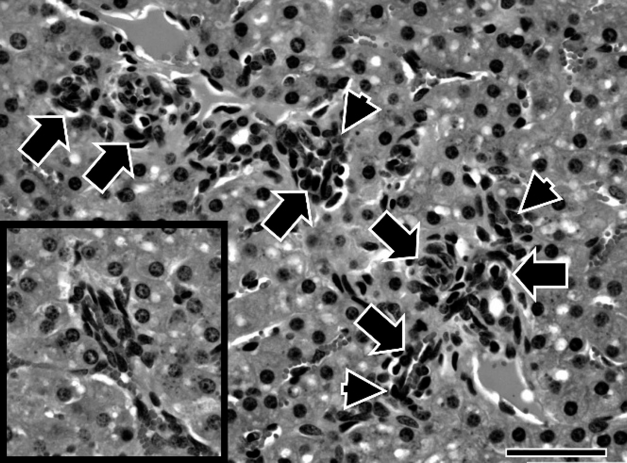
Representative liver section from goat in case 1. Two portal areas in close apposition have oval cell hyperplasia (arrowheads) and arteriolar duplication (arrows) that extend beyond the limiting membrane into the periportal hepatic parenchyma. Inset, increased numbers of small intralobular vessels (arterioles) are frequently identified in the affected goats. Hematoxylin and eosin. Bar = 50 μm. 400X.
The goat in case 1 had a female twin, and the goat in case 2 had a male twin. At the time of the present report, both twins to the affected goats were alive and were older than 2 years of age, with no clinical abnormalities. There are 31 other clinically unaffected, variably aged male and female goats (6 of which were sired by the buck in question) on the goat farm described in cases 1 and 2. The buck that sired the affected goats was never noted to have had any neurologic signs, although it was only used for a single breeding season and has since been sold; its present location is unknown.
Postmortem evaluation of the goat in case 1 revealed a subjectively small liver, although organ weight was not measured. No gross abnormalities were noted in the liver of the goat in case 2, and no other significant gross lesions were identified in either animal. All major organs, including liver and brain were collected in 10% neutral buffered formalin, routinely processed, and stained with hematoxylin and eosin. Grossly and microscopically normal liver from a 9-week-old, intact, female Nubian goat was similarly stained and used as an age-matched control.
On microscopic examination, the livers of the goats in cases 1 and 2 exhibited mild-to-moderate centrilobular and midzonal hepatocytic vacuolar change. Changes in the portal microvasculature were present in both goats but were more severe in case 1. The changes included small-to-indistinct portal veins, oval-cell hyperplasia, mild portal fibrosis, and increased numbers of portal arterioles that frequently extended beyond the limiting plate (Fig. 1). Arteriolar profiles were also increased within hepatic lobules (intralobular arteriolar hyperplasia; Fig. 1). In addition, hepatocytes and hepatic lobules from the goats in cases 1 and 2 were found to be significantly smaller in size (P < 0.0001) than those of the age-matched control. For hepatocellular size, 100 hepatocytes from zone 1 in the liver sections of case 1, case 2, and the age-matched control were measured across their widest points. a The mean zone 1-hepatocyte diameter was calculated to be 13.40 μm (standard deviation [SD]: 2.00 μm) in case 1, 13.70 μm (SD, 2.21 μm) in case 2, and 17.04 μm (SD, 2.88 μm) in the control goat. For the lobular size, the distance between the mid point of the portal area to the center of the central vein (lobular radius) was measured at 50 different sites in the liver sections from each of the 3 goats. a The mean lobular radius was calculated to be 365.7 μm (SD: 90.7 μm) in case 1, 412.2 μm (SD: 112.46 μm) in case 2, and 565.9 μm (SD: 161.12 μm) in the control goat. Because the variances of these measurements were significantly different (Levene's test), multiple Satterthwaite t-tests were used to compare the lobular and hepatocyte sizes between the 3 groups. b There was no statistically significant difference in hepatocyte or hepatic lobular size between the goats in cases 1 and 2 when using the Bonferroni-corrected significance level (P < 0.017). When considered in tandem, the hepatocellular atrophy and microvascular changes noted in both cases are characteristic for portal vein hypoperfusion. 15
On histologic examination, the brains of the goats in cases 1 and 2 had bilaterally symmetrical, marked spongiform change, most prominent at the white and gray matter junctions of the cerebral and cerebellar cortex and within the myelinated fibers of the internal capsule and medulla oblongata (Fig. 2). The spongiform change consisted of individual and coalescing discrete, clear vacuoles that ranged from 20 to 60 μm in diameter (Fig. 3). Single and small clusters of Alzheimer type II cells were also present within the neurophil and as neuronal satellites, and were most easily identified adjacent to areas of microcavitation in the cerebral cortex (Fig. 3). Inflammatory change to the CNS and neuronal involvement was minimal.
Disease conditions that can present with CNS signs in the goat include infectious encephalitides, transmissible spongiform encephalopathies, hepatic and renal encephalopathies, polioencephalomalacia, heavy-metal toxicoses, organophosphate toxicosis, copper toxicosis, copper deficiency, plant toxicoses, salt intoxication, hypoglycemia, hypoxia, nervous coccidiosis, trauma, vascular accidents, congenital CNS malformations, urea and/or ammonia supplementation and/or toxicosis, or any one of numerous possible metabolic and/or storage disorders. 4,8,12,13,16 Despite an inability to identify a macroscopic shunt, the age of onset along with the cumulative clinical, laboratory, and microscopic findings from the goats in cases 1 and 2 are consistent with the etiologic diagnoses of congenital PSS and HE, as previous described in the goat. 4,9,13
Although both macroscopic and microscopic congenital PSS have been described in several veterinary species, macroscopic PSS are far more commonly reported. 6,10,15 Congenital macroscopic PSS are vascular bypasses (usually singular) of the portal vein to the systemic circulation and are typically categorized as intrahepatic or extrahepatic shunts. 3,4,15 Intrahepatic shunts occur within the hepatic parenchyma and primarily result from the persistence of some embryologic structure, most commonly the ductus venosus. 3,15 Extrahepatic shunts exist outside of the hepatic parenchyma and most commonly involve an anastomosis (or anastomoses) between the portal vein and caudal vena cava or the portal vein and azygous vein. 3,4,15 Large, singular extrahepatic and intrahepatic shunts are frequently recognized on gross examination, whereas smaller, peripheral, and branching shunts are often best appreciated through the use of antemortem imaging modalities, such as portography, computed tomography, ultrasound, and nuclear scintigraphy. 1,3,10,11,14,16 Microscopic changes to the liver typically associated with congenital macroscopic PSS in veterinary species consist of portal arteriolar hyperplasia (duplication), hepatocellular hypoplasia or atrophy, and portal venous hypoplasia or atrophy. 3,15,16
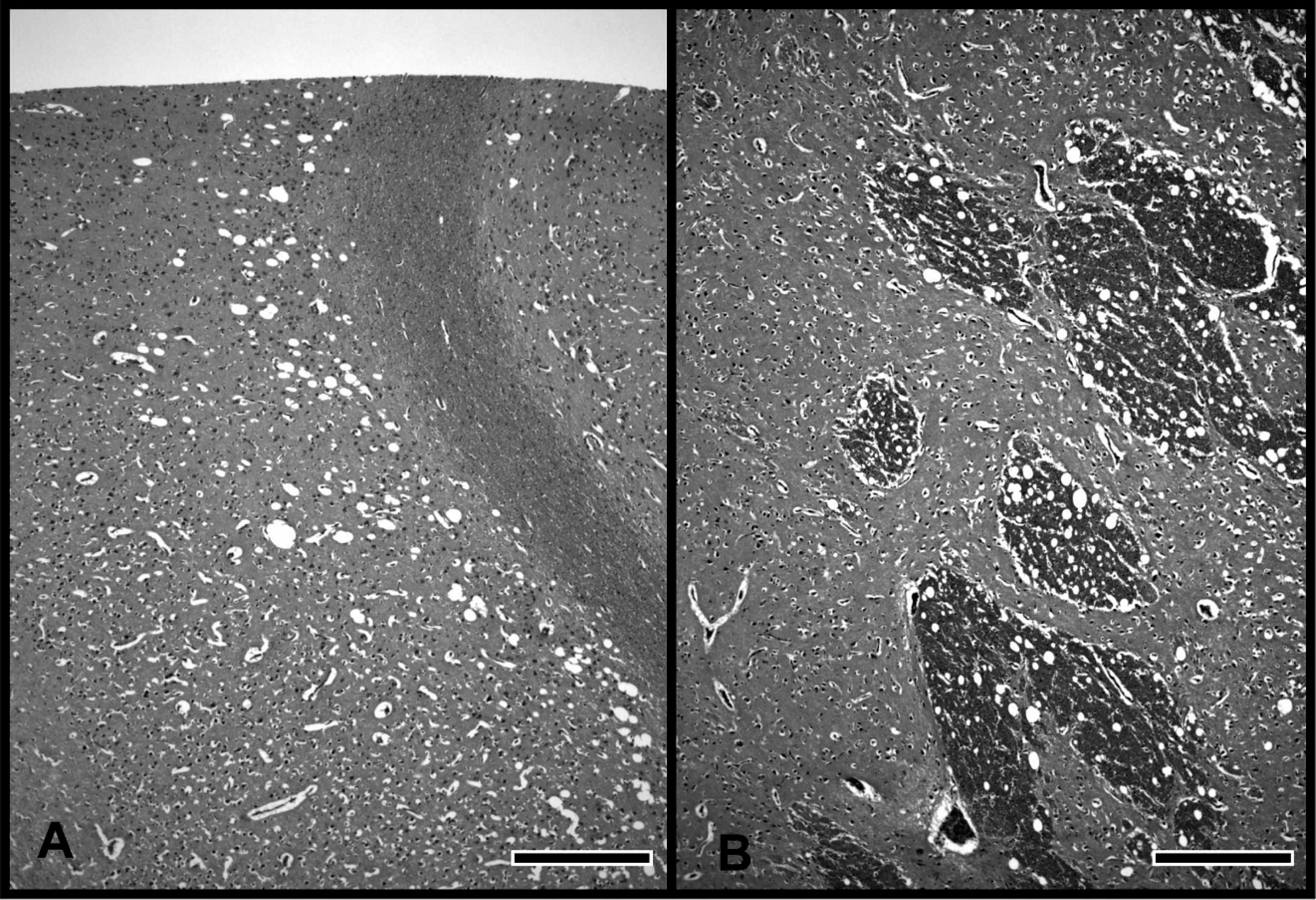
Representative brain section from goat in case 1.
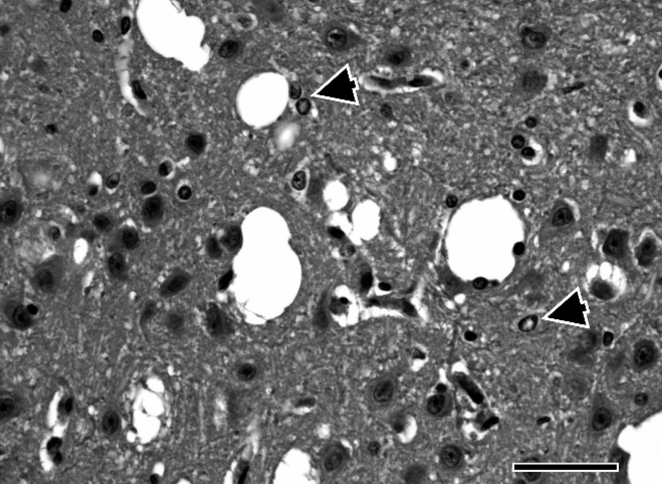
Representative brain section from goat in case 1. Spongiform change consists of single and coalescing vacuoles, along with the presence of Alzheimer type II astrocytes (arrowheads). Hematoxylin and eosin. Bar = 50 μm. 400X.
Congenital microscopic PSS, alone or in conjunction with macroscopic PSS, are not well described in the veterinary literature. Within agricultural animal species, only a single report that involved a calf and one report that involved a foal could be identified in which the diagnosis of PSS was solely attributed to microvascular anomolies. 10 In both, the calf and the foal, no macroscopic PSS could be identified by using antemortem imaging diagnostics or on postmortem examination. In addition, the livers from both these animals were similarly described as having high numbers of small, thin-walled intrahepatic vessels, which may have functioned as microscopic shunts. 10
In the companion animal species, congenital microscopic PSS have been most commonly reported as a hepatic microvascular dysplasia (HMD) of the dog. 1,2,5,11,14,15 Hepatic microvascular dysplasia, as identified in the literature, consists of microscopic hepatic changes similar to those of macroscopic PSS, with the additional key feature of prominent intralobular “juvenile-like” vessels, which have themselves been suggested to represent the microscopic shunts in these animals. 1,2,11
The standing of HMD as a distinct pathologic disorder, however, was recently rebuked by a veterinary liver standardization group after a thorough reexamination of the original literature that documented the disease. 3 Based on their collective findings, this group concluded that HMD in the dog actually represents the congenital condition of primary portal vein hypoplasia (PPVH) in which the subtle lesions of the portal vein have gone unappreciated. Whereas PPVH has as yet only been recognized in the dog, cat, and laboratory rat, 3,7,15 the findings of this standardization group perhaps bring into question all previous veterinary diagnoses of microscopic PSS in which portal-vein measurement was not performed. Consistent with this statement is the observation that the diagnosis of microscopic PSS has often been made as a rule-out diagnosis through the identification of specific microscopic liver changes in the absence of a demonstrable macroscopic PSS on antemortem and/or postmortem examination. 2,11 Further caution with regard to the diagnosis of PSS is perhaps best illustrated by the fact that some animals diagnosed with microscopic PSS through rule-out methods have subsequently been reexamined by using more contemporary modalities only to find no demonstrable shunts. 1,11,14
Hematologic and biochemical abnormalities often observed in animals with portosystemic shunts include anemia, increased liver enzyme activities, hypoglycemia, hypocholesterolemia, and low blood urea nitrogen concentration. Elevated concentrations of bile acids or ammonia in the brain, cerebral spinal fluid, and serum support the diagnosis of portosystemic shunt. 2,4,13 The clinical pathology of animals diagnosed with HMD and microvascular shunts has generally been less striking than that of animals diagnosed with macroscopic PSS, and, in some cases, only mildly increased concentrations of serum bile acids or ammonia levels were reported in the absence of other abnormalities. 1,2,10,11
Hepatic encephalopathy, as mentioned earlier, can result from various causes of hepatic insufficiency. In some reports, the age of clinical presentation and severity of neurologic signs has correlated well with the degree of hepatic compromise. 15 Hepatic encephalopathy varies in microscopic appearance between species, although it is generally described to be a bilaterally symmetrical spongiform change in the CNS white matter, most prominent within myelinated tracts and along white and gray matter junctions. Swollen astrocytes, termed Alzheimer type II cells, can also be associated with HE, depending on the species. 4,9,16 Clinical signs of HE vary with species as well, although most signs localize to the prosencephalon (i.e., changes in mentation, cortical blindness, and ataxia), despite the widespread distribution of spongiform lesions. 13,16
Although the pathogenesis of HE is not yet fully elucidated, substances thought to play key roles in the development of HE include ammonia, mercaptans, phenols, aromatic amino acids, and short- and medium-chain fatty acids, with other monoamines and manganese mentioned less frequently. Although many of these compounds are known to be directly cytotoxic, much of the CNS pathology in HE is thought to occur through more convoluted mechanisms, including alteration of the blood-brain barrier, disturbance of osmotic regulation, and production of false neurotransmitters. 4,6,12,16
In summary, 2 goat kids from a common sire demonstrated similar neurologic signs and a generalized loss of body condition beginning between 6 and 10 weeks of age. Of interest, of the 2 goats examined, the more severely affected goat presented at a younger age (case 1). The clinical, laboratory, and postmortem findings identified in these cases suggested HE that resulted from diffuse portal vein hypoperfusion as the underlying cause of morbidity in both goats. Given that the microscopic changes identified in the livers of both goats were consistent with the histopathologic lesions reported in caprine PSS, 4 it was tempting to propose PSS as an etiologic diagnosis for both of these animals. However, because no macroscopic shunts were identified on gross examination of the 2 goats and no antemortem imaging diagnostics were used in either case, the term PSS was precluded from the final diagnoses. The fact that the portal vein was not exhaustively examined in either of the affected goats perhaps further warrants a conservative diagnostic approach in these 2 cases, especially in light of the proposed misdiagnoses of canine PPVH for HMD, as previously described. As such, whereas the ultimate cause of the liver lesions in these 2 goats must be limited to the generic diagnosis of portal vein hypoperfusion, it, nonetheless, is noted that the changes seen are consistent with PSS in the goat. 4
Based on the epidemiologic, clinical, and postmortem findings described in both goats, it is proposed that the origin of the hepatic abnormalities identified in these animals is likely to be congenital in nature. Furthermore, based on the observation that both of the affected kids shared a common sire, it is also suggested that this hepatic condition may be genetic as well. Although little can be formally concluded from the limited number of cases examined, what can be stated is that the distribution pattern of clinically affected animals in the current report is not inconsistent with those of PSS and HMD (PPVH) in some breeds of dog for which an autosomal and polygenic mode of inheritance has been suggested. 2,14,17,18
In conclusion, HE that results from congenital portal vein hypoperfusion, and perhaps even congenital PSS, should be considered as a differential diagnosis in young goats that present with chronic, periodic CNS deficits and may, in fact, remain a viable diagnosis even in cases where multiple, interrelated goat kids are affected.
Acknowledgements. The authors wish to thank all Colorado State University contributors, including Dr. Barbara Powers at the Veterinary Diagnostic Laboratory, Brad Charles and Dr. E.J. Ehrhart at the Animal Cancer Center, Charlie Kerlee at the C.A.T.S. Laboratory, and Zonglin He at the Department of Statistics for their assistance with this project.
Footnotes
a.
Zeiss AxioVision® 4.6 microscope with Axioplan® 2 software, Carl Zeiss IMT Corp., Maple Grove, MN.
b.
SAS® statistical package version 9.1, SAS Institute, Cary, NC.