
Abstract
Between 1999 and 2012, 11 cases of congenital portosystemic shunts (cPSS) resulting in hepatic encephalopathy were diagnosed in goat kids necropsied at the California Animal Health and Food Safety Laboratory System and at the Department of Pathology, Immunology & Microbiology, School of Veterinary Medicine, University of California–Davis. Affected animals included 6 females and 5 males of various breeds including Boer (5/11), Nigerian Dwarf (1/11), Saanen (1/11), Toggenburg (1/11), and mixed-breed (3/11) aged between 1.5 months and 11 months, submitted live (2/11) or dead (9/11) for necropsy. The most frequent clinical signs in these goats were ataxia, blindness, tremors, head bobbing, head pressing, seizures, circling, weakness, and ill thrift. Bile acids were measured in 2 animals, and were elevated in both cases (134 and 209 µmol/l, reference interval = 0–50 µmol/l). Necropsy findings were poor to fair body condition. Grossly, the livers of 4 animals were subjectively small. Microscopic lesions included portal spaces with increased numbers of arteriolar profiles and hypoplastic or absent portal veins, diffuse atrophy of the hepatic parenchyma with the presence of small hepatocytes and, in some cases, multifocal hepatocellular macrovesicular vacuolation. In the brain and spinal cord of all animals, there was bilateral and symmetric spongy degeneration affecting the cerebrum, mesencephalon, cerebellum, brainstem, and cervical spinal cord. In all cases, the brain lesions were consistent with hepatic encephalopathy. Congenital portosystemic shunts should be considered in the differential diagnosis of young goats with a history of ill thrift, and nonspecific neurological signs.
Portosystemic shunts are congenital or acquired vascular anomalies that allow portal blood to enter the systemic circulation without passing through the liver. 4 Congenital portosystemic shunt (cPSS) is the most common cause of hepatic encephalopathy (HE) in young dogs and cats.6,14,18 In large animals, spongy degeneration (status spongiosus) of the central nervous system (CNS) due to cPSS/HE has been sporadically reported in calves,10,18,20,26 piglets, 26 foals,3,5,10,16 1 alpaca cria, 13 goat kids,2,12,29 and a neonatal roan antelope. 17 The present report describes the clinical history and pathological findings of 11 cases of cPSS/HE in goat kids.
Between 1999 and 2012, 11 cases of cPSS causing liver failure and HE were diagnosed in goat kids submitted to the California Animal Health and Food Safety (CAHFS) Laboratory System and to the Department of Pathology, Immunology & Microbiology (PMI), School of Veterinary Medicine, University of California (UC Davis), Davis, California for necropsy. Affected animals included 2 live and 9 dead goat kids, 6 females and 5 males, of different breeds including Boer (5/11), Toggenburg (1/11), Saanen (1/11), Nigerian Dwarf (1/11), and mixed-breed (3/11), mean age 3.8 months (range = 1.5–11 months), and mean body weight of 11.5 kg (range = 6.5–18 kg). The frequency of clinical signs seen in these animals were ataxia (4), head pressing (4), blindness (3), general weakness (3), head bobbing (3), ill thrift (3), inappetence (2), seizures (2), stargazing (2), tremors (2), dyspnea (2), circling (2), drooling (2), opisthotonus (1), paddling (1), recumbency (1), and teeth grinding (1). At necropsy, the general body condition ranged from poor to fair. In 4 animals livers were subjectively small.
In the 2 animals in which bile acids were measured, these were elevated (134 and 209 µmol/l, reference interval = 0–50 µmol/l). 11 All major organs, including liver and brain, were collected from all animals at necropsy and fixed in 10% neutral buffered formalin, routinely processed for histopathology, and embedded in paraffin wax; 4-µm-thick sections were cut and stained with hematoxylin and eosin. Microscopic findings in the liver included disruption and disorganization of lobular architecture and lobules with undersized portal tracts containing multiple arteriolar profiles or duplication of arterioles. The distance (in µm) from the center of the central vein to of the adjacent mid portal area was measured in 10 lobules each from 7 goat kids aged 3–4 months. Three kids were affected with cPSS, and 4 were age- and breed-matched (Saanen and Boer) controls who were not affected with cPSS. Liver lobule size in affected versus unaffected goat kids was compared a using mixed-model linear regression, with case (yes/no) as the independent variable and animal ID as a random effect to account for the lack of independence among liver lobule measurements from the same animal. Liver lobules from affected goats did not significantly differ from those of unaffected goats (P = 0.50). The average distance from central vein to adjacent midportal area was 333.2 µm (standard error [SE] = 23.6 µm) in unaffected goats and 312.2 µm (SE = 31.2 µm) in affected goats.
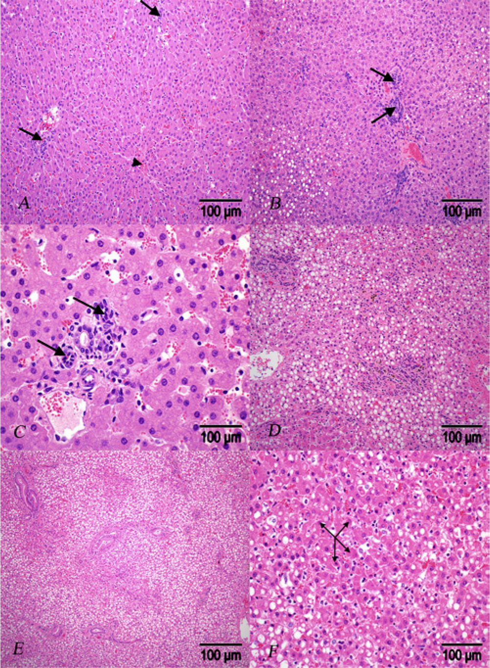
In 3 of the 11 goat kids, there was mild to moderate bile duct hyperplasia and mild to moderate periportal fibrosis. In 3 goats, portal venules were indistinguishable, and, in all animals, there was mild to severe hepatocellular macrovesicular vacuolar change interpreted as fatty change (Fig. 1B–F). In the brain and cervical spinal cord of all 11 animals there were varying degrees of widespread, bilaterally symmetrical multifocal to coalescing clear, discrete vacuoles up to 50 microns in diameter. In the cerebral cortex, this vacuolation was consistently present in the deep gray matter adjacent to the white matter (gray and white matter interface; Fig. 2A, 2B, 2D–F). In 4 goats, astrocytes within the cerebral cortex and in the adjacent white matter had undergone Alzheimer type II change (Alzheimer type II astrocytosis), arranged in single or pairs, with open-faced, enlarged clear nuclei, and contained a rim of chromatin and a tiny eccentric nucleoli (Fig. 2C). The Alzheimer II change is associated with a loss of immunohistochemically detectable glial fibrillary acid protein.
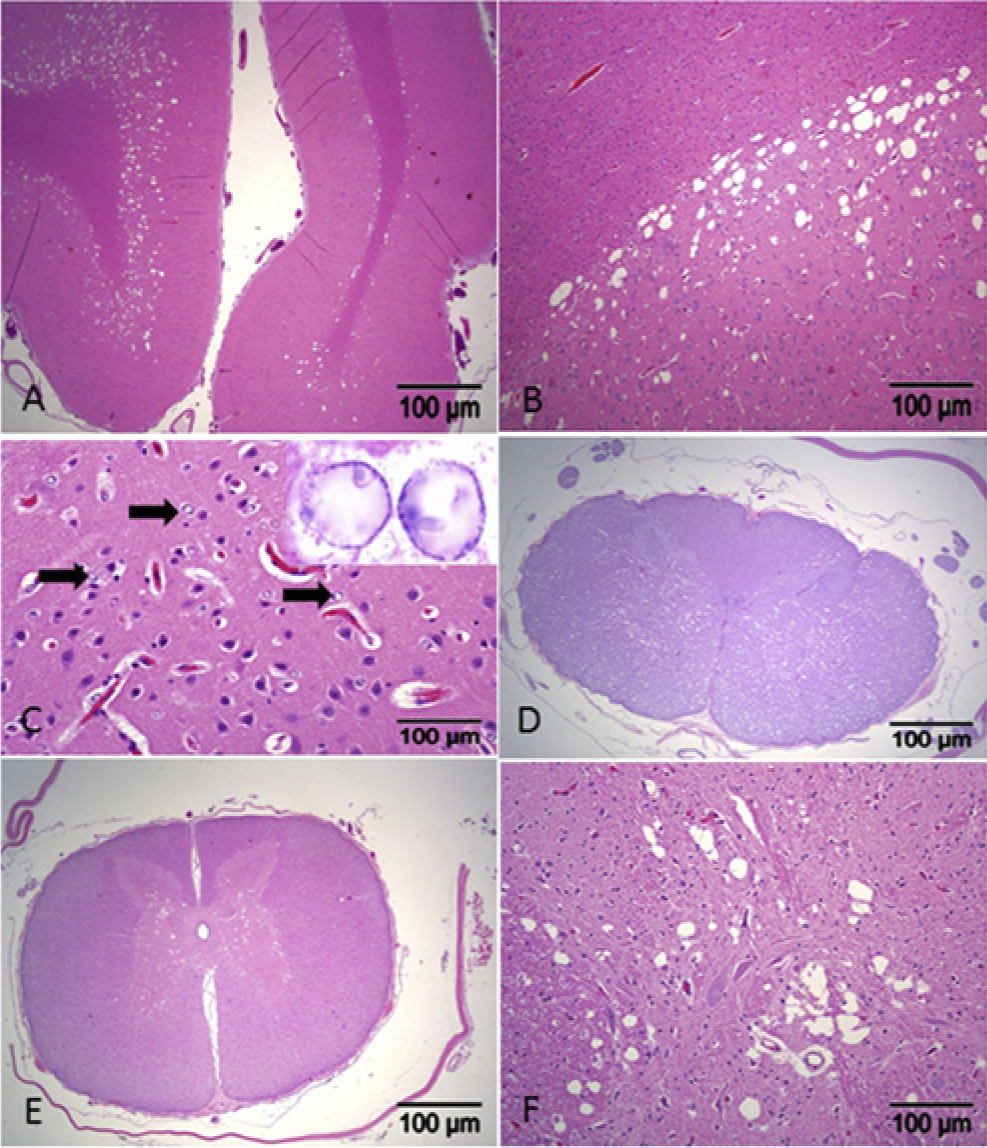
In 5 animals, there were variable degrees of symmetric and bilateral vacuolation of the white matter of the cerebellum and gray matter of the caudal brainstem. In 3 of these 5 animals there was axonal swelling with dilation of axon sheaths and occasional empty, swollen axon cylinders, some of these containing foamy, plump macrophages (gitter cells). Vacuolation of the spinal cord was of variable distribution and severity. It was severe, multifocal, affecting both white and gray matter in 1 animal; it involved only the gray matter in 2 animals; and it affected the gray matter and the gray–white matter interface in 2 other animals.
Incidental histological findings included segmental, mild enteritis in the small intestine of 7 animals associated with small numbers of intracytoplasmic coccidia (Eimeria spp.) in enterocytes. In the lungs of 3 animals, there was mild to moderate lymphocytic perivascular and peribronchiolar infiltrates, and alveolar spaces were filled with eosinophilic material interpreted as protein rich edema fluid, scattered macrophages, and small numbers of intralesional short bacterial rods. Mannheimia haemolytica and Bibersteinia trehalosi were isolated from the affected lungs. The liver copper and selenium levels by wet weight basis in 7 of the 11 animals were determined. Copper levels were low in 3 goats (2.3, 12, and 13 ppm; reference range = 25–150 ppm 22 ); and selenium levels were marginal in 2 goats (0.14 and 0.24 ppm; reference range = 0.25–1.5 ppm 22 ).
To evaluate whether cPSS was associated with breed, data (including breed and presence or absence of cPSS) on 1,067 carcasses of goat kids less than 1 year of age submitted at CAHFS for necropsy between 1999 and 2012 were analyzed. Breeds of goat kids submitted for necropsy included Alpine (107), Boer (245), LaMancha (39), Nigerian Dwarf (43), Nubian (39), Pygmy (74), Saanen (57), Toggenburg (46), cross-breed (152), breed not reported (186), and other purebreds (39). Congenital portosystemic shunt was found in Boer (5), Nigerian Dwarf (1), Saanen (1), Toggenburg (1), and cross-breed (3) kids. The breed categories Nubian and Anglo-Nubian were combined into a single category (Nubian). Breed categories with fewer than 20 accessions were combined into a single category (Other). The hypothesis that occurrence of cPSS differed significantly according to the breed was tested using a Fisher’s exact test. Analysis indicated that occurrence of cPSS did not differ significantly by breed (P = 0.26). The category “other” included the following breeds: Angora (n = 12), cashmere (n = 1), Kiko (n = 1), Kinder (n = 3), Oberhasli (n = 15), other (n = 3), Pygora (n = 2), Spanish (n = 1), and Tennessee Fainting (n = 1).
In general, HE results in episodic prosencephalic signs such as seizures, ataxia, depressed mentation, aimless walking, and head pressing. 30 In all species, the microscopic lesions consist of spongy changes due to intramyelinic edema resulting in splitting and vacuolation of myelin sheaths, and the presence of Alzheimer type II astrocytes, the latter being found only in horses, goats, and llamas with hepatic failure.9,21,29,30 Though one would expect to observe episodic prosencephalic signs in HE, there is only a single report of hepatic encephalomyelopathy in a calf exhibiting only cervical spinal cord signs with portosystemic shunts and CNS lesions characterized by extensive dilation of the myelin sheaths. 9 In the current study, while spongy change is a common feature in all goats, abnormal astrocytes were seen in only 4 animals. It is possible that, similar to calves, goats may have a variation in the clinical presentation and CNS lesions in cases of HE due to cPSS.
The pathogenesis of HE is not well elucidated but is generally accepted that in cases of liver failure, bypass, or injury to hepatocytes, ammonia, and other toxic substances in the systemic circulation are allowed to reach the CNS. 27 Hepatic encephalopathy is caused either by a portosystemic shunt or as a result of an infectious, metabolic, toxic, or degenerative liver disease. Ammonia is produced in the gastrointestinal tract by the bacterial degradation of amines, amino acids, purines, and urea. In healthy animals, ammonia is detoxified in the liver by conversion of ammonia to urea by the ornithine citrulline arginine urea cycle. Ammonia and other toxic substances increase the permeability of the blood–brain barrier leading to vasogenic edema resulting in status spongiosus (spongy degeneration of the CNS). Some of the neurotoxic effects of ammonia are altering the transit of amino acids, water, and electrolytes across the neuronal membranes, and inhibition of the generation of both excitatory and inhibitory postsynaptic potentials in neurons. The major role of astrocytes is to regulate fluid and electrolyte balance in the CNS, hence the primary cell type to be affected in HE and the component of the pathogenesis of the disease; Alzheimer type II astrocytosis develop in those cases. 30
Though the clinical signs associated with portosystemic shunts in large animals are usually nonspecific, the general consensus regarding its pathogenesis is that hyperammonemia affects the astroglial cells leading to brain edema causing neurological signs.13,18 Spongy degeneration of the CNS due to hepatocerebral disease and/or hyperammonemia, characterized by diffuse or focal vacuolation, particularly of the white matter, has been reported in a number of young domestic animal species, including sheep, cattle, pigs, and goats.2,12,29
In this study, serum bile acids were measured in 2 goats and these were elevated. Bile acids have been reported to be elevated in cases of severe liver disease 28 and in juvenile goats with a hepatic portosystemic shunt. 29 Bile acids are produced by liver cells and secreted into the bile ducts, which carry these bile acids to the small intestine; and then are reabsorbed into the systemic circulation via the lymphatic system and hepatic uptake from the portal vein as well. Portosystemic shunts bypass the liver and deliver bile acids directly to the systemic circulation. 7
In this study, copper levels were assessed in 7 of the 11 goats. Of those 3 animals copper levels were low in the liver and there were no microscopic lesions consistent with copper deficiency in the CNS. In a retrospective study of 27 goats with spinal cord lesions, 13/27 (48%) of the goats were diagnosed with degenerative myelopathy. 1 The researchers attributed that 8 of the 13 goats to have lesions suggestive of copper deficiency; and in the remaining 5, 2 had vertebral malformations, and, in the other 3, no underlying cause for the degenerative myelopathy was found. The study did not include the histological examination of the liver so it was not possible to rule out hepatic vasculopathy as a cause of degenerative myelopathy.
Portosystemic shunts are thought to be inherited in certain purebred dogs and cats but, to date, it is not known if this disease is also inherited in farm animals and if it affects particular breeds of ruminants.6,14 In the current study, although breed was not significantly associated with the occurrence of cPSS, the majority of the cases of cPSS were in the Boer breed of which 5 out of 11 (45%) were purebred while 3 out of 11 (27%) were mixed (Boer-cross, LaMancha-cross, and unspecified) breed. Two of the 5 Boer goat kids had the same sire and were bred artificially. The family history of these 2 goat kids from this study and that of a previous study in which cPSS was diagnosed in 2 Nubian-cross kids were sired by the same buck suggest an hereditary basis for this condition. 29
Spongiform myelinopathy affecting the central nervous system of African Dwarf goats characterized by extensive vacuolation predominantly of the white matter of the diencephalon, mesencephalon, and cerebellar peduncles as well as of the spinal white matter has been reported. 19 The authors suggested a hereditary basis for this disease because all affected dwarf goats from their study were half-brothers or half-sisters, and partly descended from the mating of adult females with the same sire. However, in contrast to the present study, these researchers made the diagnosis of presumptive hereditary primary spongiform encephalomyelopathy as liver lesions were absent in their cases.
Hepatic atrophy and intra- or extrahepatic vascular anomalies are the main pathological findings reported in farm animals with cPSSs.15,26 Ascites due to persistent portal hypertension is commonly associated with acquired portosystemic shunts but is not present in cases of cPSS. 8 The findings of portal arteriolar hyperplasia, portal venule hypoplasia (indistinguishable portal venules), and the lack of ascites in the present study are similar to those of previous reports of cPSS in goats.12,29
The differential diagnosis of HE includes infectious and parasitic encephalitides, transmissible spongiform encephalopathy, hepatic and renal encephalopathies, polioencephalomalacia, heavy metal toxicoses, organophosphate poisoning, plant toxicoses, salt poisoning, copper deficiency, hypoxia, trauma, congenital malformations, urea and/or ammonia supplementation and/or toxicosis, metabolic disorders (e.g., pregnancy toxemia and hypoglycemia), and lysosomal storage diseases.12,23–25 In the current study, there was no indication of an infectious disease process or other toxic condition to explain the clinical signs and the pathological findings noted.
There is a wide spectrum of mechanisms that can produce vacuolation of the white or gray matter. 27 The vacuoles in the CNS of these goats were variable in size, sharply defined, symmetrical, with specific topography, and often coalesced in severe cases. In contrast, vacuoles in autolytic changes are nonsymmetrical, ill-defined, and accompanied by other changes consistent with autolysis. 27
The findings of HE/cPSS in the goats in the present study are consistent with those of previous reports of this condition except for the fact that a macroscopic shunt was absent in the current study.12,24,29 Because of the nonspecific clinical signs, it is likely that cPSS is often underdiagnosed and overlooked. Therefore, cPSS should be considered in the differential diagnosis of young animals exhibiting stunted growth and with a history of intermittent neurological deficits.
Footnotes
Declaration of conflicting interests
The author(s) declared no potential conflicts of interest with respect to the research, authorship, and/or publication of this article.
Funding
The author(s) received no financial support for the research, authorship, and/or publication of this article.
a.
Stata 10.1, StataCorp LP, College Station, TX.