
Abstract
Based on the scarcity of reports in veterinary medicine literature, it appears that Arcanobacterium spp. are rarely isolated from horses. Recently, a single isolate from vaginal discharge in a mare was characterized as a new species, Arcanobacterium hippocoleae. The present report describes a case of necrosuppurative placentitis and stillbirth in an American Quarterhorse mare. Numerous colonies of irregular, Gram-positive, rod-shaped bacteria were observed by histological examination within fibrin lattice associated with placental lesions. Arcanobacterium hippocoleae was isolated in large numbers from the placenta, lung, and stomach contents. To the authors' knowledge, this is the first report of placentitis associated with Arcanobacterium spp. in a horse and the third reported isolation of A. hippocoleae from a horse.
Keywords
Members of the genus Arcanobacterium are facultatively anaerobic, non-spore forming, irregularly shaped (diphtheroid), Gram-positive rods. 5 Each of the 8 species within the genus has been recovered solely from animals. 8,9 Arcano-bacterium haemolyticum (the type species) and Arcanobacterium pyogenes are well-known opportunistic pathogens of man and domestic animals, respectively. Published literature on Arcanobacterium infections in horses and other companion animals is rare. 2,7,10 Based on the characterization of a single isolate (Culture Collection of the University of Goteborg, Sweden strain CCUG 44697) from vaginal discharge in a horse, the new species, Arcanobacterium hippocoleae, was assigned to the genus. 6 According to the case report, the strain was isolated from a 3-year-old Arabian cross mare with vaginal discharge. The vaginitis was considered to be bacterial in origin; however, it was not possible to attribute pathological significance to the strain because it was isolated in mixed culture with a Corynebacterium sp. and coagulase-negative staphylococci. The habitat and host range of A. hippocoleae are not known. In another report A. hippocoleae was also isolated in low numbers and in pure culture from horse urine; however, clinical significance and descriptive characteristics of the isolate were not reported. 3 As with other species of Arcanobacterium, A. hippocoleae may be regularly associated with mucosal tissues of healthy and diseased horses. To the authors' knowledge, this is the first report of an Arcanobacterium spp. associated with placentitis in a horse and the third reported isolation of A. hippocoleae from a horse.
A late-term stillborn American Quarterhorse foal and placenta were presented to the Department of Pathobiology at the University of Tennessee College of Veterinary Medicine (Knoxville, TN) for postmortem examination. The foal was within the chorioallantois, which was dried and adhered to the body. The carcass was in good condition and there was little evidence of postmortem decomposition. No gross morphologic abnormalities were noted on the foal except that the lungs were inflated. The placenta contained multifocal to coalescing ecchymotic hemorrhages with raised tan to yellow plaques (30 × 30 × 1 mm in size) present in a polar region (exact orientation of torn placental tissue was undetermined). Samples of placenta, foal lungs, and stomach contents were submitted for aerobic and anaerobic bacterial culture. Multiple tissues, such as lung, synovium, jejunum, esophagus, skeletal muscle, adrenal, heart, bone marrow, liver, spleen, kidney, thyroid, umbilicus, brain, and stomach, from the foal and placenta were fixed in 10% neutral buffered formalin and processed for routine paraffin embedding, sectioning, and staining with hematoxylin and eosin (HE) and Brown and Brenn Gram stains. Significant microscopic lesions were not observed in tissues from the foal, except that the lung was fully inflated and contained occasional squamous cells within the alveoli and larger airways.
A mild inflammatory infiltrate, composed predominantly of lymphocytes, plasma cells, and macrophages, with fewer neutrophils, was distributed within subepithelial regions of the placenta. There was focally distributed necrosis of placental villi with loss of epithelium and fibrin deposition (Fig. 1). Karyorrhectic debris and bacterial colonies were embedded within the fibrin lattice. The bacteria were small (≤0.5 μm in diameter), irregular, rod-shaped, and had a predominantly Gram-positive staining reaction (Fig. 2).
Placenta and foal lung surfaces were seared, and subsurface samples were aseptically removed for culture. Impression smears of placental tissue and stomach contents contained many irregularly stained (beaded), Gram-positive rods. From these samples many small (<1 mm), gray, nonhemolytic colonies were observed on Columbia agar a plates, supplemented with 5% sterile defibrinated sheep blood, after 48 hr of incubation at 35°Cin 7% CO2. After 96 hr of incubation, the colonies were opaque and white with a creamy consistency. Greater than 1,000 colonies of this isolate were observed on the primary plate of the placenta culture, which was a mixed culture also composed of a fastidious, nonfermentative, Gram-negative rod and a gamma hemolytic Streptococcus spp., both isolated in lower numbers (>100 colonies each) and not further identified.
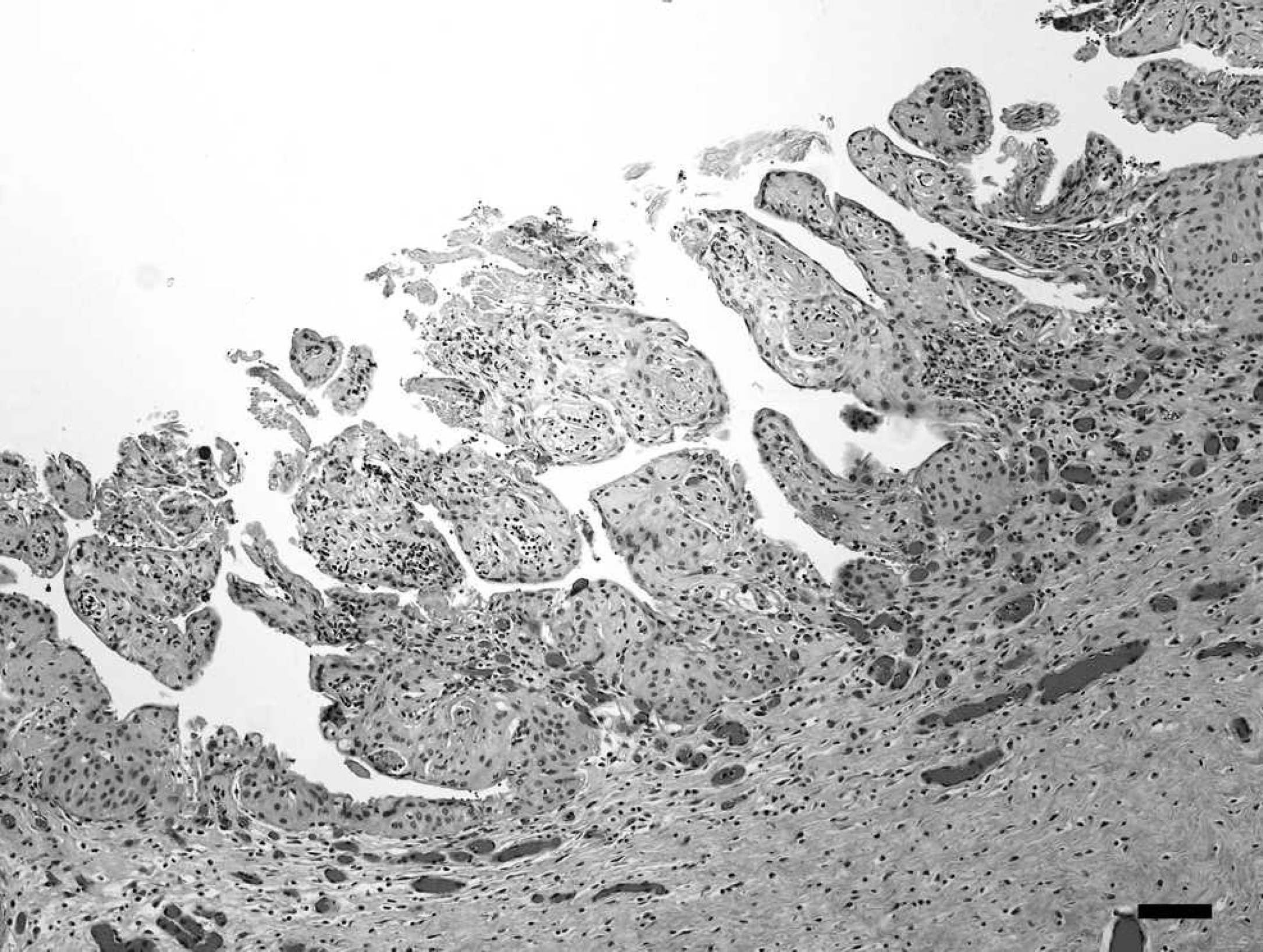
Placenta; horse. Chorioallantois, allantoic surface with blunted villous structure, areas of epithelial necrosis, and moderate subepithelial infiltrates of predominantly lymphocytes, plasma cells, and monocytes, with fewer neutrophils. Hematoxylin and eosin. Bar = 50 μm.
However, since the strain with colonies isolated in greater numbers contained irregular, Gram-positive rods, similar in appearance to those observed in the histopathological examination and direct impression smears, it was considered to be of clinical significance. In addition, isolates with morphological phenotypes identical to that of the suspected pathogen were obtained in pure culture from lung tissue (> 100 colonies) and stomach contents (> 100,000 colony-forming units/ml) of the foal.
The significant isolates recovered from placenta, lung, and stomach contents were catalase negative and nonhemolytic on sheep blood agar. The isolate from stomach contents was selected for identification to the species level since it was recovered in high numbers and in pure culture. The isolate was presumptively identified as A. hippocoleae with conventional biochemical tests. Acid was produced from glucose and lactose. Hippurate hydrolysis was strongly positive. The isolate was negative for gelatin hydrolysis, acetoin production, and nitrate reduction. Reactions in a commercial biochemical test kit designed for coryneform bacteria b identified the isolate as A. haemolyticum, with 99.8% and 99.9% probabilities in 2 repeated tests. Positive test results included the following: alkaline phosphatase, β-glucuronidase and glucose, maltose, and lactose fermentation. Negative test results were observed for nitrate reduction, pyrazinamidase, pyrolidonyl arylamidase, α-glucosidase, β-glucosidase(esculin), urease, gelatin hydrolysis, and xylose, mannitol, sucrose, and glycogen fermentation. Three of these test reactions (pyrazinamidase, usually positive; β-glucuronidase, usually negative, but this reaction can be delayed; α-glucosidase, usually positive for A. haemolyticum) were repeatedly nonsupportive of the identification of the isolate as A. haemolyticum. However, reactions in 2 of these 3 tests (pyrazinamidase, reported negative; β-glucuronidase, reported positive) were consistent with those reported for the type strain of A. hippocoleae. α-Glucosidase was reported positive for both A. haemolyticum and A. hippocoleae. Results of 3 biochemical tests (β-galactosidase, N-acetyl-β-glucosaminidase, and ribose fermentation, reportedly positive, positive and negative for A. hippocoleae, respectively) were variable on repeat testing in this assay. Reactions in another commercial biochemical test kit designed for Grampositive bacteria c did not result in an acceptable identification of the isolate. Neither commercial identification system contained reactions for A. hippocoleae in its database.
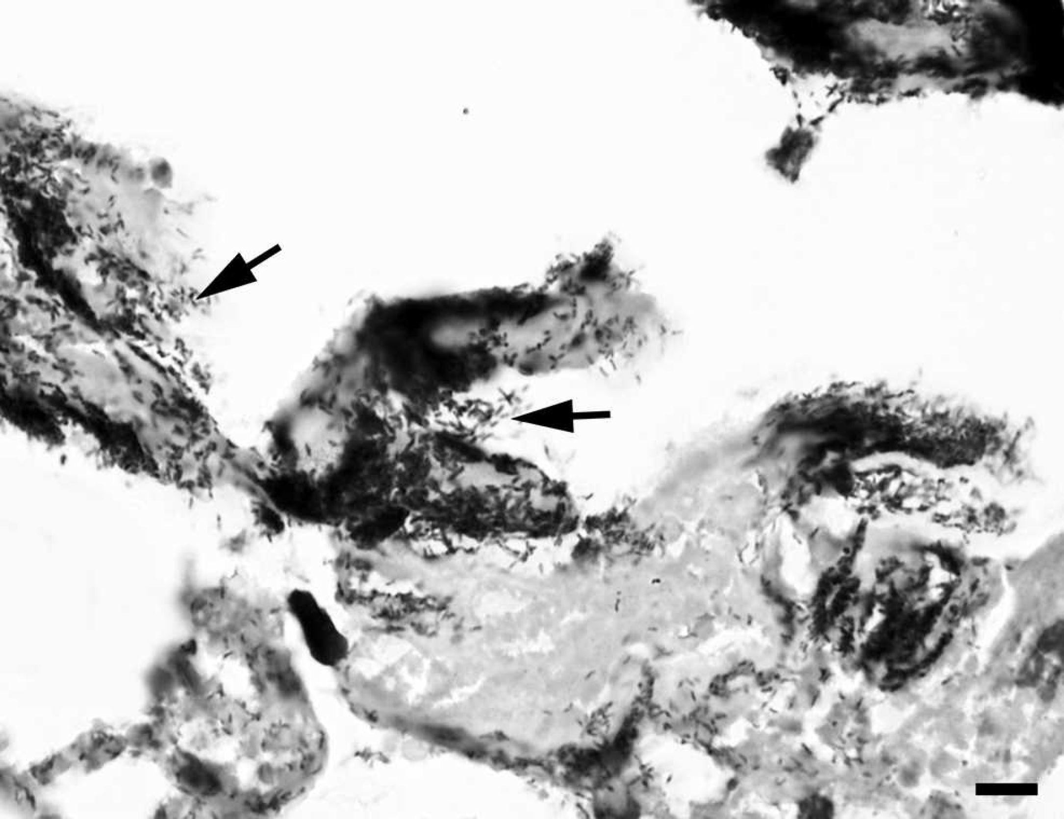
Placenta; horse. Clusters of irregular, predominantly Gram-positive, rod-shaped bacteria (arrows) associated with fibrin matrix and degenerated epithelial cells. Gram stain. Bar = 20 μm.
Species identity of the isolate obtained from stomach contents was confirmed by DNA sequencing. Template DNA was extracted from individual colonies by physical disruption with glass beads d and subjected to polymerase chain reaction (PCR) with universal eubacterial primers designed to amplify a portion of the 16S ribosomal RNA (rRNA) gene. 11 The primers, Broad 1 (GCGGATCCTGCAGAGTTTGATCCTGGCTCAG) and Broad 2 (GGCTCGACCGGGTTACCTTGTTACGACTT), correspond to bases 8–27 and 1492–1510 of the Escherichia coli small ribosomal subunit gene. Polymerase chain reaction was performed with a commercial Taq polymerase premix e containing reaction buffer and nucleotides. Thermocycler conditions included 1 cycle of denaturation and enzyme activation at 95°C for 1.5 min, 30 cycles of annealing at 50°C for 30 sec, extension at 72°C for 2.5 min, and denaturation for 1 min at 94°C. This was followed by a final cycle of annealing at 50°C for 2 min, extension at 72°C for 5 min, and holding at 4°C. The PCR amplification product was subjected to electrophoresis in 1 % agarose gel containing 1 mg/ml ethidium bromide and was visualized by ultraviolet transillumination. The PCR product was of expected size and was purified and concentrated by filtration f before submitting to the University of Tennessee core DNA sequencing facility. Nucleotide sequence data were analyzed using a commercial software program. g The 16S rRNA gene sequence of the isolate was compared to sequences in the GenBank database using the BLASTn algorithm. 1 The highest match, with 98% nucleotide similarity over 1,002 nucleotides, was to A. hippocoleae.
The A. hippocoleae type species was described as weakly hemolytic on Columbia agar with 5% horse blood. 6 Horse blood agar may not be as readily available as sheep blood agar in many diagnostic laboratories. Hemolysis was not observed with the present isolates on Columbia agar with 5% sheep blood, even after prolonged incubation and close examination of the agar surface beneath isolated colonies. It was previously noted that, with the exception of A. hippocoleae and Arcanobacterium pluranimalium, hemolysis was obtained with all other Arcanobacterium species on sheep blood agar (Euzeby JP: 2008, Dictionnaire de bacteriologie veterinaire: principaux caracteres bacteriologiques permettant de differencier les six especes du genre Arcanobacterium [Veterinary dictionary of bacteriology: principal bacteriological characters allowing to differentiate the six species from the genre Arcanobacterium]. Available at http://www.bacterio.cict.fr/bacdico/aa/arcanobacterium.hl. Accessed May 26, 2008.). It was not specifically stated that A. hippocoleae was nonhemolytic on sheep blood agar. The isolate from stomach contents was nonhemolytic on inhouse and commercially h prepared Columbia agar with 5% horse blood and on cation-adjusted Mueller Hinton agar a with 5% horse blood.
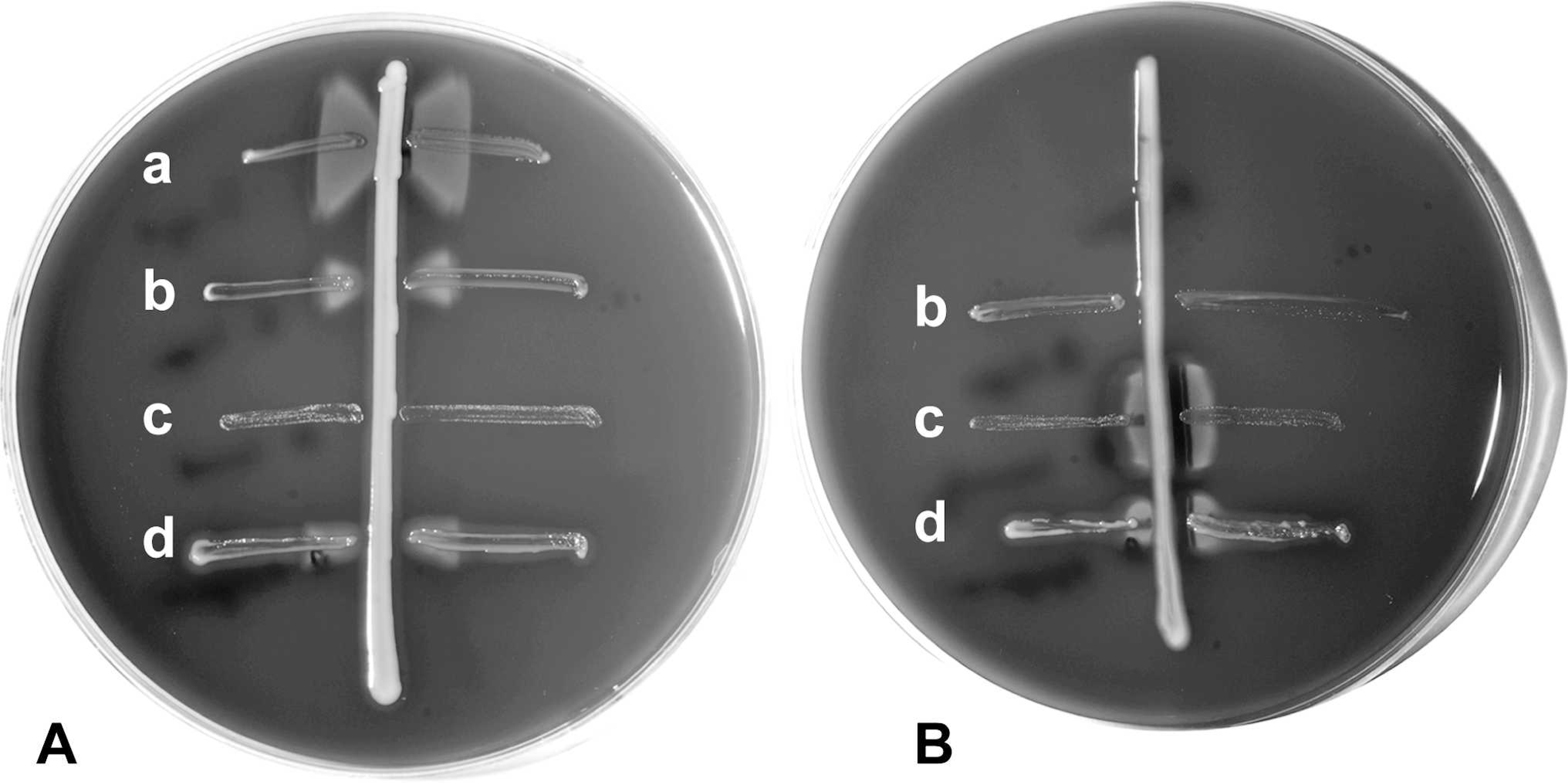
Hemolytic activity was further evaluated by the Christie-Atkins-Munch-Peterson (CAMP) test, on sheep blood agar, 4 a PCR assay for the cholesterol-binding, sulfhydryl-activated pyolysin of A. pyogenes, 2 and a phospholipase activity detection method. Staphylococcus aureus (American Type Culture Collection strain ATCC 25923) or clinical isolates of Rhodococcus equi, Streptococcus agalactiae, A. pyogenes, or A. haemolyticum were used as controls in these tests. Identities of the control isolates were confirmed by interlaboratory comparisons in 2 or more laboratories. The CAMP test is commonly used for differentiation of other Arcanobacterium spp., but results of this test have not been reported for A. hippocoleae. Enhanced hemolysis (positive reaction) was observed in the equine isolate of A. hippocoleae with the CAMP test with S. aureus, but not with R. equi (Fig. 3). Since the equine isolate of A. hippocoleae did not produce the reverse CAMP reaction characteristic of the phospholipase D family of hemolysins, PCR was performed to detect potential gene homology with the well-characterized pyolysin of A. pyogenes.
Polymerase chain reaction assays resulted in amplification products of the expected size from A. pyogenes but not from A. hippocoleae or A. haemolyticum DNA extracts. A heavy inoculum of A. hippocoleae, but not A. haemolyticum or A. pyogenes, produced a chromogenic reaction typical of phospholipase activity on a selective differential agar for rapid identification of Listeria spp. i This is a commonly used, non-blood containing medium that has proprietary selective ingredients for Listeria spp.; neither the equine A. hippocoleae isolate nor the A. haemolyticum or A. pyogenes isolates grew on this medium. Further studies of hemolysis and hemolysin genes in A. hippocoleae are warranted.
The extent of natural divergence within A. hippocoleae is not known because only the type strain has been reported in the literature, and reference sequences available in GenBank for this species are solely from this strain. Isolates are presumptively identified as belonging to the genus Arcanobacterium through their colony appearance, growth characteristics, catalase reaction, and Gram-staining coupled with type of hemolysis and fermentative activity. As demonstrated by the isolate in the present report, recognition of hemolytic activity may require use of the CAMP test or other tests, such as hemolysin gene detection or demonstration of phospholipase activity, as they are further developed for this purpose. In a taxonomic study, chemical analysis of a conserved component (menaquinone) of the respiratory electron transport chain among members of the genus Arcanobacterium placed A. hippocoleae and A. haemolyticum together in a different phylogenetic subgroup than A. pyogenes. 9 However, unlike A. haemolyticum, which is known to produce a phospho-lipase D-like hemolysin, the A. hippocoleae isolate produced a positive, rather than a reverse, hemolytic reaction in the CAMP test, perhaps as a result of the presence of a phospholipase C-like hemolysin. Hippurate hydrolysis and the absence of gelatin hydrolysis appeared to be useful biochemical indicators of the species A. hippocoleae.
Whether isolation from the equine reproductive tract is a consistent feature of A. hippocoleae awaits further confirmation. The source of A. hippocoleae infection in the present case is unknown. The mare was not examined, but, according to history provided by the owner, the mare was producing milk, and there was no suggestion of systemic illness. The farm history was also sparse. It was noted that an additional mare failed to foal during the same season, but abortions, stillbirths, or neonatal deaths of foals from other mares had not been observed. In the present case, the point at which the foal's lungs became inflated was undetermined. The foal was nonviable and still within the chorioallantois when found by the owner. Arcanobacterium hippocoleae was found in stomach contents and lungs (confirmed by subsequent DNA sequencing; this isolate was also shown to be nonhemolytic on horse blood agar) of the foal, but the observed pathological lesions (inflated lungs, in what was initially presumed to be an aborted foal, and occasional squamous cells in alveoli and larger airways) were not highly suggestive of fetal bacterial infection. Stillbirth may have been the result of bacterial placentitis; however, other causes were not ruled out, and a causal role for A. hippocoleae was not determined. The placental isolate, suspected to be A. hippocoleae, was inadvertently discarded prior to completion of the histopathological examinations and was therefore not available for species confirmation. Attempts to demonstrate bacterial DNA sequence in extracts from fixed, paraffin-embedded placental tissue were also unsuccessful. However, considering the close anatomical relationship between fetal stomach contents, lung, and placenta, it is likely that the Gram-positive, rod-shaped organism isolated from placental tissue and the similar rods associated with placental lesions were A. hippocoleae. The present report and the previous association of A. hippocoleae with vaginal discharge in a mare indicate that A. hippocoleae may be a significant bacterial pathogen in diseases of the equine reproductive tract. Microbiologists should be alerted to the possible identification of A. hippocoleae in clinical samples and should report their occurrence to allow further analyses of host range and pathogenicity.
Acknowledgements. The authors wish to thank Rupal Brahmbhatt for technical assistance.
Footnotes
a.
BBLTM, BD Diagnostic Systems, Sparks, MD.
b.
API® Coryne, bioMérieux Inc., St. Louis, MO.
c.
BBLTM CrystalTM Identification Systems Gram-Positive ID Kit, BD Diagnostic Systems, Sparks, MD.
d.
BioSpec Products Inc., Bartlesville, OK.
e.
Premix Taq TM, Takara Bio Inc., Otsu, Shiga, Japan.
f.
Montage® Centrifugal Filter Device, Millipore Corp., Billerica, MA.
g.
Lasergene® v7.2, DNASTAR Inc., Madison, WI.
h.
Veterinary Medicine Biological Media Service, University of California, Davis, CA.
i.
Rapid' L. Mono Agar®, Bio-Rad Laboratories, Hercules, CA.