
Abstract
Glanders, caused by Burkholderia mallei, is a zoonotic disease of equids. Serologic testing for glanders is required by disease-free countries before international movement of equids. The World Organisation for Animal Health Terrestrial Manual recommends the complement fixation test (CFT) for clearance of individual animals for movement, but the CFT is prone to false-positive results. A colorimetric western blot (WB) assay was developed and validated to resolve false-positive CFT results; however, that assay is relatively time-consuming, and the interpretation is subjective. We present here a procedurally similar chemiluminescent WB assay that performs comparably to the validated colorimetric WB assay and offers noticeable benefits of decreased time-to-result and greater ease of interpretation.
Glanders, which is caused by infection with the bacterium Burkholderia mallei, is a zoonotic disease affecting mainly horses, mules, and donkeys. Glanders has been eradicated from Europe and North America; but countries in Southeast Asia, the Middle East, and South America have experienced outbreaks within the past 25 y.2,6,7,9,12,13,18,20,22,24 Disease-free status is maintained through stringent testing of all equids prior to importation. Although the “gold standard” test is culture and isolation of the bacteria from infected animals, this method lacks sensitivity and is not appropriate for the high-speed and high-throughput testing demanded for international trade of equids. 23 For these reasons, serologic assays are preferred for classifying animals as free of disease prior to importation into a disease-free country.
International movement of horses requires highly sensitive and specific serologic assays to maintain the disease-free status of countries accepting these horses. High sensitivity is needed to prevent the introduction of disease from areas experiencing outbreaks and to exclude inapparent carrier horses. Conversely, high specificity is essential to resolve false-positive results from high-throughput screening tests without causing undue delays in animal movement. To date, the complement fixation test (CFT) is the only recommended assay for equine movement according to the World Organisation for Animal Health (WOAH) 25 ; however, the sensitivity and specificity of the CFT are dependent on the antigen used and the incubation temperature.11,12,14 The CFT has been used for decades and has diagnostic sensitivity as high as 1.0. 11 Additionally, the CFT can detect inapparent carriers of disease, which account for as many as 90% of cases. 15 However, antigens for the CFT are crude, whole-cell preparations; consequently, the CFT may yield false-positive results, possibly due to cross-reactions with other bacterial species. 16
In one study, 2 CFT antigens had specificities of 0.965 and 0.975, with 3.5% and 2.5% false-positive rates, respectively. 11 Although these false-positive rates are relatively low, they can still result in an undesirable number of false-positives in countries that import thousands of horses per year. In another study, false-positive rates were as high as 24.3%. 10 False-positives can be expensive and stressful to owners and shippers, may lead to delays in meeting event or competition deadlines, or can result in the euthanasia of high-value animals.
Other serologic assays, including ELISA, agglutination, and polysaccharide microarray, have been developed for glanders and have undergone various degrees of validation, but none are recommended by the WOAH over the CFT.3,8,14,17,19,21,23 A commercial ELISA has been evaluated as a replacement for the CFT because it exhibited considerably fewer false-positives; ELISAs are well-suited for high-throughput laboratories. 4
A colorimetric western blot (WB) assay was developed as a highly specific confirmatory assay to resolve false-positive CFT results. 5 Diagnostic sensitivities for that assay have been reported as 1.0 and 0.97, with corresponding specificities of 1.0 and 0.99.3,5 Interpretation of the colorimetric assay is subjective, requiring visual judgment of bands and banding patterns. The assay requires ~5–6 h to perform, which may not be suitable for fast-paced laboratories providing results for timely release of horses from quarantine. Colorimetric WBs can be overdeveloped, resulting in bands that are difficult to discern or are interpreted as false-positive results. We present here a procedurally similar WB assay with shorter incubation times and a chemiluminescent development system that overcomes the issues with overdevelopment and enhances band visualization.
Group I samples comprised sera from naturally infected glanders-positive animals (n = 224) collected as described previously. 4 Infection with B. mallei was confirmed either by observation of clinical signs paired with positive CFT, or isolation and/or molecular detection of B. mallei. 23 Horses (n = 185), mules (n = 22), ponies (n = 13), and donkeys (n = 4) were represented in the panel of positive sera. Group II samples were negative field sera (n = 100) selected from horse serum samples submitted to the National Veterinary Services Laboratories for routine testing. Geographic origin of the group II horses spanned North America (n = 70), Europe (n = 24), and South America (n = 6). Glanders-negative status of sera was either presumed due to glanders-free status of the country of origin, or the samples tested negative for glanders by CFT.
Lipopolysaccharide (LPS) antigens for the WB were prepared as described previously and lyophilized for storage. 5 Lyophilized antigen was reconstituted with 1 mL of sterile water. Reconstituted B. mallei strains Bogor, Mukteswar, and Bahrain1 were combined in equal parts. Antigen was prepared for SDS-PAGE (NuPAGE LDS sample buffer; Thermo Fisher) according to the manufacturer’s instructions. Gel electrophoresis was performed with 200 µL of prepared antigen loaded in a NuPAGE precast 10% polyacrylamide 2D or 4–12% Bis-Tris ZOOM gel (Thermo Fisher) in 3-(N-morpholino)propanesulfonic acid (MOPS) buffer (Thermo Fisher) according to the manufacturer’s instructions. Either gel percentage will produce the expected protein banding pattern. Separated antigen was transferred to a nitrocellulose membrane (iBlot 2 blotting system; Thermo Fisher), then the membrane was blocked for 30 min in 10% (w/v) non-fat dry milk (NFDM) in PBS with 0.2% (v/v) Tween-20 (PBST). The membrane was removed from the blocking solution and washed 3 times for 20 min using PBST. The membrane was cut into ~3-mm wide strips and stored at −20°C if not used immediately for serum analysis.
For immunoblot analysis, the strips were incubated individually for 30 min in the test serum diluted 1:20 in 10% NFDM-PBST, followed by 5 washes of 5 min in PBST. Next, strips were incubated for 30 min in horseradish peroxidase–conjugated goat anti-horse secondary antibody (SeraCare) diluted 1:5,000 in 10% NFDM-PBST, followed by 5 more 5-min PBST washes. Finally, strips were incubated with chemiluminescent substrate (Pierce ECL Plus; Thermo Fisher) for 5 min and visualized in an imaging system (iBright; Thermo Fisher) by using the “smart exposure” feature.
A serum sample was classified as positive if the characteristic LPS ladder pattern was observed between 20 and 60 kDa, and classified as negative if no banding pattern was observed. If a weak signal was observed that did not meet the definition of positive or negative, the serum was considered suspect (Fig. 1). The difference between positive and suspect can be very subtle, and may differ from technician to technician. The distinction between positive and suspect is not as important as the distinction between negative and non-negative.
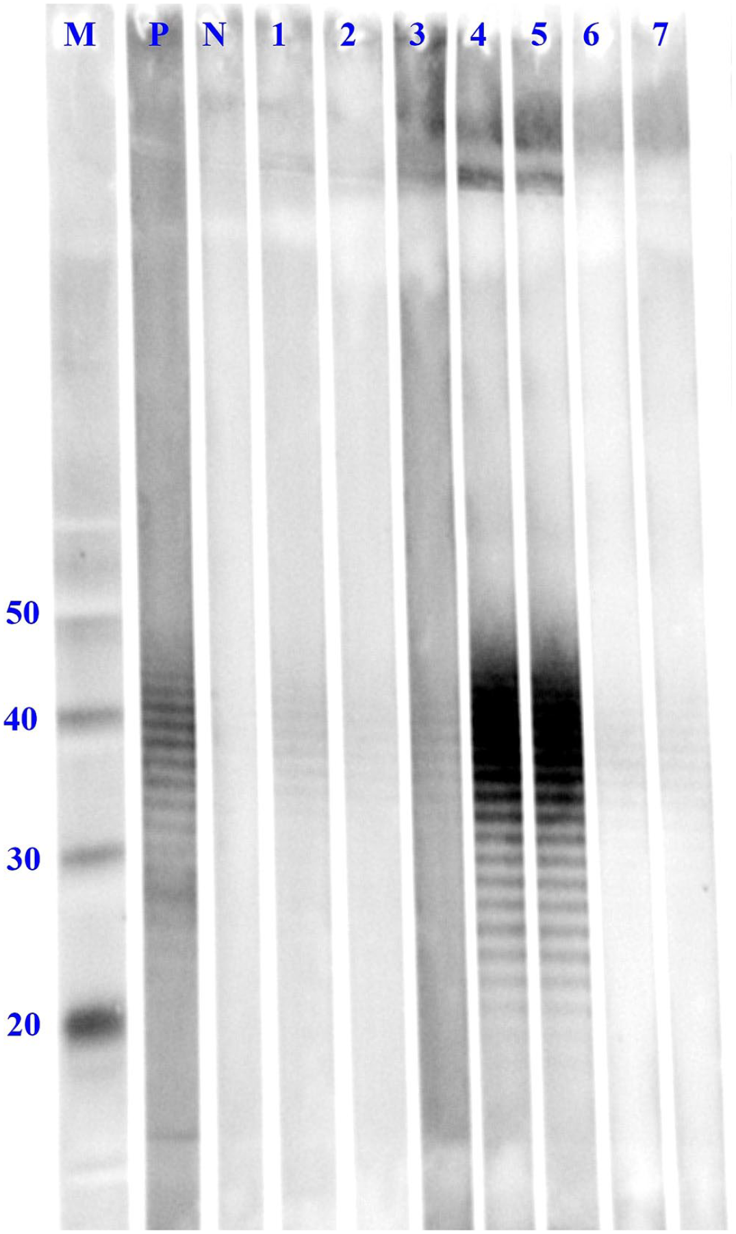
Interpretation of chemiluminescent western blot assay of known Burkholderia mallei seropositive equine samples. M = marker; N = negative control; P = positive control; 1 = positive; 2 = suspect; 3 = positive; 4 = positive; 5 = positive; 6 = positive; 7 = positive. Numbers to the left indicate molecular weights in kDa.
The colorimetric WB assay was performed as described previously. 5 Briefly, the LPS antigen was separated by gel electrophoresis and transferred to a nitrocellulose membrane. Membrane strips were incubated with serum diluted 1:50, washed, then incubated with alkaline phosphatase–conjugated anti-horse secondary antibody, and washed again. Finally, an enzyme substrate was added to induce color development to visualize the characteristic ladder pattern. The assay was interpreted as described for the chemiluminescent assay.
Agreement between assays was calculated using the Cohen kappa. 1 Calculations were performed using R v.3.5.3 (https://www.r-project.org/).
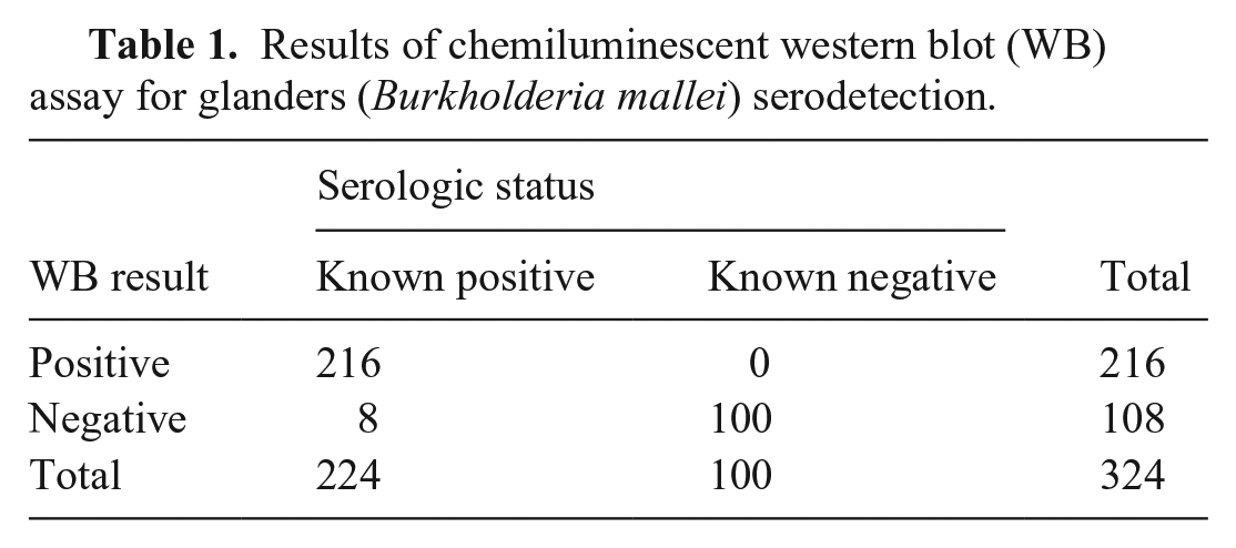
Both the colorimetric and chemiluminescent WB assays correctly classified 216 of 224 group I samples as positive or suspect, resulting in a diagnostic sensitivity of 0.96 (95% CI: 0.93, 0.98) for both assays (Tables 1, 2; Fig. 1). All 100 group II samples were negative on the chemiluminescent WB assay, resulting in a diagnostic specificity of 1.0 (95% CI: 0.96, 1.0). Two of the samples in group II were positive on the colorimetric WB assay, which resulted in a diagnostic specificity of 0.98 (95% CI: 0.93, 1.0). Both blots had almost perfect agreement, with a Cohen kappa value of 0.986 (95% CI: 0.967, 1.000).
Results of chemiluminescent western blot (WB) assay for glanders (Burkholderia mallei) serodetection.
Results of colorimetric western blot (WB) assay for glanders (Burkholderia mallei) serodetection.
WB assays for serodetection of glanders antibodies have been evaluated extensively.3,5,10 Our chemiluminescent WB had almost perfect agreement with the colorimetric WB developed and validated previously, indicating that the assays could be used interchangeably. 5 Both WB assays had relatively high diagnostic sensitivity and thus can be trusted to identify glanders positive sera. The chemiluminescent assay demonstrated a slightly higher diagnostic specificity than the colorimetric assay, with no false-positives.
Although both assays performed comparably, the chemiluminescent assay has technical advantages over the colorimetric assay. Notably, the chemiluminescent assay has variable and adjustable exposure times (i.e., the amount of time light is collected by the camera sensor) to avoid overexposing the blot strips. A difficulty of using the colorimetric development system is allowing the color development to proceed for too long, which causes excessive staining that makes the result of the blot difficult to decipher. Additionally, the shorter incubation time (30 min vs. 90 min) of the chemiluminescent assay translates to shorter turnaround times for confirmatory results in a diagnostic laboratory setting. An imaging system is required for visualization of a chemiluminescent WB, which may be a drawback for some laboratories. Imaging equipment may also be needed for documentation of a colorimetric WB, depending on policies of the laboratory.
Although a single assay for glanders testing would be ideal, a 2-assay protocol is presently the best option in terms of sensitivity and specificity. In fact, the CFT and WB assay used in series is the testing algorithm used by the WOAH Reference Laboratory (Friedrich Loeffler Institute, Jena, Germany). In a recent study, using the WB assay to confirm positive or suspect CFT results increased specificity from 0.96 to 0.99; in another study, specificity was increased from 0.97 to 0.99 by addition of the WB assay.3,4
Although the current recommendation of the WOAH for individual animal freedom from infection is CFT alone, CFT in conjunction with one of the described WB assays in series when confirming a suspect or positive result appears to have increased diagnostic sensitivity and specificity compared to the CFT alone. 23
Footnotes
Acknowledgements
We thank Amber Gustafson and Peggy Marten for their conceptualization of the project and excellent technical work on this study.
Declaration of conflicting interests
The authors declared no potential conflicts of interest with respect to the research, authorship, and/or publication of this article.
Funding
The authors received no financial support for the research, authorship, and/or publication of this article. The findings and conclusions in this publication are those of the authors and should not be construed to represent any official USDA or U.S. Government determination or policy.