
Abstract
Anaplasma marginale and Anaplasma centrale are rickettsial pathogens responsible for acute disease and mild infections, respectively, in cattle herds. A duplex real-time polymerase chain reaction (PCR) assay with probes labeled with different fluorophores was developed for simultaneous detection and quantification of A. marginale and A. centrale DNA in bovine blood samples. The assay was able to detect as few as 101 and 102 DNA copies for A. marginale and A. centrale, respectively, with optimal specificity and reproducibility. Analysis by real-time and nested PCR carried out on 54 samples previously tested by reverse line blot hybridization showed that the established duplex real-time PCR assay can detect and quantify the 2 Anaplasma spp., even if present simultaneously in the same blood samples. Such an assay could be used in pathogenesis studies on bovine acute anaplasmosis.
Anaplasma marginale and Anaplasma centrale (order Rickettsiales, family Anaplasmataceae) are 2 intraerythrocytic rickettsiae that are closely related at the antigenic level but that exhibit differences in pathogenicity for cattle. Although A. marginale is responsible for severe hemolytic anemia associated with fever, weight loss, abortion, decreased milk production, and, in some cases, death of the infected cattle, 16 A. centrale causes asymptomatic or paucisymptomatic infections and is used for extensive vaccination of cattle against A. marginale infection in endemic areas (Callow LL, Dalgliesh RJ: 1980, The development of effective safe vaccination against babesiosis and anaplasmosis in Australia. In: Tick and tick-borne diseases, ed. Johnston LAY, Cooper MG, pp. 4–8. Proceedings of a Symposium held at the 56th Annual Conference of the Australian Veterinary Association, Townsville, Australia). However, clinical cases of acute anaplasmosis associated with A. centrale infection were reported. 4 A diagnosis of bovine anaplasmosis is commonly made by combining clinical findings with the examination of Giemsa-stained blood smears for the presence of hemoparasites. Identification of Anaplasma sp. by microscopy is based on the position of inclusion bodies within the erythrocyte, with A. marginale and A. centrale inclusions marginally and centrally located, respectively. However, there is difficulty in microscopically differentiating these bacteria in mixed infections so that discrimination through microscopic examination is not definitive.
Polymerase chain reaction (PCR) based methods were developed for specific detection of these Anaplasma species, but they do not provide quantitative information with regard to rickettsemia levels. 1,13,14,17 Recently, real-time PCR was successfully applied to the detection of A. marginale DNA in bovine blood. 3 In the present study, the A. marginale real-time PCR was modified by adding a primer/probe set specific for A. centrale, thus obtaining a duplex assay for simultaneous detection of both Anaplasma spp. in the same reaction.
For standardization of the duplex real-time PCR assay, the same field samples and DNA extraction protocols were used as described in the monoplex A. marginale assay 3 (i.e., 54 blood samples collected in southern Italy from 51 cattle, 2 sheep, and 1 goat [Table 1]) and previously tested by reverse line blot (RLB) assay for detection of hemoparasites of ruminants. 1,11 Both sheep samples contained Anaplasma ovis, whereas the single goat sample had tested positive for A. ovis and Anaplasma bovis. Oligonucleotides for A. marginale have already been described. 3 In the current study, primers and a TaqMan probe specific for A. centrale were designed by taking into account the nucleotide differences encountered in the groEL sequences between the 2 Anaplasma spp. The reporter and quencher dye fluorophores for the A. centrale probe were Texas Red and Black Hole Quencher 2 (BHQ2), respectively (Table 2). For the construction of an A. marginale standard curve, a plasmid that contained a 729 base pair (bp) fragment of the msp1b gene 3 was used, whereas standard DNA for A. centrale was obtained ex novo by amplifying a 488-bp fragment of the groEL gene by using primers groEL-ACF (TCTTCTTCTGACTACGACAAGGAAAAACTG) and gro-EL-ACR (GTCATGAATACARCTGCGAGTGA-CACAGCC). The PCR fragment was cloned into a vector a and amplified in competent cells. b Subsequent purification and quantification steps were done as previously described. 3 Tenfold dilutions of each plasmid, which represents 100–108 copies of DNA per 10 μl of template, were carried out in a blood sample collected from a calf that had tested negative by RLB for both Anaplasma spp. Aliquots of each dilution were frozen at −70°C and were only used once. Real-time PCR for simultaneous detection and quantification of A. marginale and A. centrale DNA was performed on a real-time machine. c The reaction mixture contained a total volume of 25 μl, including 12.5 μl of iTaq Supermix, d primers at a concentration of 600 nmol/l (A. marginale) or 900 nmol/l (A. centrale), and each probe at a concentration of 200 nmol/l (Table 1) and 10 μl of template (1:10 dilution) or plasmid DNA. The thermal cycling consisted of activation of iTaq DNA polymerase at 95°C for 10 min, 40 cycles of denaturation at 95°C for 1 min, and annealing extension at 60°C for 1 min. The TaqMan assay was carried out in duplicate for each unknown and standard sample, and a no-template control was included in each assay. Extracted DNA was tested for PCR competency by adding and quantifying an exogenous internal control (IC) represented by Canine parvovirus-2 (CPV-2) DNA, 8 as previously described. 3 Briefly, the CPV-2 DNA was added to the lysis buffer e at the concentration of 10,000 DNA copies per ml of buffer before blood digestion. The fixed amount of the IC added to each sample had been calculated to give a mean cycle threshold (Ct) value in a real-time PCR assay 8 of 36.26, with an SD of 0.81 as calculated by 50 separate runs. Real-time PCR for IC detection was carried out in a separate run, as previously described. 3 Samples in which the Ct value for the IC was > 37.88 (average +2 SD) were excluded from the analysis.
The following species of ruminant hemoparasites were used to assess the analytical specificity of the duplex realtime PCR assay: A. marginale, A. centrale, A. bovis, A. ovis, A. phagocytophilum, Babesia bovis, Babesia bigemina, Theileria annulata, and Theileria buffeli. No-template controls and Anaplasma-negative blood samples were also tested. Reference strains for all hemoparasites were obtained from bovine blood samples containing single infections, as assessed by RLB assay 1,11 and sequence analysis of the 16S or 18S ribosomal RNA (rRNA) and groEL genes. 12,13,15 By sequence analysis, the A. centrale strains used for specificity assessment displayed a nucleotide identity to the LT strain and vaccine isolates of 99.92–100% and 97.72–98.82% in the 16S rRNA and groEL genes, respectively. 4 Sensitivities and dynamic ranges were calculated as previously described, 3 by using 10-fold dilutions of standard DNA, which ranged from 108 to 100 copies, made in a blood sample collected from an Anaplasma-negative calf. The intra- and interassay coefficients of variation (CV) of the quantitative assay were assessed by multiple measurements of the absolute copy numbers obtained from high, intermediate, and low concentrations of standard DNA within the same run and between runs, respectively. 3
To determine the ability of discriminating between and quantifying correctly A. marginale and A. centrale DNA contained in the same blood samples, artificially generated DNA mixtures composed of DNA from the 2 Anaplasma spp. in various concentrations (102, 104, and 106 copies, with all possible combinations between A. marginale and A. centrale) were performed in an Anaplasma-negative blood sample and subsequently tested by the duplex real-time PCR assay; the absolute copy numbers for each Anaplasma sp. were calculated.
Two nested PCR (nPCR) assays based on ethidium-bromide staining for specific detection of A. marginale and A. centrale DNA in blood samples were chosen as reference tests and were performed as previously described, 14,17 with minor modifications. The 25-μl reaction volume for the first amplification contained 1X AccuPrime PCR Buffer II, f 1 μl of AccuPrime Taq DNA polymerase, f 1 μmol/l of external primers AM456/AM1164 (A. marginale) or AC1826/AC2367 (A. centrale), and 10 μl of 1:10 diluted template DNA. After a 2-min step at 94°C for template denaturation and enzyme activation, amplification was obtained with 40 cycles of 94°C for 30 sec, 60°C (A. marginale), or 65°C (A. centrale) for 30 sec, and 68°C for 1 min. The 25-μl reaction for the second-round amplification (nPCR) was prepared as for the first amplification, by using 1 μl of the primary PCR product as template and 1 μmol/l of internal primers AM100/AM219 (A. marginale) or CIS1925/CIS2157 (A. centrale). The thermal conditions were as for the first amplification, except that the annealing temperatures were 54°C and 60°C for A. marginale and A. centrale, respectively. The PCR products were detected by electrophoresis through a 1.5% agarose gel and visualized under ultraviolet light after ethidium bromide staining.
As a result of the fluorescence signals (6-carboxyfluorescein [FAM] or Texas Red) generated during the amplifications, each dual-labeled probe recognized its specific target. FAM fluorescence was exclusively generated by blood samples that contained A. marginale, whereas Texas Red signals were detected only by A. centrale-positive samples. No template controls, negative blood samples, and other hemoparasites of ruminants produced any detectable fluorescence signal, which confirmed that the assay was highly specific for the detection of and discrimination between A. marginale and A. centrale. To establish that the test had an acceptable sensitivity, its performance was compared with nPCR assays for specific detection of A. marginale and A. centrale DNA. 14 The detection limit of the duplex real-time PCR assay was shown to be 101 and 102 standard DNA copies for A. marginale and A. centrale, respectively. In contrast, the sensitivity of nPCR was the same as the duplex real-time PCR for A. marginale and even 1 log higher (101 copies of plasmid DNA) for A. centrale. The dynamic range of the TaqMan assay was analyzed by generating standard curves by using the standard DNA dilutions prepared in an Anaplasma-negative blood sample, to mimic natural conditions. The resulting standard curves demonstrated a strong linear correlation between 101 (A. marginale) or 102 (A. centrale), and 108 copies. Slope values were −3.337 and −3.405 for standard curves generated for A. marginale and A. centrale, respectively, whereas coefficients of correlation were 0.998 for both hemoparasites. The CVs of the within-run precision experiments were 4.42–10.23% and 10.77–24.30% for A. marginale and A. centrale, respectively, whereas the CVs of the between-run experiments were 11.32–23.73% (A. marginale) and 17.84–34.52% (A. centrale). The CVs for A. marginale were generally lower than those observed for A. centrale in both intra- and interassay experiments. Samples spiked with low (102 copies), intermediate (104 copies), and high (106 copies) concentrations of DNA from A. marginale and A. centrale showed no remarkable interference during detection and quantification of the 2 Anaplasma spp. contained in the same sample, with DNA titers calculated correctly for both rickettsiae. Interference did not occur, even when analyzing samples with high concentrations of A. marginale and low concentrations of A. centrale and vice versa (data not shown).
Comparison between reverse line blot hybridization, duplex real-time polymerase chain reaction (PCR), and nested PCR assays for detection of Anaplasma marginale and Anaplasma centrale in blood of ruminants.
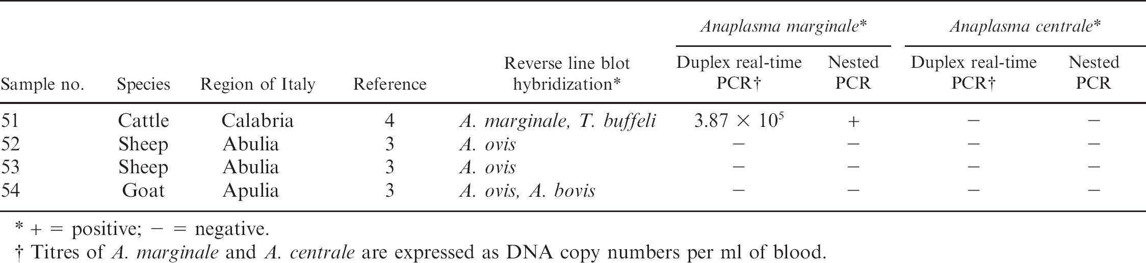
+ = positive; -= negative.
Titres of A. marginale and A. centrale are expressed as DNA copy numbers per ml of blood.
Results of the duplex real-time PCR and nPCR assays carried out on field samples are reported in Table 1. The results for A. marginale detection and quantification were similar to those obtained with the monoplex assay previously developed. 3 Anaplasma centrale was detected by both real-time PCR and nPCR assays in 18 bovine samples, with rickettsemia levels that ranged from 6.30 × 10³ to 3.24 × 10⁸ DNA copies per ml of blood. A 100% concordance was found between duplex real-time PCR and nPCR for both Anaplasma spp. As previously observed, 3 no A. marginale-positive sample was detected by RLB, whereas 3 samples that had been found to contain A. centrale by RLB did not give any signal by real-time PCR and nPCR, and 1 additional sample negative by RLB was found positive for A. centrale by both real-time and conventional PCR assays. As expected, the sheep samples and the single goat sample did not produce either FAM or Texas Red fluorescence, thus confirming the specificity of the duplex TaqMan assay. The IC was detected in all the examined samples, with Ct values below the threshold value of 37.88.
Because of its natural attenuation and cross-protection against A. marginale, A. centrale is used as a live vaccine for routine vaccination of cattle in Israel, Australia, Africa, and South America (Callow LL, Dalgliesh RJ: 1980, The development of effective safe vaccination against babesiosis and anaplasmosis in Australia). 2 Several molecular assays were established to detect A. centrale in bovine blood, including RLB 1 and PCR assays, 12–14,17 but none of those methods is quantitative. A real-time PCR assay for detection and quantification of A. centrale could provide useful information about the vaccination status of cattle. In addition, when taking into account the recent report of a clinical case of bovine anaplasmosis from A. centrale, 4 such an assay may be applied to discriminate between avirulent and virulent infections based on the A. centrale DNA titers detected in the blood. In a previous study, a real-time PCR assay was established for specific detection and quantification of A. marginale DNA in blood samples of cattle. The assay was found not to recognize the closely related A. centrale. 3 Duplex real-time PCR has proven to be a powerful tool for differentiating between closely related infectious agents 5,6 and between vaccine and field strains of the same genotype. 7,10 In the present study, a duplex Anaplasma TaqMan assay was developed for sensitive and specific detection, discrimination, and quantification of A. marginale and A. centrale that may be contained in the same samples. In comparison with nPCR protocols, the established assay is equally sensitive (A. marginale) or 1 log less sensitive (A. centrale). However, the duplex real-time PCR is faster and can be completed in about 2 hr. In contrast, nPCR is a 2-step assay based on 2 independent runs, thus requiring 4–5 hr for completion. 3,9 Moreover, cross-contaminations are considerably reduced in real-time PCR, because no postamplification manipulations are required, and the use of 2 differently labeled probes (FAM and Texas Red) allows the performance of the assay in 1 tube, which leads to lower cost as less master mix is used for detection of 2 pathogens. The duplex Anaplasma real-time PCR assay correlated well with the nPCR assays for detection of A. marginale and A. centrale, whereas less concordance was found with the RLB assay, which produced 1 false-negative and 3 false-positive results. A limit of the established real-time PCR assay is that all tested samples had the same geographic origin (southern Italy), although they were collected from cattle herds of different regions that, in some cases, were more than 500 km apart. However, during the primer/probe design, a highly conserved region of the A. centrale groEL sequences, which contains enough nucleotide differences with A. marginale to prevent binding of the probe to this rickettsia, was chosen. Based on the primer and probe homology to the sequences of reference strains available in the GenBank database, the established assay would be specific for A. centrale identification and its discrimination from the highly related rickettsia A. marginale. Therefore, it is the authors' opinion that strain variability should not affect the ability of the assay to detect A. centrale isolates from other geographic areas. In conclusion, a powerful tool for the detection and differentiation of the closely related A. marginale and A. centrale has been established for improved diagnosis of these rickettsial hemoparasites. This quantitative assay would be useful for studying the pathogenesis of A. marginale infection in cattle.
Sequence, position, and specificity of the oligonucleotides used in the current study.
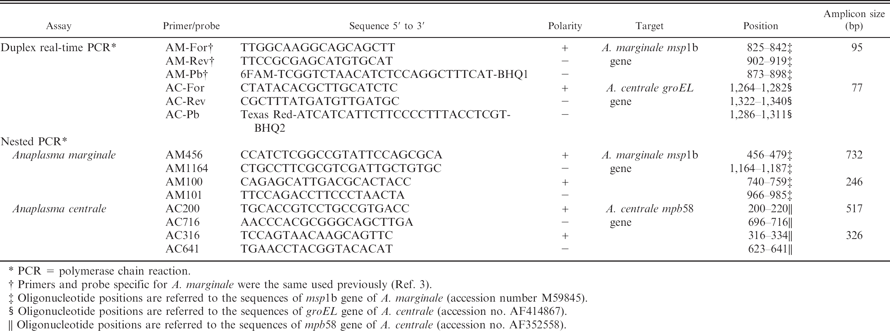
PCR = polymerase chain reaction.
Primers and probe specific for A. marginale were the same used previously (Ref. 3).
Oligonucleotide positions are referred to the sequences of msplb gene of A. marginale (accession number M59845).
Oligonucleotide positions are referred to the sequences of groEL gene of A. centrale (accession no. AF414867).
Oligonucleotide positions are referred to the sequences of mpb58 gene of A. centrale (accession no. AF352558).
Acknowledgements. The authors thank Donato Narcisi for his excellent technical assistance. Undergraduate student Graziana Borracci is also thanked for her help with the laboratory work.
Footnotes
a.
pCR® T7/NT-TOPO, Life Technologies, Invitrogen, Milan, Italy.
b.
Escherichia coli TOP10F' cells, Life Technologies, Invitrogen, Milan, Italy.
c.
7500 Real-time PCR System, Applied Biosystems, Foster City, CA.
d.
iTaqTM Supermix added with ROX, Bio-Rad Laboratories Srl, Milan, Italy.
e.
AL buffer, Qiagen SpA, Milan, Italy.
f.
AccuPrimeTM Taq Supermix II, Invitrogen SRL, San Giuliano Milanese, Italy.