
Abstract
African horse sickness is an arthropod-borne disease of the equine included in the World Organization for Animal Health (OIE) list with important economic consequences for horse trade. The disease is caused by African horse sickness virus (AHSV; family Reoviridae, genus Orbivirus), which is transmitted by Culicoides midges. It is endemic in sub-Saharan Africa, spreading occasionally outside this area where the occurrence of Culicoides vectors allows virus transmission. Currently, only conventional (gel-based) reverse transcription polymerase chain reaction (RT-PCR) protocols are available for its detection; however, these methods are cumbersome and difficult to apply when large numbers of samples are to be tested, as in the case of epizootics. To overcome this problem, a real-time RT-PCR method has been developed, based on a 5'-Taq nuclease-3′-minor groove binder-DNA probe (TaqMan MGB) for detection of a wide range of AHSV serotypes and strains designed to the highly conserved region of the VP7 gene (segment 7). The method was able to detect all prototype strains from the 9 known serotypes of the virus, with a high analytical sensitivity; no cross-reactions were observed with other orbiviruses or with other viruses affecting horses. The diagnostic sensitivity was assessed using a panel of AHSV-positive tissue samples from an epizootic that occurred in Spain between 1987 and 1990. This method, which can be performed in 96-well format, is suitable for large-scale surveillance of AHSV in areas where it can potentially spread.
African horse sickness virus (AHSV) is a member of the genus Orbivirus, within the family Reoviridae. As with other members of this genus, namely, Bluetongue virus (BTV) and Epizootic hemorrhagic disease virus (EHDV), AHSV is transmitted by biting midges of the genus Culicoides, but distinctively, it affects only Equidae. African horse sickness is endemic in sub-Saharan Africa but occasionally spreads beyond that area when the presence of competent vector allows viral transmission and persistence in the environment for a variable period of time. 12 That was the case in a number of epizootics that occurred in northern Africa (1965, 1989–1990), the Middle East (1959–1961), and the Iberian Peninsula (1966, 1987–1990). 12,18
The genome of AHSV consists of 10 double-stranded RNA segments, encoding 7 structural proteins (VP1–7) and 4 nonstructural proteins (NS1, NS2, NS3, NS3A). African horse sickness virus exhibits high antigenic and genetic variation, with 9 known serotypes (AHSV1–9) 8 and substantial strain variation within each serotype. Most variation concentrates in the outer capsid proteins VP2 and VP5, whereas highly conserved sequences within the serogroup are found in the VP7 coding region. 17
African horse sickness is included in the World Organization for Animal Health (OIE) list comprising diseases of utmost impact and causing severe economic losses in livestock. In disease outbreak situations, rapid detection of AHSV is crucial to prevent the spread of the disease. The virus can readily be detected by both antigen enzyme-linked immunosorbent assay (ELISA) and reverse transcription polymerase chain reaction (RT-PCR) methods. 6,7,11,14–16,20 Virus isolation through inoculation in tissue culture or newborn mice remains the reference technique to confirm RT-PCR results as well as to provide a reproducible source of virus suitable for further characterization and reference.
In recent years, real-time RT-PCR methodology has become a widely used tool in the diagnosis of animal diseases for various reasons. First, it enables the highly specific and highly sensitive amplification of nucleic acids and detection of the amplified product at the same time, which, in addition to saving time, makes unnecessary the electrophoretic analysis at the end of the process, simplifying the technique. Second, this technology, coupled with robotized nucleic acid extraction methods, allows full automation in both 96- and 384-well formats, 1,10 enabling high-throughput detection of virus and making thousands of samples per day a realistic option. This methodology relies upon fluorogenic 5'-nuclease (TaqMan) probes, molecular beacons, fluorescence resonance energy transfer (FRET) probes or SYBR green fluorescent dyes. 2 Of these, TaqMan-based assays are the most widely used in real-time PCR methods for virus detection because they combine high specificity with relative easy design, particularly when using minor groove binder (MGB) groups attached to the TaqMan probe, which enables shortening the length of the probe, thus facilitating selection of suitable target regions with conserved sequences in highly variable genomes. 9 Another great advantage of MGB probes is their high specificity, allowing the discrimination of single nucleotide polymorphisms, which not only enables rapid identification of viral variants 5 but also the discrimination between vaccinal and field strains 4 and between closely related viruses. 3
Analytical sensitivity of the African horse sickness virus (AHSV) real-time reverse transcription polymerase chain reaction (RT-PCR) using AHSV reference strains, and comparison with conventional RT-PCR.
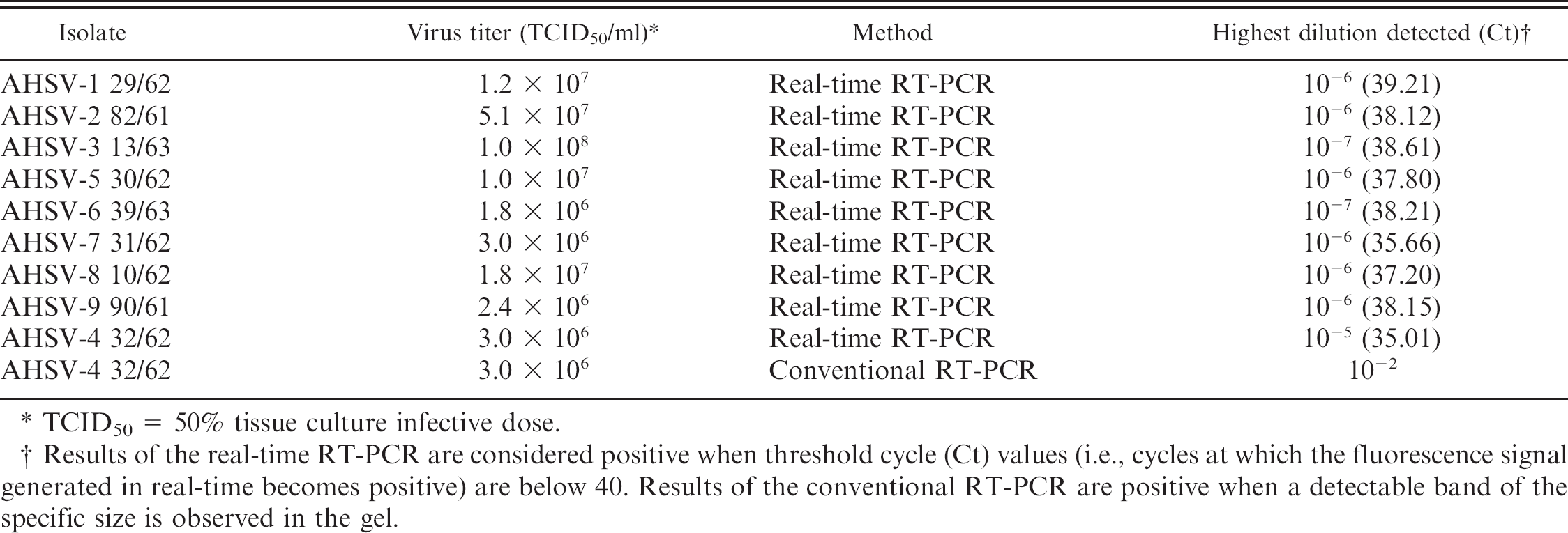
TCID50 = 50% tissue culture infective dose.
Results of the real-time RT-PCR are considered positive when threshold cycle (Ct) values (i.e., cycles at which the fluorescence signal generated in real-time becomes positive) are below 40. Results of the conventional RT-PCR are positive when a detectable band of the specific size is observed in the gel.
To date, there is no real-time RT-PCR described for the detection of AHSV. The current OIE terrestrial manual 18 references a conventional (gel-based) RT-PCR method for this purpose. 20 With the aim of bringing up to date the repertoire of techniques available for AHSV detection, a real-time fluorogenic RT-PCR using a TaqMan–MGB probe was developed. In the current study, the performance of this test is compared with the method referenced in the OIE terrestrial manual. 18
The primers and probe set were directed to a highly conserved sequence within the VP7 (segment 7) region of the AHSV genome, using nucleotide sequence data on this region available in GenBank under the following accession numbers: AF021238 (AHSV-6); A27209, X56676, NC_006011, D12533 (AHSV-4); S69829, U90337 (AHSV-9); and AF545433 (AHSV-3). The sequences were aligned using ClustalW 1.8 software. a Once identified, the target sequence, the primers, and TaqMan–MGB probe were designed using Primer Express (version 2.0.0) software. b Primer forward (nucleotide positions 1,032–1,051, numbered according to GenBank D12533) consisted of sequence 5′-CCAGTAGGCCAGATCAACAG-3′. Primer reverse (nucleotide positions 1,114–1,095, numbered as above) consisted of 5′-CTAATGAAAGCGGTGACCGT-3′, and the probe (nucleotide positions 1,061–1,078, numbered as above) consisted of 5′-FAM–GCTAGCAGCCTACCACTA–MGB-3′ (probe was labeled in 5′ with 6-carboxyfluorescein [FAM], and in 3′, with a nonfluorescent quencher bound to an MGB group c ).
The following viruses were analyzed in the current study: prototype strains of AHSV serotypes 1–9 d ; AHSV serotype 4 strain Spain 1987 (AHSV-4/1694–1-MS/1987) e , isolated from spleen of a horse infected during the epizootic occurring in Spain in 1987 by intracerebral inoculation in suckling mice; prototype strains of BTV serotypes 1–24, f as well as BTV-4 Spain 2004, e BTV−1 Algeria 2006, f and BTV-8 Belgium 2006 g ; North American prototype strains of EHDV serotypes 1–8 h and EHDV serotype 318 from Morocco (2006) I ; Equine influenza virus (A/equine/Kentucky/81 H3N8) e ; Equid herpesvirus 1 e (EHV-1 21/8/00 IRK); Equid herpesvirus 4 (EHV-4 V1999) e ; Equine arteritis virus (strain Bucyrus) e ; and West Nile virus (strain Eg101). j All viruses were grown in Vero cells, k except EHV-1 and EHV-4, which were propagated in RK-13 k cells, and equine influenza virus, which was propagated by inoculation in embryonated chicken eggs following the protocol described in the OIE manual. 19 Virus preparations were titrated following a standard limiting dilution assay 13 in the same system used for propagation.
Field samples consisted of 10 spleens from AHSV-infected horses from an African horse sickness outbreak that occurred in Spain between 1987 and 1990. Since that time, the samples were conserved frozen at −70°C and were positive by antigen ELISA. l Four spleens from horses were received at the laboratory for diagnosis of diseases other than AHSV and tested negative by conventional RT-PCR for AHSV. 20 Before RT-PCR or real-time RT-PCR, clarified viral preparations, viral dilutions, or spleen samples homogenated in phosphate buffered saline (PBS) by MagNA Lyser m following manufacturer's instructions, in a volume of 200 μl, were subjected to nucleic acid extraction using an automated procedure in 96-well format n according to the manufacturer protocol, eluting in a final volume of 100 μl. The fluorogenic real-time RT-PCR was carried out in a volume of 20 μl using a commercial kit. n Briefly, 2 μl of isolated RNA was mixed with forward and reverse primers (2.5 μl of each primer at 8 μM; final concentration: 1 μM) and ribonuclease (RNase)-free water up to 7 μl. This mixture was denatured by heating at 95°C for 5 min, followed by rapid cooling on ice. Then the following mixture was added per reaction well: 10 μl 2X QuantiTect Probe RT-PCR Master Mix, 0.2 μl of QuantiTect RT-mix, 0.1 μl of the fluorogenic MGB–TaqMan probe at 50 μM (final concentration: 0.25 μM), and 2.7 μl of RNase-free water. Amplification conditions consisted of a first reverse-transcription step at 48°C for 25 min, followed by 10 min at 95°C (“hot start”), and 40 cycles of 15 sec at 95°C, 35 sec at 55°C, and 30 sec at 72°C. Fluorescence data was acquired at the end of the 55°C step. The reaction was carried out in ABI Prism 7500 equipment and software. o The same samples were analyzed in parallel with the conventional reference RT-PCR method, 20 using 2 μl of each isolated RNA, primers at 0.2 μM final concentration, denaturation by heating at 95°C for 5 min, followed by rapid cooling on ice. A commercial kit p was used for the reverse-transcription amplification steps in 25 μl final volume of each reaction.
Specificity analysis in African horse sickness virus (AHSV) strains, and assessment of the performance of the real-time reverse transcription polymerase chain reaction (RT-PCR) for detection of AHSV in field samples.*
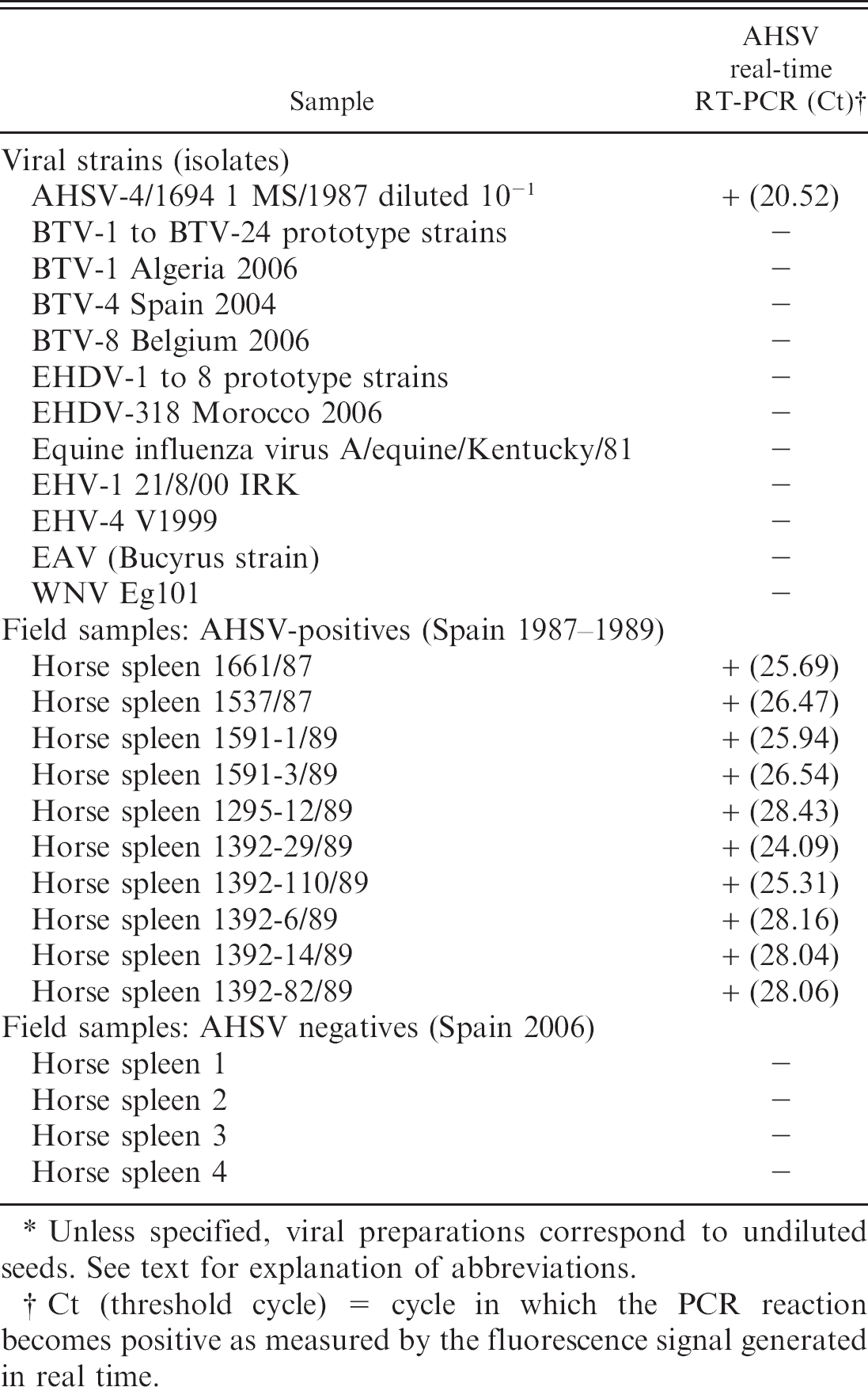
Unless specified, viral preparations correspond to undiluted seeds. See text for explanation of abbreviations.
Ct (threshold cycle) = cycle in which the PCR reaction becomes positive as measured by the fluorescence signal generated in real time.
The analytical sensitivity of the method was assessed by analyzing 10-fold dilutions of titrated cell-cultured viral supernatants from the prototype strains of AHSV serotypes 1–9. As shown in Table 1, the real-time RT-PCR method could detect between 10-5 and 10−7 dilutions of the viral preparations. Furthermore, whereas the real-time RT-PCR method could detect up to 10-5 dilution of the AHSV serotype 4 reference strain, the conventional RT-PCR method barely detected 10−2 dilution. Consequently, the analytical sensitivity of the new real-time RT-PCR method is at least 1,000-fold higher than that of the conventional RT-PCR method. The detection achieved by the real-time RT-PCR assay showed a linear relationship between the signal and the concentration of template present in the sample (correlation coefficient: 0.9970; slope: −3.296) over a dynamic range of 101.5 to 106.5 50% tissue culture infective dose (TCID50) per ml, using 10-fold dilutions of an infection supernatant of the AHSV-4 reference strain (virus titer: 3.0 × 10 6 TCID50/ml).
The current study also determined whether the real-time RT-PCR method cross-reacted with other members of the Orbivirus genus (i.e., BTV, EHDV) or with other viruses that could infect horses and, therefore, interfere in AHSV diagnostics (i.e., West Nile virus [WNV], EHV-1 and EHV-4, Equine influenza virus, and Equine arteritis virus [EAV]), using a field isolate (AHSV-4/1694 1 MS/1987) as control. The real-time RT-PCR assay did not cross-react with any other orbivirus and did not react with other horse-infecting viruses while detecting the field isolate (Table 2). The nucleic acids isolated from the non-AHSV viruses gave positive results when analyzed by specific PCR or RT-PCR methods (data not shown), thus confirming both the integrity of the nucleic acid material as well as the absence of PCR inhibitors.
To assess the performance of the new AHSV detection method with field samples, 10 horse spleen samples, from a historical collection corresponding to the AHSV epizootic that occurred in Spain in 1987–1990 and maintained in the authors' laboratory at −70°C (previously identified as AHSV positive by an antigen ELISA), were analyzed by real-time RT-PCR. At the same time, 4 spleen samples from horses not affected by AHSV, which were sent to the laboratory for diagnosis of other diseases and tested negative by RT-PCR for AHSV, were also analyzed. As shown in Table 2, all results obtained with the real-time RT-PCR assay in these samples were consistent with previous assays; all AHSV-positive samples gave positive results, whereas the 4 AHSV-negative samples remained negative. To ascertain that the negative results were not due to the presence of PCR inhibitors, 1 AHSV-negative spleen was spiked with detectable amounts of AHSV-4. The real-time RT-PCR analysis gave results similar to those obtained in the absence of spleen homogenate (data not shown).
The authors have developed a fluorogenic real-time RT-PCR method for AHSV diagnosis that improves the OIE referenced method for detection of AHSV genome, increasing the sensitivity by 1,000-fold. To the authors' knowledge, this is the first real-time RT-PCR method for AHSV diagnosis described in the literature. In addition, the newly developed assay is highly specific for AHSV, and neither other orbiviruses nor other viruses infecting equines give any cross-reaction in the assay. Furthermore, it provides the advantages of speed of analysis, suitability for high-throughput, and the capability for automation. Moreover, the technique has proven its ability for analysis of samples conserved frozen (−70°C) for at least 18 years. However, it will be necessary to analyze more field samples belonging to different AHSV serotypes to validate this technique. In addition, for validation and standardization of this type of assay, the use of internal controls is mandatory. Although the advantages of this methodology are clear-cut, it is important to bear in mind that its use in the control of this disease in endemic areas, comprising developing countries in sub-Saharan Africa, will require extensive international cooperation to allow affected countries to afford this still expensive technology.
Acknowledgements. The authors are indebted to B. Erasmus for providing AHSV isolates, K. de Clerq for providing isolate BTV-8 Belgium 2006, P. Mertens for providing BTV-1 Algeria 2006 and the collection of reference isolates BTV-1 to BTV−24, W. C. Wilson for providing EHDV-1 to EHDV-8, M. El Harrak for providing EHDV-318, and H. Zeller for providing the WNV Eg101 isolate.
Footnotes
b.
Primer Express (version 2.0.0) software, Applied Biosystems, Branchburg, NJ.
c.
TaqMan–MGB Probe, Applied Biosystems, Branchburg, NJ.
d.
AHSV prototype strains: AHSV-1 (29/62); AHSV-2 (82/61); AHSV-3 (13/63); AHSV-4 (32/62); AHSV-5 (30/62); AHSV-6 (39/63); AHSV-7 (31/62); AHSV-8 (10/62); AHSV-9 (90/61), Onderstepoort, Pretoria, South Africa.
e.
AHSV-4/1694 1 MS/1987, BTV-1 Spain 2004, Equine influenza virus (A/equine/Kentucky/81 H3N8), EHV-1 21/8/00 IRK, EHV-4 V1999, EAV strain Bucyrus, Laboratorio Central de Veterinaria, Algete, Madrid, Spain.
f.
BTV prototype strains and BTV-1 Algeria 2006, Institute for Animal Health, Pirbright, Surrey, UK.
g.
BTV-8 Belgium 2006, Veterinary and Agrochemical Research Centre, Brussels, Belgium.
h.
EHDV North American prototype serotypes 1–8, Arthropod-Borne Animal Disease Research Laboratory, Laramie, WY.
i.
EHDV serotype 318, Biopharma, Morocco.
j.
WNV Eg101, Institut Pasteur, Lyon, France.
k.
Vero cells (ATCC CCL-81), RK-13 cells (ATCC-CCL-37), American Type Culture Collection, Manassas, VA.
l.
ELISA-Ag AHSV, INGENASA, Madrid, Spain.
m.
MagNA Lyser, Roche Diagnostics, Indianapolis, IN.
n.
BioSprint DNA Blood Kit, BioSprint 96 Workstation, and QuantiTect Probe RT-PCR, Qiagen, Inc., Valencia, CA.
o.
ABI Prism 7500, Applied Biosystems, Branchburg, NJ.
p.
SuperScript One-StepTM RT-PCR System with Platinum® Taq, Invitrogen, Inc., Carlsbad, CA.