
Abstract
Isolation of Porcine reproductive and respiratory syndrome virus (PRRSV) from oral fluids was first reported in 1997. The objective of the present study was to determine whether PRRSV and/or anti-PRRSV antibodies were present in oral fluids at diagnostic levels. The level and duration of PRRSV and anti-PRRSV antibodies in serum and oral fluids was evaluated in 3 age groups of pigs (4,8, or 12 weeks of age) inoculated with a type 2 (North American) PRRSV isolate. Serum, buccal swabs, and pen-based oral fluid samples were collected for 63 days following inoculation. Specimens were assayed for PRRSV by real-time quantitative reverse transcription polymerase chain reaction (qRT-PCR), and for anti-PRRSV antibodies by enzyme-linked immunosorbent assay (ELISA) and indirect fluorescent antibody test (IFAT). Porcine reproductive and respiratory syndrome virus was detected by real-time qRT-PCR in serum for approximately 5 weeks and in oral fluids for approximately 4 weeks postinoculation. Pig age at the time of inoculation had no effect on the quantity or duration of virus in oral fluid samples. Low levels of anti-PRRSV antibody were detected in oral fluid samples by ELISA and IFAT. Although the approach remains to be validated in the field, the results of this experiment suggest that pen-based oral fluid sampling could be an efficient, cost-effective approach to PRRSV surveillance in swine populations.
Introduction
“Oral fluid” is a mixture of saliva and mucosal transudate. Saliva is produced by the parotid, submandibular, and sublingual salivary glands, as well as the minor salivary glands located on the lips, tongue, palate, cheeks, and pharynx. 14 Mucosal transudates originate from the gingival and buccal mucosa and contain serum-derived antibodies. 24
The presence of Porcine reproductive and respiratory syndrome virus (PRRSV; order Nidovirales, family Arteriviridae, genus Arterivirus) in oral fluids was first reported in 1997, in which virus was isolated from buccal swabs collected from experimentally inoculated young pigs on 7,14, 21, 28, 35, and 42 days postinoculation (DPI). 36 The isolation of PRRSV was also reported from oropharyngeal (“tonsil scraping”) samples, in which virus was recovered from 3 of 4 pigs sampled on 56, 70, and 84 DPI, and 1 pig sampled on 157 DPI. 37 These reports suggested the possibility that porcine oral fluid samples could be used to monitor PRRSV infection. The purpose of the present study was to determine if PRRSV and/or anti-PRRSV antibodies were present in oral fluids at consistently detectable levels and, if so, how long PRRSV and/or anti-PRRSV antibodies were present and whether this was affected by pig age.
Materials and methods
Experimental design
The level and duration of PRRSV and anti-PRRSV antibodies in serum and oral fluids was evaluated in 3 age groups of pigs (4, 8, or 12 weeks of age at time of inoculation). Each age group consisted of 16 pigs (12 PRRSV-inoculated, 4 negative controls) housed in pens of 4 pigs each, with the exception of 4–week-old PRRSV-inoculated pigs, which were housed in 2 pens of 6 pigs each. Pigs were randomly assigned to treatment groups. Serum samples collected 8 days before the start of the experiment and on day 0 were assayed by enzyme-linked immunosorbent assay (ELISA) to confirm the absence of PRRSV infection. The pigs were intramuscularly inoculated on day 0 with 2 ml of a preparation containing 1 × 101.7 50% tissue culture infective dose (TCID50) of PRRSV per ml. After PRRSV inoculation, serum, buccal swabs, and oral fluids were collected at regular intervals for 63 days. At the end of the experiment, all samples were randomized, relabeled, and submitted for testing. Samples were assayed for the presence of PRRSV by real-time quantitative reverse transcription polymerase chain reaction (qRT-PCR) and for the presence of anti-PRRSV antibodies by ELISA and indirect fluorescent antibody test (IFAT).
Animals and animal care
Pigs were obtained from a commercial swine herd known to be free of PRRSV. Pigs were received at 3, 7, or 11 weeks of age and housed in the Livestock Infectious Disease Isolation Facility at Iowa State University (Ames, IA). Animals were housed on the floor in pens that were cleaned daily. Feeder space, square footage per animal, ambient temperature, and room air exchanges all met or exceeded guidelines and requirements set forth in the Guide for the Care and Use of Agricultural Animals in Agricultural Research and Teaching. 8 Animals were fed ad libitum an age-appropriate commercial feed that met or exceeded the nutritional requirements for swine as determined by the National Research Council. 26
Porcine reproductive and respiratory syndrome virus
A PRRSV-ISU-P (type II [North American] prototype) isolate was used in this study. The isolate was initially recovered from a homogenate prepared from a pool of lung tissues collected in October 1990 from young pigs in a herd experiencing an acute outbreak of PRRSV in Illinois. As described elsewhere, 39 the virus was initially isolated on porcine alveolar macrophage (PAM) cultures. Subsequently, the isolate was cloned by 3 rounds of limiting dilutions in PAMs and twice by plaquing in MA-104 cells. The working stock of virus used in the present study represented the fourth passage in MA-104 cells.
Preparation of PRRSV inoculum
The PRRSV inoculum was prepared as previously described. 12 Each dilution was run in duplicate, and the means were used to calculate the virus titer using the Spearman-Karber method. 13 The titer of the inoculum was estimated at 1 × 101.7 TCID50 per ml.
Collection of biological samples
Blood samples were collected from all pigs twice weekly through 14 DPI, then weekly through 63 DPI. Samples were collected using a single-use blood collection system. a Blood samples were centrifuged at 1000 × g for 10 min, after which the serum was harvested and stored at −20°C. Buccal samples were collected from all pigs twice weekly through 21 DPI using polyester swabs. b Following collection, swabs were placed in 5-ml snap-cap tubes a containing 1 ml of PBS and stored at −20°C.
Pen-based oral fluid samples were collected twice weekly through 61 DPI by suspending a length of 3-strand twisted cotton rope c in each pen of 4 or 6 pigs. To facilitate oral fluid collection, each pen was equipped with a 90° bracket with a 2.54-cm hole in the center of the horizontal surface. Brackets were fixed in place by bolting the bracket to a back plate through the vertical bars of the pen. One bracket was located in each pen such that the rope could be placed and recovered without the need to enter the pen. For sample collection, 1 end of the rope (1.59 cm or 1.27 cm [5/8 inch or 1/2 inch]) was knotted and the opposite (unknotted) end threaded through the hole in the horizontal surface of the bracket. The rope was cut to length so that the end was at shoulder height to the animals. Ropes were left in place for 20–30 min. Pigs actively sought out and chewed the rope, leaving the strands moistened with oral fluids. To recover the oral fluid sample, the bottom 30.48 cm (12 inches) of the rope was inserted into a 17.78 cm by 30.48 cm (7 inches by 12 inches) stomacher filter bag b while still suspended from the bracket and cut from the upper portion of the rope. Oral fluids were extracted from the rope by mechanical compression (wringer), decanted into 5-ml snap-cap tubes, and stored at −20°C until assayed.
Porcine reproductive and respiratory syndrome virus real-time quantitative reverse transcription polymerase chain reaction
For purposes of comparison, oral fluid samples were submitted to 2 laboratories (A and B) for PRRSV real-time qRT-PCR analysis. Serum samples were only submitted to laboratory A.
Laboratory A. Quantitative RT-PCR to detect and quantify PRRSV RNA (ORF7) was performed as previously described. 11 In brief, PRRSV RNA was extracted from 0.14 ml of sample with a QIAamp Viral RNA Mini Kit. d Real-time RT-PCR was performed using an ABI Prism 7900 HT sequence detection system e using oligonucleotide primers f and minor groove binder (MGB) probes e specific for ORF7. The thermal profile for amplification of PRRSV viral RNA was a reverse transcription at 50°C for 30 min, followed by enzyme activation at 95°C for 15 min. then 40 cycles of denaturation at 94°C for 15 sec and a combined annealing/extension step at 60°C for 60 sec. For each assay, a standard curve was generated using virus standards (101 to 106 TCID50 equivalents per ml), and positive and negative control samples were tested with the unknowns.
Laboratory B. Oral fluids were shipped overnight on ice to laboratory B. Samples were not frozen when received, but were kept cold and processed the same day as arrival. Quantitative RT-PCR was performed as previously described. 35 This assay has minor differences in comparison to laboratory A. Specifically, an MGB 5′ nuclease probe and primers were designed from the 3′UTR PRRSV genomic region by alignment of GenBank g isolates and based on conserved areas of the 3′UTR primer and probe region. A PCR reaction was considered positive if the cycle threshold (Ct) level was obtained at ≤39 cycles. A standard curve used for qPCR, consisted of known amounts of serially diluted in vitro transcript RNA product (1 × 10⁻¹ through 1 × 10⁸ copies per μl). Copy per ml concentrations of the unknown samples were determined by linear extrapolation of the Ct values plotted against the known concentration of the 3′UTR transcript product.
Oral fluid, serum, and buccal swab Porcine reproductive and respiratory syndrome virus real-time quantitative reverse transcription polymerase chain (qRT-PCR) reaction categorical results by time postinoculation.
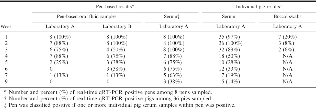
Number and percent (%) of real-time qRT-PCR positive pens among 8 pens sampled.
Number and percent (%) of real-time qRT-PCR positive pigs among 36 pigs sampled.
Pen was classified positive if one or more individual pig serum samples within pen was positive.
Indirect fluorescent antibody test
Oral fluid samples were assayed for the presence of specific anti-PRRSV IgG and IgA antibodies by IFAT using fixed MARC-145 cells infected with PRRSV as previously described, 39 with a few modifications. Known positive and negative controls were diluted 1:20 in 0.01 M PBS (pH 7.2) with 0.05% Tween 20 (PBST) while oral fluid samples were tested after being diluted 1:2 in PBST. For detection of anti-PRRSV antibodies, FITC-conjugated antiporcine IgG h and FITC-conjugated antiporcine IgA h were used as the secondary antibody for detection of anti-PRRSV IgG and IgA, respectively.
Enzyme-linked immunosorbent assay
Both oral fluid and serum samples were tested for the presence of antibodies against PRRSV using the HerdChek PRRS Antibody 2XR Test Kit. i Serum samples were diluted 1:50 and assayed according to the manufacturer's instruction. Oral fluids were assayed according to the manufacturer's instructions, with the exception that samples were centrifuged at 1000 × g for 10 min and diluted 1:2 in dilution buffer provided by the manufacturer prior to being assayed. All ELISA results were expressed as sample/positive (S/P) ratios.
Statistical methods
All statistical analyses were performed using JMP 6.0.0. j Serum and oral fluids results for ELISA and real-time qRT-PCR were analyzed using multivariate analysis of variance (MANOVA), with time as the repeated measure and age as a model effect. When statistically significant (P < 0.05) age-by-time interactions were observed, Tukey Honestly Significantly Different (HSD) test was used to determine when in time (DPI) the response by age differed.
Linear regression was used to evaluate the relationship between oral fluid samples and serum samples with respect to the quantity of viral RNA and, in a separate analysis, ELISA S/P values. Consolidation of data was necessary to conduct linear regression, despite its recognized effect on type 2 error rate. That is, oral fluid samples were collected from pens (not individual pigs) at 2- to 4-day intervals, whereas serum samples were collected from individual pigs twice a week through day 14, then weekly. Thus, the linear regression analyses (real-time qRT-PCR, ELISA) were based on a weekly average of oral fluid results for each pen and a weekly average of serum results for individual pigs within the same pen.
Results
Porcine reproductive and respiratory syndrome virus real-time quantitative reverse transcription polymerase chain reaction
Oral fluid samples were assayed by real-time qRT-PCR at 2 laboratories (A and B). All oral fluid samples collected from negative control pens were negative in both laboratories (n = 55). Overall, 88% of the qualitative results from laboratories A and B were in agreement (i.e., 37 positive samples and 147 negative samples of the 209 samples assayed). Categorical results from the 2 laboratories are given by week postinoculation in Table 1. Real-time qRT-PCR estimates of virus concentration were based on extrapolation to TCID50 standards (laboratory A) or transcript RNA product standards (laboratory B). Linear regression analysis showed a positive correlation (r2 = 0.60) between the quantitative estimates from the 2 laboratories. Age at the time of inoculation had no effect on the quantity of virus present in oral fluid samples (MANOVA: laboratory A, P = 0.21; laboratory B, P = 0.42; Figs. 1, 2; Tables 2, 3).
Given that real-time qRT-PCR results from the 2 laboratories were comparable, buccal swabs and serum samples (reported below) were only assayed at laboratory A. Buccal swab samples were collected from individual pigs twice weekly through 21 DPI.
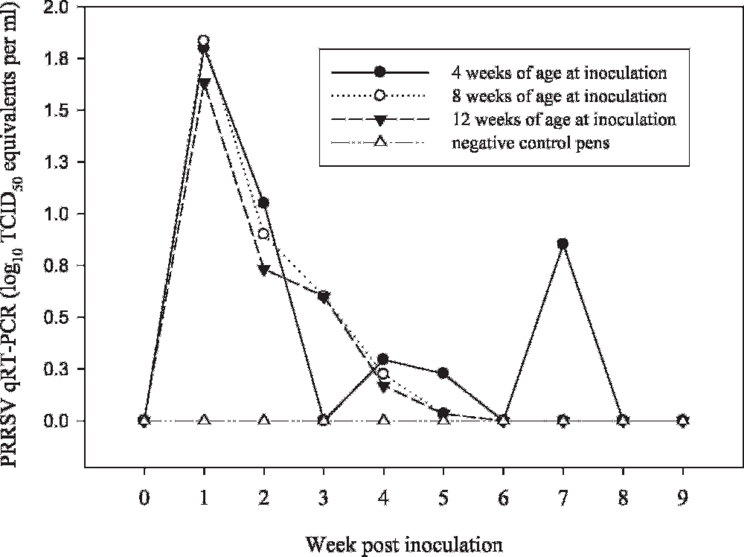
Laboratory A: Oral fluid Porcine reproductive and respiratory syndrome virus (PRRSV) quantitative reverse transcription polymerase chain (qRT-PCR) least square means by pig age and week postinoculation.
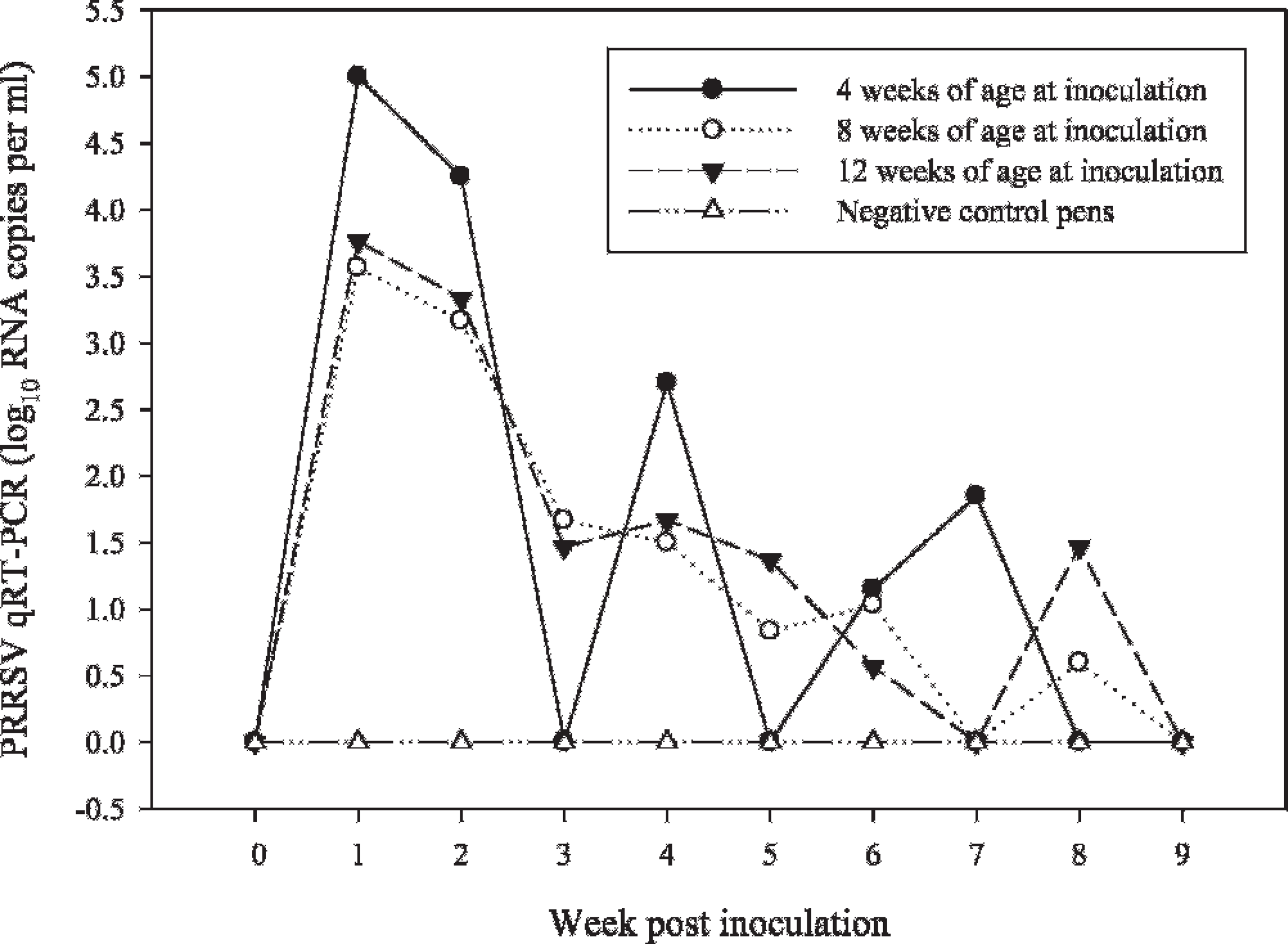
Laboratory B: Oral fluid Porcine reproductive and respiratory syndrome virus (PRRSV) real-time quantitative reverse transcription polymerase chain (qRT-PCR) least square means by pig age and week postinoculation.
All samples collected prior to inoculation (n = 48) were real-time qRT-PCR negative. Of 216 buccal swabs collected from PRRSV-inoculated pigs on 3, 7, 10, 14, 17, and 21 DPI, 29 were positive by real-time qRT-PCR. One of 72 buccal swab samples collected from negative control pigs during the observation period tested positive. All other specimens (serum, oral fluid, buccal swab) collected from negative control pigs tested negative by qRT-PCR and ELISA. This supports the interpretation of this 1 result as a false positive. Pig-matched serum and buccal swab samples were collected from PRRSV-inoculated pigs on 7, 14, and 21 DPI. Of 108 paired samples, 12 were real-time qRT-PCR-positive for both specimens; 92 were serum-positive and buccal swab-negative; and 4 were negative for both samples.
Serum samples were collected from all pigs twice weekly through 14 DPI, then weekly through 63 DPI. Analysis (MANOVA) of the real-time qRT-PCR data detected a difference (P = 0.0001) in the level of viremia by the age of the pig at the time of inoculation. Overall, the youngest pigs had the highest level of viremia followed by the next youngest group (Fig. 3; Table 4). Statistically significant differences in the level of PRRSV viremia by age group were identified (Tukey HSD) at 7, 10, and 42 DPI (Table 4).
Comparison showed that the levels of virus in serum and oral fluid samples followed a similar pattern, with oral fluid consistently containing a lower concentration of virus (Fig. 4). Both serum and oral fluid samples were real-time qRT-PCR positive from 3 DPI to 4 to 5 weeks postinoculation, with sporadic positives thereafter. Linear regression analysis estimated the correlation (r2) between virus concentration in serum and oral fluids at 0.56 (Fig. 5).
The diagnostic sensitivity of real-time qRT-PCR for serum and oral fluid samples corresponded to the percentage of inoculated animals (or pens) that tested positive (Table 1). For oral fluid samples, the mean diagnostic sensitivity for the first 4 weeks postinoculation was 88% for laboratory A and 81% for laboratory B. By comparison, the mean diagnostic sensitivity of laboratory A on individual pig serum samples for the same 4-week period was 89%. Neither laboratory reported positive real-time qRT-PCR results for serum or oral fluid samples from negative controls (i.e., diagnostic specificity was 100%).
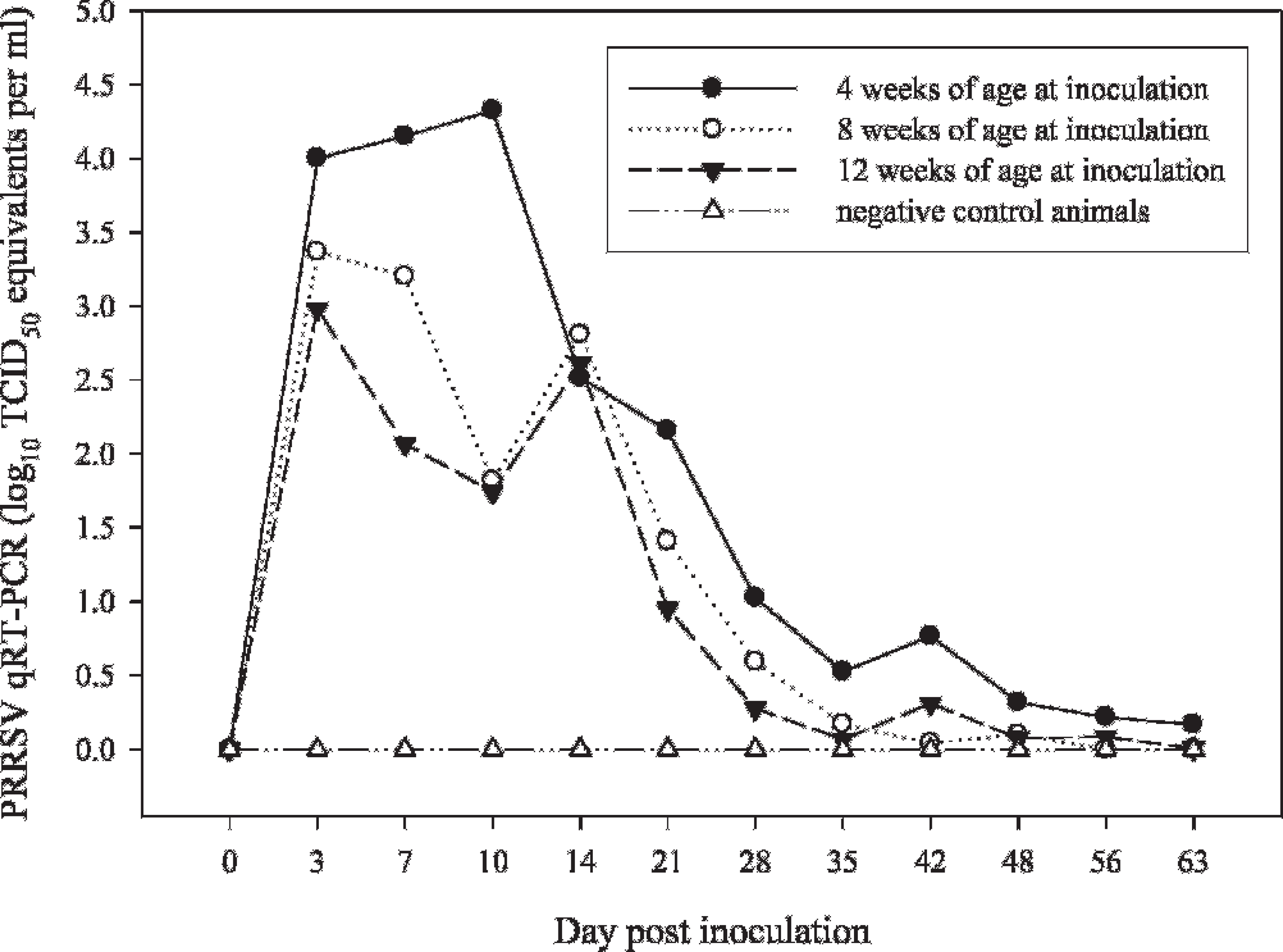
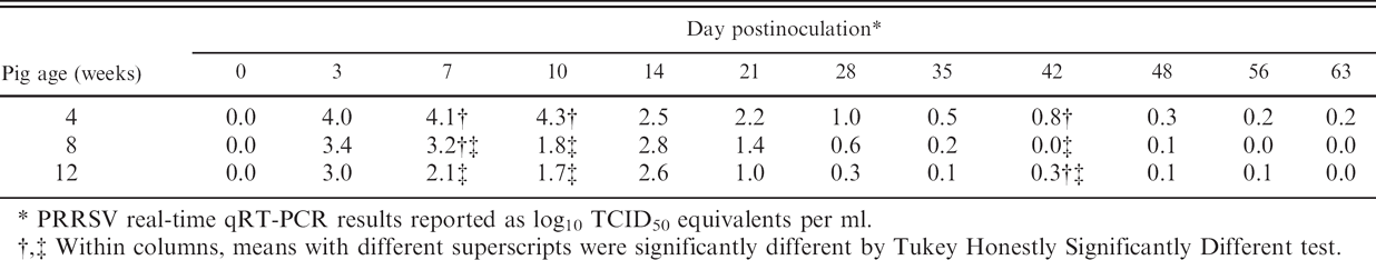
Laboratory A: Oral fluid Porcine reproductive and respiratory syndrome virus (PRRSV) real-time quantitative reverse transcription polymerase chain (qRT-PCR) least square means by pig age and week postinoculation.
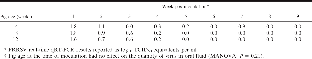
PRRSV real-time qRT-PCR results reported as log10 TCID50 equivalents per ml.
Pig age at the time of inoculation had no effect on the quantity of virus in oral fluid (MANOVA: P = 0.21).
Laboratory B: Oral fluid Porcine reproductive and respiratory syndrome virus (PRRSV) real-time quantitative reverse transcription polymerase chain (qRT-PCR) least square means by pig age and week postinoculation.
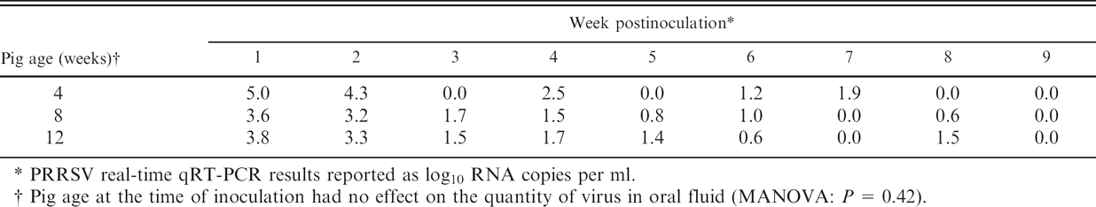
PRRSV real-time qRT-PCR results reported as log10 RNA copies per ml.
Pig age at the time of inoculation had no effect on the quantity of virus in oral fluid (MANOVA: P = 0.42).
Porcine reproductive and respiratory syndrome virus enzyme-linked immunosorbent assay
Statistical analysis of the serum antibody response found a significant difference in ELISA S/P values by pig age at the time of inoculation (MANOVA: P = 0.0001). The youngest pigs had the highest S/P response, followed by the next youngest group (Fig. 6; Table 5). Statistically significant differences in S/P values by age group were identified (Tukey HSD) from 14 to 63 DPI, with the 4-week-old group consistently showing the highest S/P response.
In contrast, a comparison of oral fluid ELISA results found no significant difference in least square means between PRRSV-inoculated and negative-control pens (MANOVA: P = 0.72). Likewise, no significant difference was detected in the ELISA response within the inoculated groups by pig age at time of inoculation (MANOVA: P = 0.15).
Serum Porcine reproductive and respiratory syndrome virus (PRRSV) real-time quantitative reverse transcription polymerase chain (qRT-PCR) least square means by pig age and day postinoculation.
Indirect fluorescent antibody test
Anti-PRRSV IgG was detected by IFAT in 20 of 154 oral fluid samples from inoculated pens, with positives sporadically distributed across the postinoculation observation period. No anti-PRRSV IgA was detected in oral fluid samples with the protocol described. Samples from negative control pens were negative for both IgG and IgA.
Discussion
In humans, viral infections in which the agent is present in oral fluids include Hepatitis A virus, 22 Hepatitis B virus, 18 Hepatitis C virus, 30 Hepatitis G, 38 Human herpesvirus I, 7 Human herpesvirus 6, 5 Human immunodeficiency virus, 10 Measles virus, 17 Mumps virus, 17 Mycobacterium tuberculosis, 18 Rubella virus, 17 Severe acute respiratory syndrome coronavirus, 34 Transfusion-transmitted virus, 38 and Human herpesvirus 3 (commonly known as Varicella-zoster virus 1). 9 Some viral infections were also demonstrated to produce detectable levels of specific antibodies in oral fluids: Dengue virus, 6 Hepatitis A virus, 28,29 Hepatitis B virus, 28,29 Hepatitis C virus, 29 Human immunodeficiency virus, 28 Measles virus, 27 Norwalk virus, 25 Rubella virus, 27 and others. In human diagnostic medicine, the presence of pathogens and/or antibodies has stimulated important developments in diagnostic medicine. For example, in 2004, the FDA approved a rapid assay (20 min) for detection of antibodies against HIV-1 in oral fluid, blood, or plasma samples (Anonymous: 2004, FDA approves oral fluid rapid HIV test. Dentistry Today 23:42). Likewise, IgG-based assays for oral fluid have been developed for Epstein-Barr virus, Hepatitis A and B viruses, Human parvovirus B19, and Rubella virus, 24 and assays based on the IgM antibodies have been developed to detect recent infections by Hepatitis A and B viruses, Measles virus, Mumps virus, Human parvovirus B19, and Rubella virus. 24
Serum Porcine reproductive and respiratory syndrome virus (PRRSV) real-time quantitative reverse transcription polymerase chain (qRT-PCR) least square means by age and day postinoculation.
PRRSV real-time qRT-PCR results reported as log10 TCID50 equivalents per ml.
Within columns, means with different superscripts were significantly different by Tukey Honestly Significantly Different test.
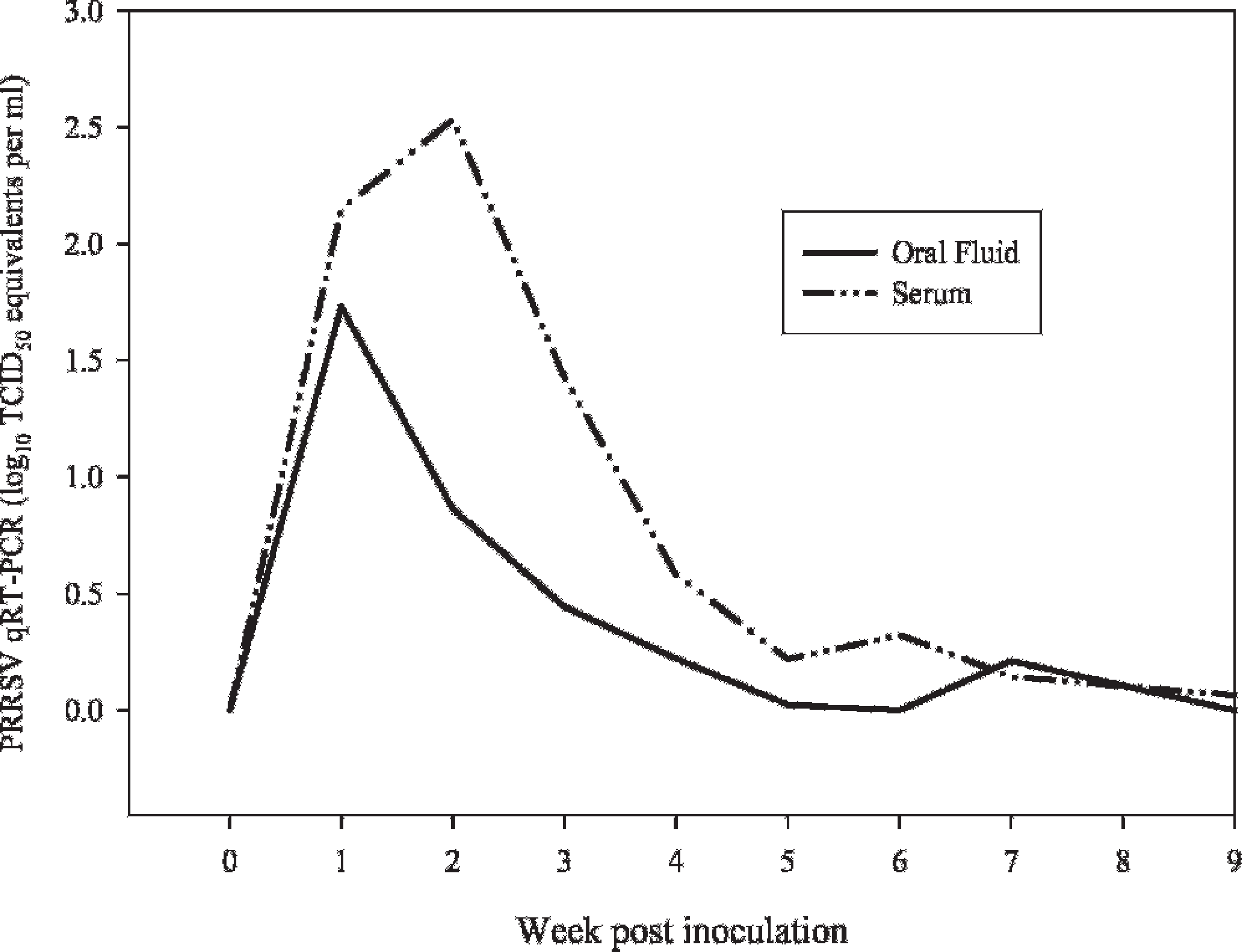
Serum and oral fluid Porcine reproductive and respiratory syndrome virus (PRRSV) quantitative reverse transcription polymerase chain (qRT-PCR) results by week postinoculation.
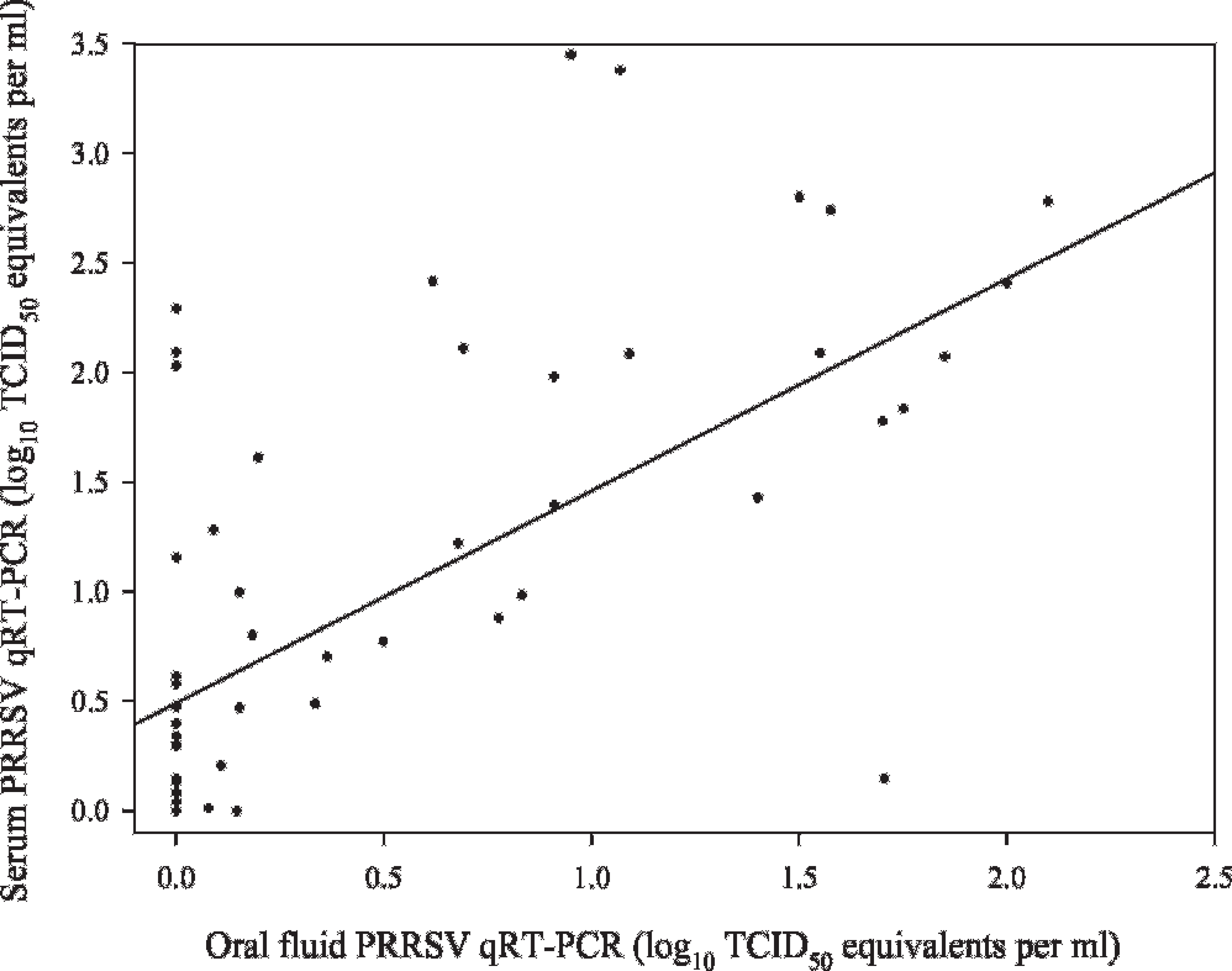
Correlation between serum and oral fluid Porcine reproductive and respiratory syndrome virus (PRRSV) quantitative reverse transcription polymerase chain (qRT-PCR) results: (r2
In animals, the presence of pathogens in oral fluid has generally been described in the context of transmission (e.g., Rabies virus). 2 In swine, examples of viral pathogens present in oral fluids include Bovine viral diarrhea virus 1 and 2, 33 Foot-and-mouth disease virus, 1,4 Porcine circovirus−2, 32 Suid herpesvirus 1 (commonly known as Pseudorabies virus or Aujeszky's disease virus), 3 and Vesicular stomatitis viruses. 21 Pathogen-specific antibodies in oral fluids have also been described in animals, although the research is extremely limited. In swine, the appearance of specific antibodies in oral fluids was shown following inoculation with Actinobacillus pleuropneumoniae, 19,20 cholera toxin B subunit, 15 and group E Streptococcus spp. 16
The use of oral fluids in veterinary diagnostic medicine has been minimal, for the most part limited to the diagnosis of Feline immunodeficiency virus. 30 Recently, oral fluid samples were reported as a method for the detection of Escherichia coli 0157:H7 in feedlot cattle (Renter DG, Visser A, McFall M et al: 2004, Rapid pen-level surveillance of E. coli 0157:H7 in finished feedlot cattle. Animal Health Forum 91:3).
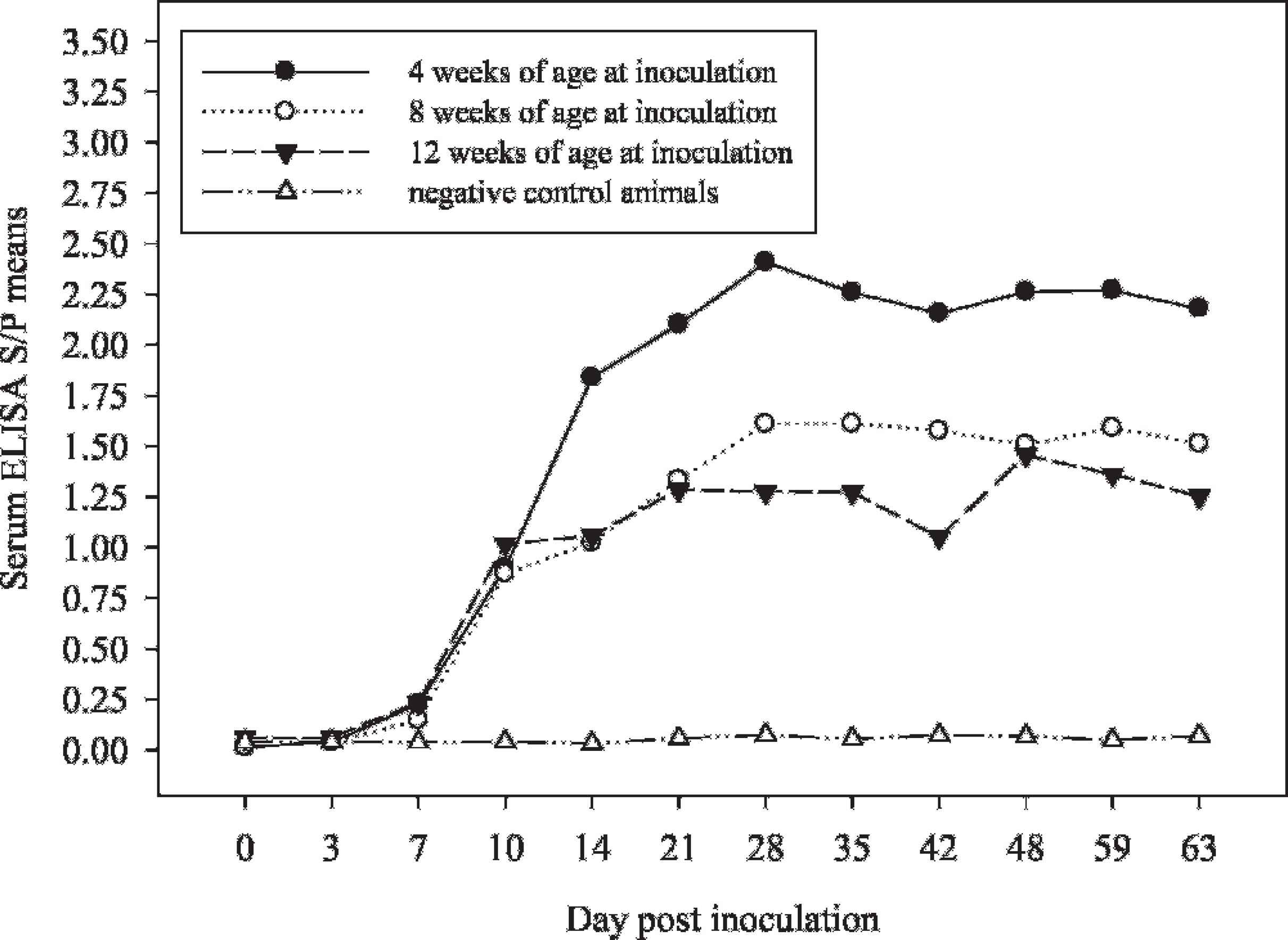
Serum Porcine reproductive and respiratory syndrome virus (PRRSV) enzyme-linked immunosorbent assay (ELISA) least square means by pig age and Day postinoculation.
Serum Porcine reproductive and respiratory syndrome virus enzyme-linked immunosorbent assay sample/positive least square means by pig age and Day postinoculation
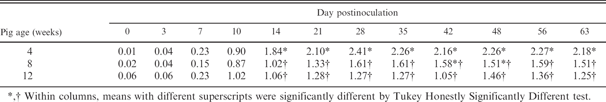
Within columns, means with different superscripts were significantly different by Tukey Honestly Significantly Different test.
Within columns, means with different superscripts were significantly different by Tukey Honestly Significantly Different test.
The research reported here represents a further investigation of the application of oral fluids to veterinary diagnostic medicine. In this experiment, the collection of oral fluid samples from pens of pigs was determined to be easy and efficient. Normal pig behavior was conducive to sample collection, that is, pigs naturally investigate and chew on new objects within the pen (i.e., rope). Porcine reproductive and respiratory syndrome virus was detected by real-time qRT-PCR in oral fluid samples for approximately 4 weeks. The fact that 2 laboratories independently arrived at similar results (88% agreement) suggests that oral fluid samples could be used to monitor PRRSV infection in commercial swine herds using currently available PCR-based assays by testing at ≤4-week intervals. Specific anti-PRRSV antibody was detected in oral fluids, but additional research will be required to develop diagnostically sensitive assays. Oral fluid sampling must still be validated in the field, but preliminary data suggest that this approach could offer a significant improvement in the ease, timeliness, and cost of disease surveillance in commercial swine populations.
Acknowledgements
The study was supported in part by Pork Checkoff funds distributed through the National Pork Board, PO Box 9114, Des Moines, IA 50306. The authors thank the faculty and staff of the Iowa State University Veterinary Diagnostic Laboratory for advice and technical support.
Footnotes
a.
Becton Dickinson, Franklin Lakes, NJ.
b.
Fisher Scientific, Logan, UT.
c.
Ace Hardware Corporation, Colorado Springs, CO.
d.
QIAGEN, Inc., Valencia, CA.
e.
Applied Biosystems, Foster City, CA.
f.
Integrated DNA Technologies, Coralville, IA.
g.
GenBank, Bethesda, MD.
h.
Bethyl Laboratories, Montgomery, TX.
i.
IDEXX Laboratories, Inc., Westport, ME.
j.
SAS Institute, Cary, NC.