
Abstract
A real-time polymerase chain reaction (PCR) assay was used for quantifying Leishmania infantum DNA in urine samples from naturally infected dogs. Forty-one infected dogs were divided into 3 groups: 22 dogs showing only cutaneous signs (group 1), 12 dogs showing hematuria (group 2), and 7 dogs affected by severe nephropathy (group 3). Groups 2 and 3 dogs showed altered laboratory parameters related to an impairment of renal function. The real-time PCR analysis showed higher levels of Leishmania DNA in the lymph node aspirates from all groups of infected dogs versus those measured in their blood or urine. Interestingly, urine samples from dogs belonging to groups 2 and 3 contained a higher Leishmania DNA load than that detected in their blood. This finding suggests that a real-time PCR analysis of urine from infected dogs could be a useful and noninvasive tool for monitoring the severity of leishmaniasis.
Leishmaniasis is a zoonotic disease caused by different species of the protozoal genus Leishmania. In the Mediterranean area, the disease is endemic and is mainly due to Leishmania infantum. 6 Dogs are the main reservoir of L infantum parasites, and dogs play a central role in the transmission of the disease to humans through phlebotomine sand flies. 1 To successfully control the disease, efficient and reliable diagnosis of canine visceral leishmaniasis (CVL) is essential. Conventional procedures for diagnosing CVL include serological tests such as indirect fluorescent antibody test (IFAT), enzyme-linked immunosorbent assay, immunoblot analysis, and direct agglutination test. 11 However, serological tests show a high degree of cross-reactivity and cannot discriminate between subclinical infections and current and past infections. Therefore, detection of parasites by direct microscopic examination of smears and cultural isolation is required to reliably confirm the diagnosis.
In the last decade, polymerase chain reaction (PCR) has proved to be a sensitive and powerful tool to detect Leishmania DNA, as well as for parasite characterization. 14,18,20 Polymerase chain reaction assay solves several problems, such as the low sensitivity issue often shown by the microscopic examination and the low predictive value of serology, whose results may be influenced by either persistent antibodies or immunosuppression. However. classical PCR protocols do not allow a quantitative analysis of the parasite load in clinical specimens.
A recent approach to estimate the parasitic load from mouse livers and naturally infected dogs with L. infantum has been reported. 3,10 This approach is based on the use of real-time quantitative PCR to target the parasite DNA polymerase gene. Furthermore, real-time PCR assays that detect, quantify, and/or differentiate Leishmania organisms have been developed. 5,10,16,19 It was recently demonstrated that the simultaneous evaluation of Leishmania DNA load and cytokine levels in different kinds of tissues by real-time PCR assay can be used to predict disease development in asymptomatically infected dogs. 9 This study compares the use of urine versus lymph node and blood to detect and quantify Leishmania DNA by real-time PCR.
Biological samples were collected from naturally infected dogs with leishmaniasis examined at the Department of Veterinary Internal Medicine (University of Naples Federico II, Napoli, Italy) over a period of 1 year. Leishmaniasis diagnosis in all subjects was assessed by IFAT and confirmed by conventional PCR in lymph node aspirates, as previously described. 8 Three uninfected dogs living in the Abruzzo region of Italy where the disease is not endemic were selected as negative controls. The infected dogs of different breeds and sex, ranging in age from 1 to 10 years, were classified into 3 groups using the following criteria: 22 dogs showing cutaneous signs (group 1), 12 dogs showing hematuria but no cutaneous signs (group 2), and 7 dogs affected by severe nephropathy (group 3). Renal biopsy was performed to achieve diagnosis of glomerular disease in group 3 dogs. Biochemical profiles were determined for all dogs. The biochemical test profile included serum total proteins, creatinine, urea, globulins, and albumin and urine analysis including chemical and physical assessment, as well as sediment evaluation, and determination of urine protein-to-creatinine ratio (UP/C). Furthermore, differential diagnosis for infections that may cause the same clinical and laboratory signs in the study area, such as ehrlichiosis, babesiosis, and leptospirosis, was performed by IFAT and conventional PCR plus sequencing. 8 No coinfections were detected in all dogs belonging to the 3 groups.
Urine, blood samples, and lymph node aspirates were collected from each dog. Urine samples collected by free catch were centrifugea at 1,200 × g for 10 minutes at 4°C. The pellet was stored at −80°C before DNA extraction. One part of supernatant was used to determine the creatinine and protein concentrations for calculating UP/C by using a procedure previously described. 13 Blood samples in ethylenediamine tetra-acetic acid (EDTA)-containing tubes and lymph node aspirates were stored at −80°C before DNA extraction.
The reference strain IPT1 ZMON1 was obtained from the collection of the Leishmaniosis National Reference Center of the Istituto Zooprofilattico Sperimentale della Sicilia (Palermo, Italy). The IPT1 was grown at 25°C for 7 days in Evans' modified Tobie's medium 21 with 15% rabbit blood, 5% fetal bovine serum, 250 μg gentamicin/mL, and 500 μg/ml 5-fluorocytosine.
DNA extraction from parasite cultures was carried out as previously described 18 with some modifications. The parasite culture was centrifuged for 15 minutes at 3,500 × g at 4°C and resuspended in lysis buffer (50 mM NaCL 10 mM EDTA, and 50 mM Tris-HCl, pH 7.4) with 1% sodium dodecyl sulfate. The mixture was incubated for 30 minutes at 60°C, and proteinase K (0.1 mg/ml, final concentration) was added and incubated for 3 hours at 60°C. After double extraction with buffered phenol-chloroform-isoamyl alcohol (25:24:1, v/v/v), the DNA was precipitated with 0.3 M sodium acetate, overlaid with 2.5 volumes of ice-cold ethanol, and mixed by inversion. DNA was recovered by centrifugation at 13,000 × g for 20 minutes, washed with 70% ethanol, and suspended in sterile distilled water.
DNA was extracted from urine, whole blood samples, and lymph node aspirates by the QIAamp Blood Kit a according to the manufacturer's instructions. Briefly, the pellet obtained from 5 ml of urine samples and 0.2 ml of blood or lymph node aspirate diluted with 0.2 ml sterile phosphate-buffered saline (PBS) was incubated in a suitable lysis buffer with 0.025 ml proteinase K (20 mg/ml) for 5 minutes at 70°C or overnight at 56°C. After vortexing, 0.2 ml of ethanol was added to the mixtures. The mixtures were applied into the QIAamp spin columns a and centrifuged. After 2 washings, DNA was eluted with 0.2 ml of the supplied buffer preheated at 70°C. The concentration and purity of extracted DNA were assessed by measuring spectrophotometrically the absorbance at 260 nm and 280 nm, respectively, and by gel electrophoresis.
TaqMan primers and probe for L. infantum DNA were chosen in the constant region of the kinetoplast DNA minicircle (GenBank accession number AF291093): forward primer 5′-GGCGTTCTGCGAAAACCG-3′, reverse primer 5′-AAAATGGCATTTTCGGGCC-3′, probe 5′-AAAATGGCATTTTCGGGCC-3′. The primers and probe for the housekeeping beta-actin gene were selected as previously reported. 9 Amplification was performed in 0.025 ml of reaction mixture containing 1X TaqMan Universal Master Mix, b 100 pmol/μl of the specific primer. 10 pmol/μl of the labeled probe, and 50 ng of DNA. The thermal cycling conditions included an initial incubation for 2 minutes at 50°C, followed by a 10-minute denaturation at 95°C and 45 cycles at 95°C for 15 seconds and 60°C for 1 minute each. Each amplification run contained a negative control. Each standard, sample, and negative control was analyzed in triplicate for each run.
Standard curves were prepared for both the target and the housekeeping gene. A stock solution of L. infantum DNA was obtained by extraction from 10 9 promastigotes. Ten-fold serial dilutions of the DNA stock solution were performed to obtain the points of the curve spanning from the DNA equivalent to 10 6 cells to 1 cell/μl. The standard curve, calculated by independent experiments, was linear over an at least 6-log range of DNA concentration points, with an average correlation coefficient of 0.988. The difference for each point of the curve was 1 log factor. For each experimental sample, the amount of target and housekeeping gene was determined from the appropriate standard curves. The target DNA amount was then divided by the housekeeping gene amount to obtain a normalized target value.
Statistical analysis for Leishmania load in the different tissues of each group of infected dogs was carried out with GraphPad InStat software 3.0 application. c Creatinine, urea, total serum protein, albumin, globulins, albumin-to-globulin ratio (A/G), and UP/C for each dog were evaluated using 1-way analysis of variance.
Forty-one dogs showing clinical signs of infection by L. infantum and testing positive by both IFAT analysis of serum and PCR of lymph node aspirates at the time of the diagnosis were included in this study. Twenty-two infected dogs showing only cutaneous lesions were designated as group 1. Besides disorders of the skin, these dogs showed loss of body weight, enlarged lymph nodes, and/or fever. Their serum levels of biochemical parameters were normal, with the exception of total proteins and beta globulins, which were slightly higher than normal. Urine analysis showed a slightly altered UP/C (Table 1). For group 1. Leishmania load as measured by real-time PCR was higher in lymph node aspirates and in blood than in urine samples. However, a Leishmania DNA load ranging from 1 to 21 parasite DNA/μl was still detected in the urine of these dogs (Fig. 1).
The second group of 12 dogs showing hematuria and other signs of infection, such as a severe lymphadenopathy but no cutaneous signs, had serum globulin levels slightly higher than normal, whereas albumin levels and A/G were lower than normal. Urine protein-to-creatinine ratio was also altered (Table 1). In these dogs, the quantification of Leishmania by real-time PCR showed a higher number of parasites in lymph node aspirates than in blood and urine samples. However, urine samples of these dogs contained a Leishmania DNA load ranging from 89 to 115 parasite DNA/μl, whereas the amount of parasite DNA in their blood was much lower (Fig. 1).
Seven dogs designated as group 3 at clinical examination showed severe infection symptoms, including clinical and laboratory parameters related to renal failure. The majority of serum and urine parameters were strongly altered (Table 1). In particular, their serum levels of urea, creatinine, and beta and gamma globulins were much higher than normal values, whereas albumin levels and A/G were lower than normal values. These dogs had a statistically significant higher UP/C compared with groups 1 and 2. Urine sediment findings for group 3 were compatible with renal failure, whereas group 2 was compatible with hematuria, and group 1 had normal urinary sediment (data not shown). For group 3, the estimation of Leishmania organisms by real-time PCR assay showed a much higher parasite DNA load in their urine samples (568–590 parasite DNA/μl) compared with that of blood. However, lymph node samples still contained a higher amount of Leishmania DNA than the other 2 samples. No Leishmania DNA was detected in urine, blood, and lymph node samples collected from healthy dogs selected as negative controls.
Laboratory findings of infected dogs.*
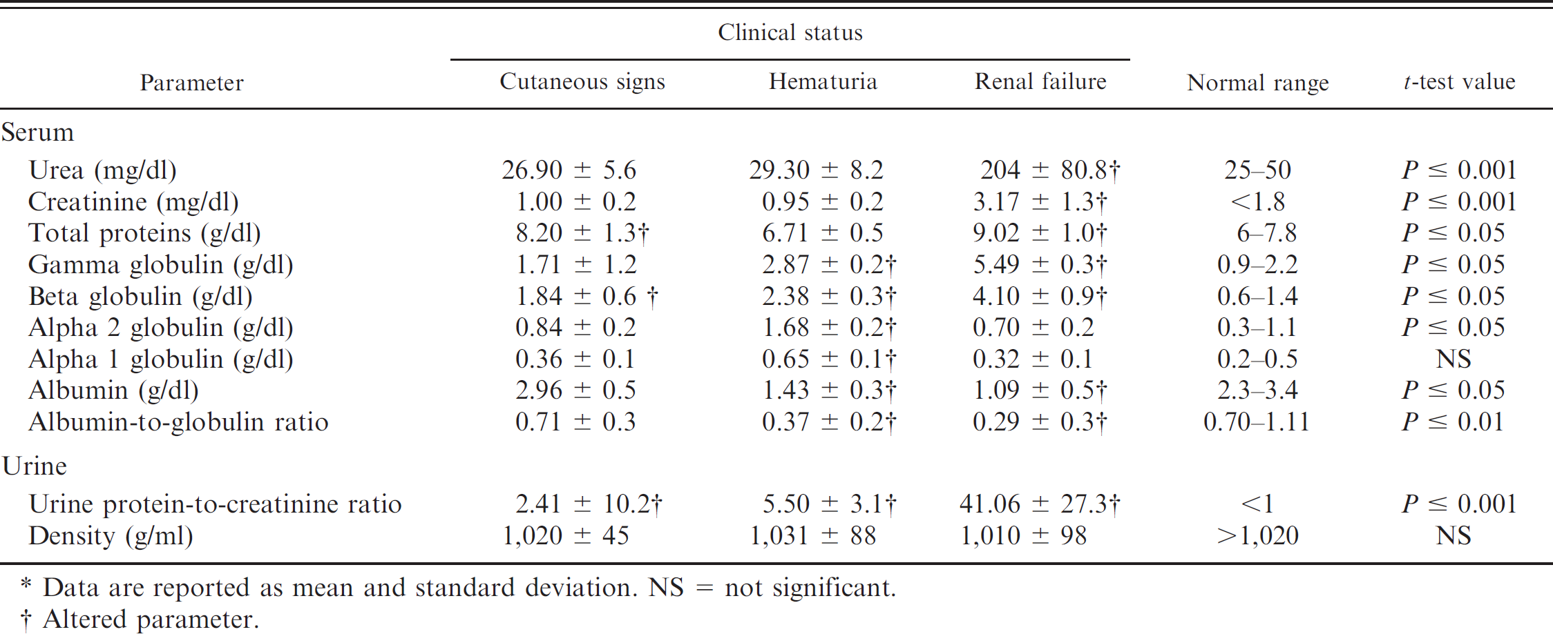
Data are reported as mean and standard deviation. NS = not significant.
Altered parameter.
The real-time PCR assay is based on the TaqMan chemistry that permits quantitative sensitive and reproducible detection in a wide range of parasite loads. 5,10 The DNA target chosen (kinetoplast minicircle elements) ensured high sensitivity levels compared with a conventional PCR because the kinetoplast DNA elements are repeated a thousand times in each parasite (∼10,000 copies per parasite). 15 In this study, the quantification of Leishmania DNA load in the different samples showed differences between the 3 groups of dogs exhibiting distinct clinical symptoms.
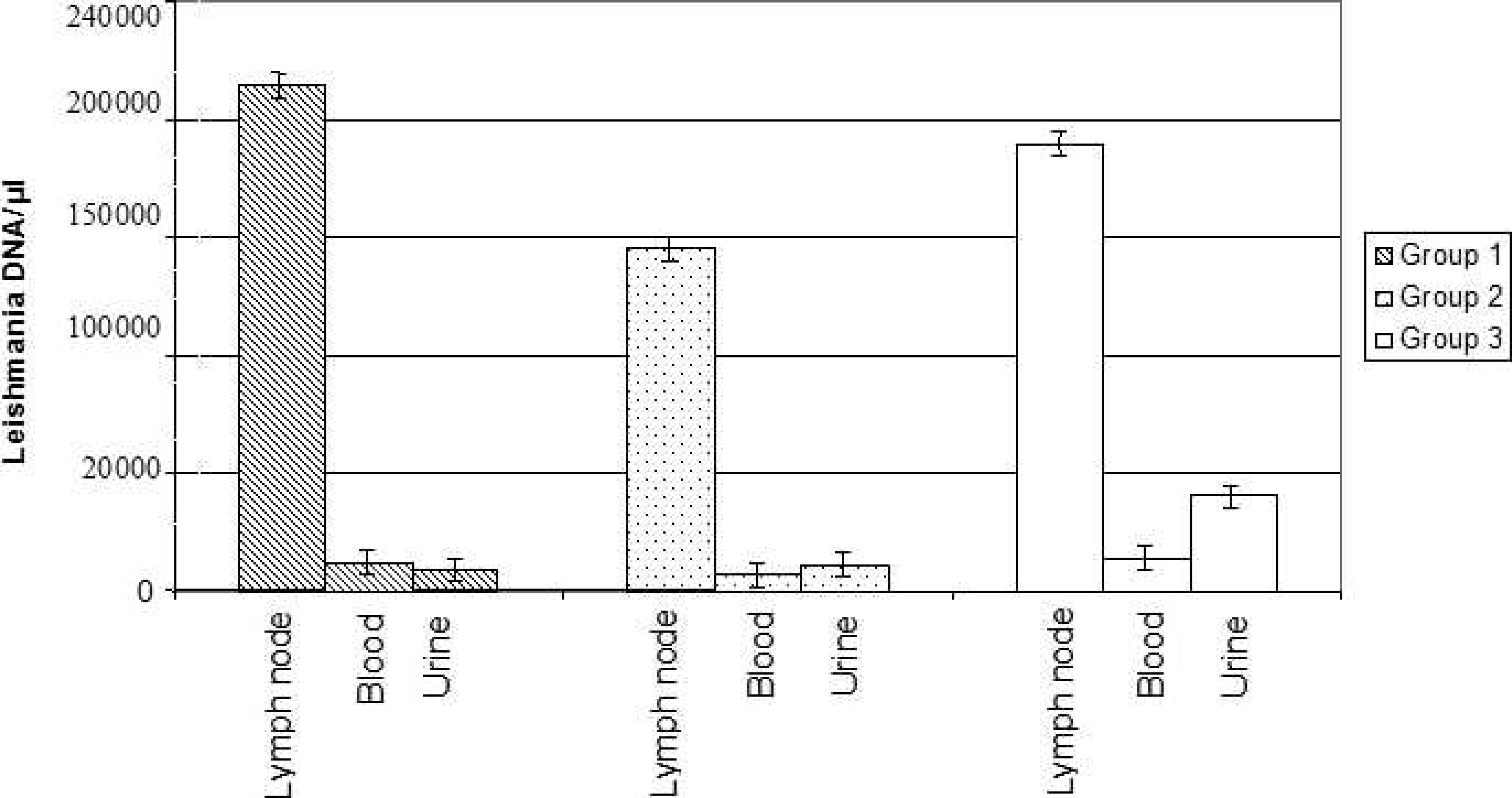
Leishmania load measured by real-time polymerase chain reaction (PCR) in tissue samples collected from naturally infected dogs. Amount of Leishmania DNA/μl detected by realtime PCR assay in the lymph node aspirates, whole blood, and urine from 3 groups of infected dogs: group 1, dogs with only cutaneous signs; group 2, dogs with hematuria; group 3, dogs with severe renal failure. Data represents the mean values for each group. Bars indicate standard deviations.
Group 1 dogs for whom skin lesions were the most noticeable clinical symptom showed the highest levels of Leishmania DNA in lymph node aspirates compared with those in blood and urine samples. These results confirm the existing evidence that lymph node is the best sample for CVL diagnosis using both conventional PCR and real-time PCR assays. 5,8 However, because lymph node collection is invasive, blood or urine appear to be reliable samples for real-time PCR-based diagnosis and monitoring of Leishmania infection in dogs with a mild clinical picture.
Lymph node aspirates proved to be the sample for detecting Leishmania load by real-time PCR in group 2 dogs. Although hemorrhages are believed to be the common cause of proteinuria in dogs, hematuria may not cause an increase in urinary albumin until it becomes macroscopic and may not increase the UP/C. 21
Leishmania infection can cause chronic renal failure in dogs, usually characterized by glomerulonephritis, interstitial nephritis, and occasionally amyloidosis. 12 Immune complex deposition may cause a secondary inflammatory reaction, and the decreased perfusion of the peritubular capillaries may lead to tubular and interstitial tissue ischemia. 7 Proteinuria is observed as a result, which can range from moderate to severe according to the evolution of the disease. 13 However, proteinuria was observed in dogs without evident morphologic renal lesions and with normal renal function as assessed by histologic analysis and by evaluation of serum concentrations of creatinine, urea, and phosphorus. 2 In the present study, all groups of infected dogs, including the dogs showing only cutaneous signs without any sign of nephropathy, showed a UP/C higher than 1, albeit to different extents.
The group of dogs affected by hematuria had decreased levels of serum albumin, a decreased A/G, and a high UP/C, whereas urea and creatinine levels were in the normal range. These laboratory findings suggest that these dogs may progress toward renal failure. In humans, renal involvement has been demonstrated in patients with visceral leishmaniasis showing hematuria and proteinuria. 17 However, renal involvement in visceral leishmaniasis can be mild and seems to subside with the cure of the leishmanial infection. 4 In endemic areas, veterinarians are usually confronted not only with cases compatible with CVL based on the symptoms but also with several diagnostic tests, sometimes with no definite results. This has led to the development and application of different PCR protocols to diagnose Leishmania infection. However, because one of the features of leishmaniasis is the presence of residual or latent parasites after treatment, quantitative approaches have been developed to elucidate the status of PCR-positive dogs and for monitoring the parasitemia in the follow up of infected dogs. The present findings demonstrate that urine sampling may represent a simple and noninvasive route for a real-time PCR assay useful for improving prognosis and clinical management of infected dogs, especially those with symptoms of renal involvement. Leishmania can be detected in differing quantities depending on the symptoms: urine might be a suitable sample in many situations, although low Leishamnia levels are present in cutaneous infections. In these cases, blood may represent a valid noninvasive option. Blood does not seem a suitable sample for hematuriac dogs, and urine would be more appropriate. In all cases, the lymph node aspirate remains the ideal sample, and the benefits of the noninvasive sampling technique versus lymph node sampling will need to be determined on a case-to-case basis.
Acknowledgements. This work was supported by a grant from Regione Campania, Legge 5/2004, Italy.
Footnotes
a.
Qiagen, Santa Ciarita, CA.
b.
Applied Biosystems, Foster City, CA.
c.
GraphPad software, San Diego, CA.