
Abstract
Chronic wasting disease (CWD) in Wisconsin was first identified in February 2002. By April 2005. medial retropharyngeal lymph node (RLN) tissues had been examined from over 75,000 white-tailed deer for the presence of CWD by either immunohistochemical (IHC) staining for the prion protein associated with CWD (PrPres) or by using enzyme-linked immunosorbent assays with confirmation of positives by IHC staining and had been detected in 469 animals. Obex tissue was also available from 438 of the CWD-positive animals and was CWD positive by IHC staining in 355 (81%). To verify whether false-negative results were possible examining only RLN, both obex and RLN samples were examined for CWD by IHC staining from 4,430 of the white-tailed deer harvested from an area in Wisconsin where the overall deer CWD prevalence was approximately 6.2%. Two hundred and fourteen of the 269 positive deer (79.6%) had deposits of PrPres in both obex and lymphoid tissues. 55 (20.4%) had deposits only in lymphoid tissue, and there were no deer that had deposits only in obex.
Keywords
Chronic wasting disease (CWD) is a progressive, fatal, transmissible, spongiform encephalopathy that has been described in mule deer (Odocoileus hemionus), 10,12 white-tailed deer (Odocoileus virginianus), 8 elk (Cervus elaphus), 13 and moose (Alces alces shirasi) 1 in North America. As in other transmissible spongiform encephalopathies, such as scrapie and bovine spongiform encephalopathy, the accumulation of an abnormal isoform (PrPres) of a host protein (PrP c ) in the central nervous system (CNS) and lymphoid tissues of CWD-affected cervids is a hallmark feature of the lesions of CWD. Detection of PrPres in tissue sections by immunohistochemical (IHC) staining is the accepted “gold standard” method for diagnosis of CWD 9 ; however, enzyme-linked immunosorbent assay (ELISA)-based methods are now a standard screening method in many laboratories.
There appears to be variation in the pathogenesis of the disease in deer and elk. 5 In infected mule deer, the dorsal nucleus of the vagus nerve is the first area of the brain in which PrPres can be detected, 11 although the palatine tonsil and medial retropharyngeal lymph node (RLN) accumulate PrPres before it can be detected in the brain. 6,11 Early accumulation of PrPres in lymphoid tissue has also been observed in the preclinical stage of infection in elk; however, a substantial proportion (12.4%) of elk with brains that test positive do not have detectable PrPres in either tonsil or RLNs. 7 The results of this brief communication demonstrate that, in white-tailed deer, PrPres is consistently present in lymphoid tissues before CNS and that, if only brain tissue (obex) is examined, then approximately 20% of animals that are CWD positive will not be detected.
Tissue samples from approximately 75,000 free-ranging, white-tailed deer collected throughout the state of Wisconsin between November 1999 and April 2005 were examined for evidence of CWD by either IHC staining alone or by a combination of an ELISA screening test a,b with IHC confirmation. Beginning in the fall of 2002, the Wisconsin Department of Natural Resources (DNR) made a decision to use only RLN for initial screening as part of its enhanced surveillance program; however, obex from the majority of deer sampled was archived and available for additional testing. Representative samples of RLN and medulla oblongata that contained obex were collected from each animal, and the tissues were preserved in 10% neutral buffered formalin and/or placed in small, labeled, plastic bags and stored at −20°C. In the first season of enhanced surveillance (2002–2003), approximately 41,000 RLN tissues were examined by IHC staining with monoclonal antibody (MAb) F99/97.6.1 and staining techniques as previously described, 4,9 with the exception of the initial formic acid treatment. Briefly, tissue was embedded in paraffin, sectioned at 5 μm, mounted on positively charged glass slides, c deparaffmized, and hydrated in preparation for IHC staining. Tissue treatment before IHC staining consisted of slide immersion in formic acid solution for 5 min followed by several rinses in APK and water. Tissue sections were then autoclaved in Target Retrieval solution d for 20 min with a 25-min cooling period. Prepared slides were stained by using an automated immunostainer e and PrPres MAb F99/97.6.1, a biotinylated secondary antibody, an alkaline-phosphatase-streptavidin conjugate, a substrate chromogen (Fast Red A, naphthol, Fast Red B), and a hematoxylin and bluing counterstain. In the second season (2003–2004), approximately 15,000 RLNs were tested: 9,500 with an ELISA assay b as a screening test with any reactors confirmed by IHC staining, and the remaining 5,500 with IHC staining only. In the 2004–2005 season, approximately 19,000 samples were tested with an ELISA, a with any reactors being confirmed with IHC staining. Of the deer harvested in Wisconsin between November 1999 and April 2005, 469 were confirmed positive by IHC staining. Suitable obex tissue was available from 438 of these animals and was subjected to IHC staining to compare PrPres presence with RLN: 355 (81%) obex samples were positive and 83 (19%) negative.
A 2×2 table comparing medial retropharyngeal lymph node and obex positivity for chronic wasting disease (CWD) in white-tailed deer in deer management units 70A-CWD and 76-CWD in southern Wisconsin from March 2002 to April 2004.
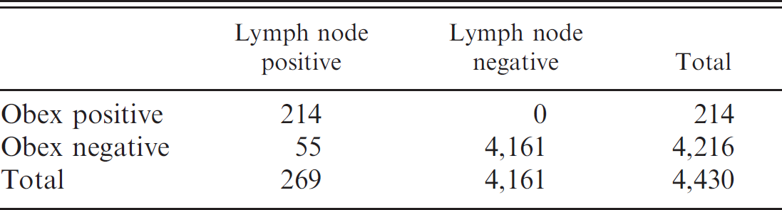
Chronic wasting disease is endemic in southern Wisconsin (http://www.dnr.state.wi.us/org/land/wildlife/whealth/issues/CWD/maps.htm). Within this area, deer management units (DMU) 70A-CWD and 76-CWD include a core area where estimated overall CWD prevalence is 6.2%. 2 To determine whether there were any false-negative results by examining only RLN, a subset of the 75,000 deer tested for which there were both obex and lymph node available for examination (4,430 deer sampled within DMUs 70A and 76 over 2 hunting seasons [March 2002–April 2004]) were reexamined and compared. Two hundred and fourteen deer had deposits of PrPres in both obex and lymph node, 55 had deposits only in lymph node, and no deer had positive staining only in obex (Table 1).
By using RLN with IHC staining as the gold standard, obex IHC diagnostic sensitivity was 79.6% (95% confidence interval [CI]: 74.2–84.2%) and specificity was 100% (95% CI: 89.9–100%). In the sampled population, the apparent prevalence of CWD when using only obex was 4.8% (95% CI: 4.2–5.5%) compared with a true prevalence of 6.1% (95% CI: 5.4–6.8%). Testing only obex would underestimate the number of CWD positive animals by 20%. This compares with a 10% underestimate when using only obex in a recent study 3 conducted in mule deer. However, in the latter study, the obex of 1 mule deer tested positive for CWD, whereas both RLN and tonsil were negative. In this current study conducted in Wisconsin, no animals were detected that were obex positive and RLN negative. Another study compared tissues from Rocky Mountain elk (Cervus elaphus nelsoni) 7 and found 68.6% of positive elk had PrPres in both obex and RLN (compared with 79.6% in white-tailed deer), 19% had PrPres in RLN only (20.4% in white-tailed deer), and 12.4% (0% in white-tailed deer) had PrPres in obex only.
To gain the best diagnostic accuracy for CWD, it would appear that different cervid species require different tissues to be tested; RLN is the tissue of choice in testing for CWD in white-tailed and mule deer, whereas elk require both obex tissue and RLN to be tested for maximum accuracy.
Acknowledgements
Numerous support personnel of the Wisconsin DNR, the Wisconsin Diagnostic Veterinary Laboratory, and deer hunters in Wisconsin made this study possible. The authors appreciate their dedication and hard work in the collection and processing of samples.
Footnotes
a.
Bio-Rad Laboratories, Inc., Hercules, CA.
b.
IDEXX Laboratories, Inc., Westbrook, MA.
c.
Superfrost/plus, Fisher Scientific, Pittsburgh, PA.
d.
Dako North America, Inc., Carpinteria, CA.
e.
Ventana Medical Systems, Inc., Tucson, AZ.