
Abstract
A 20 × 10 cm, lobulated mass was present in the perianal region of a 4-year-old Dales pony. Histopathology revealed an unencapsulated, loose arrangement of sheets and whorls of narrow mesenchymal cells, situated in the deep dermis. Intervening areas had a myxomatous appearance. The whorls were often arranged around a central structure resembling an axon or a vascular structure. Immunohistochemistry revealed that the elongated mesenchymal cells and central axon-like structures expressed vimentin, S-100, and neuron-specific enolase, but not pancytokeratin, glial fibrillary acid protein, and the neurofilament markers. NR4 and 2F11. On the basis of the histopathology and immunohistochemistry, a diagnosis of benign peripheral nerve sheath tumor (schwannoma type) was made. This case was unusual in that the concentric laminations of Schwann cells were very loosely arranged, with an intervening myxomatous stroma (Antoni type B appearance) and despite its benign histological appearance, the mass extended deeply to the proximal sacral vertebrae. Its exact origin was unclear; it may have arisen from cutaneous nerves with deep extension or from neural structures in the sacral region. Due to the incomplete surgical removal, regrowth of the mass occurred.
Peripheral nerve sheath tumors (PNSTs) are derived from Schwann cells, axons, and perineural connective tissue elements and can be benign or malignant. In humans. PNSTs are subclassified as benign (BPNSTs) or malignant (MPNSTs). Neoplasms subclassified as BPNSTs include schwannomas and neurofibromas. 15 In veterinary literature, these subclassifications are less well recognized, and mesenchymal tumors of the skin and soft tissues in domestic animals often combine malignant and benign forms of neurofibroma and schwannoma under the title of peripheral nerve sheath tumor, 10 although more recently, clearer distinctions have been proposed. 3
Benign peripheral nerve sheath tumors are relatively common in humans but occur infrequently in domestic animals, with most frequent reports in cattle and dogs. 3,5,8,16 In humans, the schwannoma type of BPNST account for an estimated 8% of intracranial 2 and 29% of primary spinal tumors. 5,18 Benign peripheral nerve sheath tumors generally present as slow growing tumors that can be located anywhere in the peripheral nervous system (PNS) but are most frequent at the intracranial segment of the eighth cranial nerve. 15 They occur very rarely in horses but have been described in the periorbital region, 12 extradurally in the cranium, 17 in the small intestine, 9 in the skin, and in the spinal cord. 15 This paper describes the histopathological and immunohistological features of an unusual BPNST affecting the perianal and sacral region of a young pony.
A 4-year-old crossbred Dales pony gelding weighing approximately 450 kg was presented with a mass located at the junction between the left lateral aspect of the tail head and the perianal region (Fig. 1). The mass had been present for approximately 3 months and had slowly increased in size during this period, without any other clinical signs. The mass was approximately 20 × 10 cm, smooth, partially alopecie, nonulcerated, firm on palpation, movable locally, and appeared to be dermal to subcutaneous. No lymphadenopathy was identified on external and internal (rectal) lymph node palpation, and no further significant clinical abnormalities were evident. Ultrasonographic examination identified a multilobulated, homogeneous structure present within the deep dermis and extending deep into the gluteal musculature. The deepest margins of the mass could not be visualized adequately on ultrasonographic evaluation.
After discussion with the owner, surgical resection was carried out under general anesthesia with the horse in dorsal recumbency, by blunt dissection of the mass from the surrounding tissues. Despite removal of the more superficial margins of the mass, further dissection revealed that the mass involved the proximal sacral vertebrae and complete excision was not possible. The wound was allowed to heal by secondary intention. Over the following 3 years, the mass has slowly regrown.
The resected tissue was fixed in 10% phosphate-buffered formalin (pH 7.4). Tissues were embedded in paraffin wax and sections (4 μm) were cut, processed by routine methods, and stained with hematoxylin and eosin and Masson's trichrome. Consecutive sections to those used for the histopathological examination were immunolabeled for vimentin, a pancytokeratin (CK), b S-100, c glial fibrillary acid protein (GFAP), d neuron-specific enolase (NSE), e and the neurofilament markers NR4 f and 2F11. g
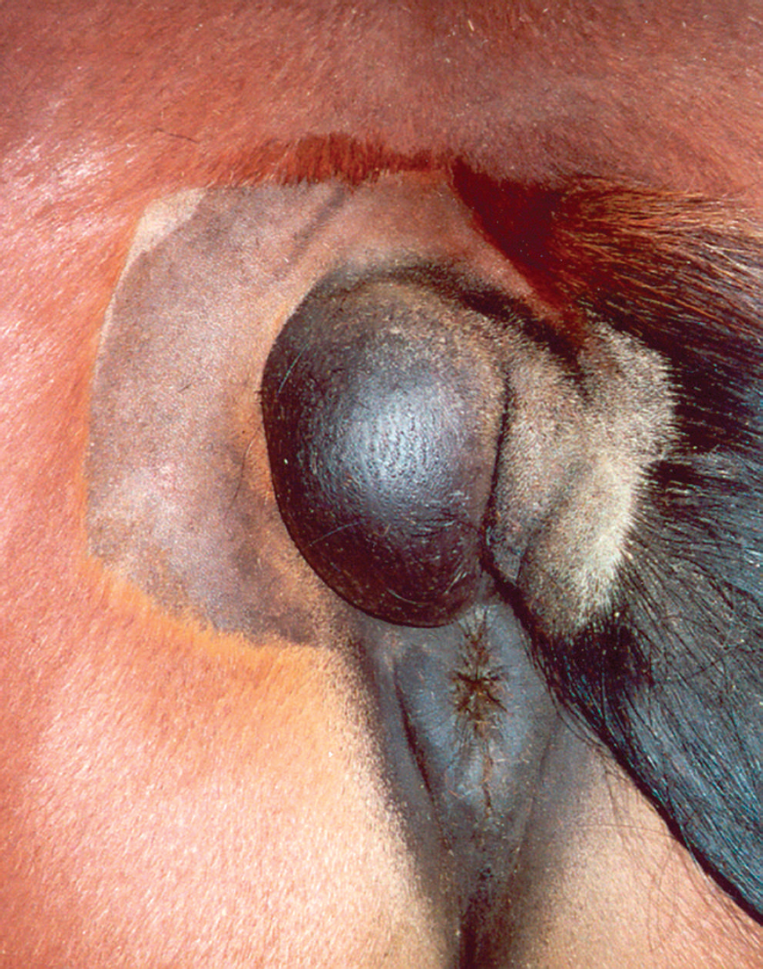
Mass located at the junction between the left lateral aspect of the tail head and the perianal region of a 4-year-old Dales pony.
Immunolabeling was carried out on fixed paraffin wax-embedded sections (4 μm). For vimentin, CK, S-100. GFAP, and NSE, sections were cut onto coated slides. h The sections were dewaxed, hydrated, and pretreated in citrate buffer for 4 minutes in a plastic pressure cooker i followed by quenching in H2O2 1.0% in methanol for 20 minutes. To prevent nonspecific binding, sections were treated with 2.5% normal horse serum for 20 minutes. They were then incubated for 1 hour with the appropriate primary antibody diluted 1/50 (vimentin), 1/100 (CK). 1/1000 (S-100), 1/50 (GFAP), or 1/6 (NSE). The secondary antibody (biotinylated horse antimouse IgG) j was then applied for 30 minutes, followed by the avidin-biotin complex j for 30 minutes. Immunoreactivity was then visualized with horseradish peroxidase using diaminobenzidine (DAB) as the substrate. k
For NR4 and 2F11 immunohistochemistry, sections were cut onto capillary gap slides, incubated overnight, dewaxed, and hydrated. Those for NR4 were pretreated by placing the slides in distilled water and gently heating in running tap water. The slides were then transferred to preheated citrate target retrieval solution, l microwaved on medium-high for 10 minutes, and then incubated at room temperature for 20 minutes. The slides were then cooled in running tap water and washed in Tris buffered saline Tween-20 (TBST). The anti-NR4 antibody was optimized at 1:50. For 2F11, no antigen retrieval was required, and the anti-2F11 antibody was optimized at 1:200. For NR4 and 2F11, peroxidase blocking m was used to prevent nonspecific staining due to endogenous peroxidase. This was followed by detection using DAB. n All immunohisto-chemical sections were counterstained with hematoxylin.
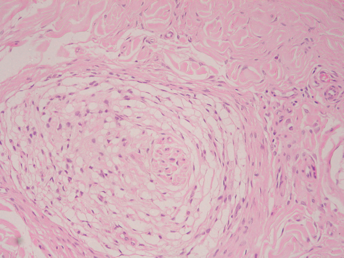
Benign peripheral nerve sheath tumor showing loose whorls of mesenchymal cells with small, elongated hyper-chromatic nuclei (Antoni type B appearance) arranged around a central structure resembling a focus of epithelioid tumor cells. Hematoxylin and eosin, 20X.
Negative controls were prepared by replacing the primary antisera (vimentin, CK, S-100, GFAP, NSE, NF4, or 2F11) with normal rabbit or mouse serum, as appropriate. This resulted in complete absence of immunolabeling. Positive controls were based on the use of tissue from normal control animals in which abundant immunoreactivity for these antigens had previously been demonstrated.
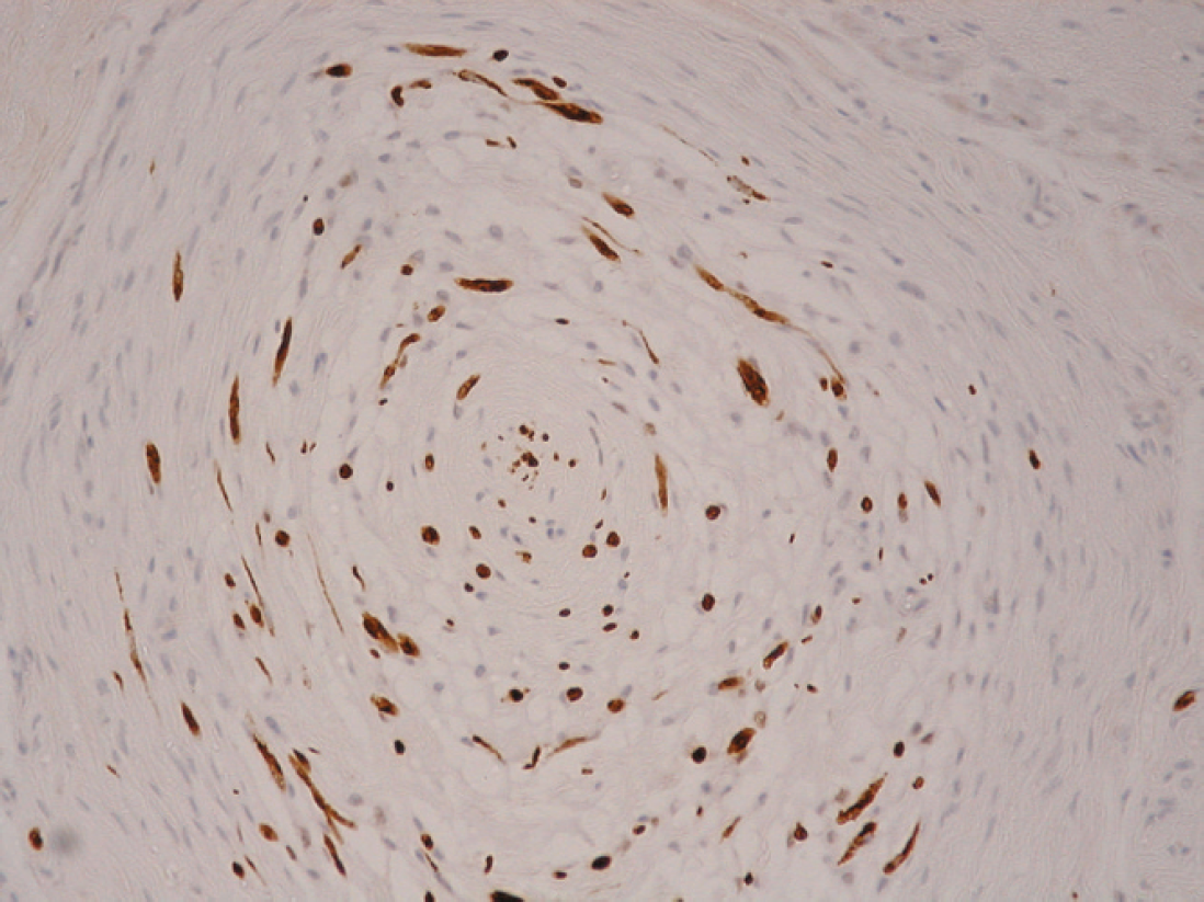
Benign peripheral nerve sheath tumor showing loose whorl of mesenchymal cells with small, elongated hyper-chromatic nuclei (Antoni type B appearance) arranged around a central structure resembling a focus of epithelioid tumor cells. S-100, 40X.
Histopathology revealed a neoplastic mass extending from the deep dermis into the subcutaneous tissues. Overlying hair follicles and adnexal glands were normal, and the epidermis was intact. The mass was unencapsulated and consisted of multiple lobules with an intervening collagenous stroma (Fig. 2). Within the lobules was a loose arrangement of sheets and whorls of mesenchymal cells with a palely eosinophilic, extended, poorly defined cytoplasm and small, elongated hyperchromatic nuclei (Antoni type B appearance). Short linear arrangements of more plump mesenchymal cells were also present. In some areas, a palely eosinophilic, finely granular material was present within the spaces formed by the loose stroma, giving a myxomatous appearance. The whorls were often arranged around a central structure resembling an axon (Fig. 2) or, in some areas, a vascular structure. No mitoses were seen. Staining with Masson's trichrome showed moderate amounts of collagen forming an irregular, loose stroma with occasional denser collagen bundles within the lobules and surrounding the nuclei of the central axon-like structures. Dense collagen was present in the interlobular connective tissue.
Immunohistochemistry showed that the elongated mesenchymal cells and the central axon-like structures expressed vimentin, S-100 (Fig. 3), and NSE, but not CK. GFAP, NR4, and 2F11. Vimentin expression was also present in the connective tissue between the lobules. On the basis of the histopathology and immunohistochemistry, a diagnosis of BPNST (schwannoma type) was made.
PNSTs are recorded in horses worldwide although their prevalence appears very low, with a wide age range of cases ranging from 1 month to 24 years old. 15 The distribution of lesions is equally variable with extradural, cervical spinal cord, mediastinal, gastrointestinal, cutaneous, cardiac, and periocular sites recorded. 1,9,14,15,17
In humans, clinical signs associated with PNSTs are variable and relate both to space-occupying or conductive effects. The latter is influenced by whether their origin is proximal or distal to the affected ganglion. 19 In horses, the clinical manifestations reflect this with records of colic, 9 central nervous signs, 15,17 or space-occupying effects. 15 The clinical progression of this case was insidious due to its ability to expand without major alteration in function of surrounding structures and without causing obvious pain. The differential diagnosis on presentation included sarcoid, fibroma, fibrosarcoma, leiomyoma, leiomyosarcoma, melanoma, and lymphoma in addition to BPNSTs and MPNSTs. 3,4,11,12,16
Macroscopically, benign schwannomas of domesticated animals are usually encapsulated, globoid, lobulated masses of variable size and shape, depending on location of the neoplasm and animal species affected. 1,13,16 Malignant schwannomas are usually nonencapsulated and infiltrative. Microscopically, the classical description of schwannomas is characterized by Antoni type A and B patterns and Verocay body formation. 4 Antoni type A pattern is characterized by a parallel arrangement of Schwann cell fusiform nuclei, giving a palisaded pattern. Antoni type B areas are more loosely arranged with fewer cells, smaller, round dark nuclei, separated by dense, closely aligned, eosinophilic cell processes 1 ; this pattern predominated in the current case. Peripheral nerve sheath tumors lacking these classical morphologic patterns are often difficult to differentiate from other spindle cell tumors without recourse to immunohistochemistry. In addition, BPNSTs and MPNSTs can be difficult to differentiate, and in 1 study in dogs, mitotic index, growth pattern, cellularity, cellular pleomorphism, and necrotic foci were found to be useful criteria. 3 In the present study, the benign histological appearance of the lesion (low mitotic index, lack of cellular pleomorphism or necrosis) occurred in conjunction with deeply infiltrative growth and recurrent behavior. This illustrates how histological assessment of recurrent potential for this type of tumor requires both assessment of the cellular characteristics and the marginal behavior, which may necessitate collection of several incisional biopsies (from the center and from the edge of the lesion) or collection of an excisional biopsy.
The criteria used in this study for diagnosis of a BPNST of the schwannoma subclassification were based on a grossly encapsulated, globoid, and lobulated mass with loosely arranged sheets and whorls of typical tumor cells (Antoni type B). Additional immunohistochemistry revealed that the tumor expressed vimentin, S-100, and NSE. consistent with previous reports. 3 Positive staining for GFAP was not present in the whorls of neoplastic cells and is known to be variable in PNSTs. 3,15 However, the central structures, which resembled axons on routine stains, were negative for GFAP and neurofilament markers. Possible explanations for this negative staining were that these structures were not axons, were axons that were not showing expected immunohistochemical characteristics, or were foci of epithelioid tumor cells seen in some PNSTs. 3 Concentric laminations of perineural cells around individual axons occurs in perineuromas, giving some histological similarities to the present case, but perineuromas are negative for S-100 6 and that diagnosis was therefore discounted.
This case was unusual in 2 respects: the concentric laminations of Schwann cells were very loosely arranged, with an intervening myxomatous stroma containing a moderate amount of collagen and despite its benign histological appearance, the mass extended deeply to the proximal sacral vertebrae. Thus its exact origin was unclear; it may have arisen from cutaneous nerves with deep extension or from neural structures in the sacral region. Due to the incomplete surgical removal, regrowth of the mass is unsurprising.
Footnotes
a.
Vimentin, mouse monoclonal antibody, NCL-L VIM-V9, Novocastra, Newcastle, UK.
b.
Pancytokeratin, mouse monoclonal antibody, MNF116, M0821, Dako, Ely, UK.
c.
S-100, rabbit polyclonal antibody, Z0311, Dako, Ely, UK.
d.
GFAP, mouse monoclonal antibody, M07651, Dako, Ely, UK.
e.
NSE, mouse monoclonal antibody, AM055–5M, BioGenex, San Ramon, CA.
f.
Mouse antineurofilament protein NR4, M0726, Dako, Ely, UK.
g.
Mouse antineurofilament protein 2F11, M0762, Dako, Ely, UK.
h.
Thermo Electron, Runcorn, Cheshire, UK.
i.
Menarini Diagnostics Ltd., Wokingham, Berks, UK.
j.
RTU Vectastain Universal Elite ABC kit, PK7200, Vector Laboratories, Burlingame, CA.
k.
Peroxidase Substrate kit, SK4100, Vector Laboratories, Burlingame, CA.
l.
REAL Target Retrieval Solution Citrate pH 6 (×10), S2369, Dako, Ely, UK.
m.
REAL Peroxidase Blocking Solution, S2023, Dako, Ely, UK.
n.
REAL Envision Detection Kit HRP Mouse/Rabbit, K5007, Dako, Ely, UK.