
Abstract
Twenty-six cattle and 7 horses were diagnosed with rabies. Samples of brain and spinal cord were processed for hematoxylin and eosin staining and immunohistochemistry (IHC). In addition, refrigerated fragments of brain and spinal cord were tested by direct fluorescent antibody test and intracerebral inoculation in mice. Statistical analyses and Fisher exact test were performed by commercial software. Histologic lesions were observed in the spinal cord in all of the cattle and horses. Inflammatory lesions in horses were moderate at the thoracic, lumbar, and sacral levels, and marked at the lumbar enlargement level. Gitter cells were present in large numbers in the lumbar enlargement region. IHC staining intensity ranged from moderate to strong. Inflammatory lesions in cattle were moderate in all spinal cord sections, and gitter cells were present in small numbers. IHC staining intensity was strong in all spinal cord sections. Only 2 horses exhibited lesions in the brain, which were located mainly in the obex and cerebellum; different from that observed in cattle, which had lesions in 25 cases. Fisher exact test showed that the odds of detecting lesions caused by rabies in horses are 3.5 times higher when spinal cord sections are analyzed, as compared to analysis of brain samples alone.
Rabies is an invariably fatal zoonosis 41 that affects the central nervous system (CNS) of humans and virtually all domestic and wild mammals.1,5 It is caused by Rabies virus (RABV), a highly neurotropic, enveloped RNA virus (order Mononegavirales, family Rhabdoviridae, genus Lyssavirus).10,35,44 The occurrence of rabies is dependent on the presence of vectors. 33 In South America, rabies in herbivores occurs as outbreaks, and is usually transmitted by the hematophagous bat Desmodus rotundus. 5
Laboratory testing is essential for the diagnosis of rabies because this disease exhibits various clinical signs, and lesions are not specific at autopsy. 6 Brain is the organ of choice for diagnosis of rabies; submission of spinal cord fragments for analysis is also recommended for diagnosis in horses. 35 In Brazil, official diagnosis is performed by means of a direct fluorescent antibody test (DFAT). 7 Fragments of cerebellum, cervical spinal cord, thalamus, and cerebral cortex are refrigerated or frozen for detection of RABV by DFAT; the remaining material is fixed in 10% formalin and submitted for histopathology. 4 Despite being highly efficient for rapid diagnosis of rabies, DFAT has some drawbacks that include cost of equipment, human laboratory exposure to live virus, and the need for fresh samples.3,17,23 Biological testing (inoculation into the brains of newborn mice or into cell cultures) is considered the most accurate method for the diagnosis of rabies 46 ; however, at least 3 weeks are required to obtain a final diagnosis by these means. 6 Immunohistochemistry (IHC) is a specific and sensitive method for detection of viral antigens, 14 and it can be considered the most sensitive test for early diagnosis of viral infection when characteristic microscopic lesions or detection of viral antigens are not possible using routine histologic analysis and DFAT.3,24,44
Histologic findings include nonpurulent meningoencephalitis and meningomyelitis with ganglioneuritis as the main lesions. 19 Intracytoplasmic acidophilic inclusion bodies, called Negri bodies, are considered pathognomonic for rabies 42 ; however, they can be absent in 20–60% of cases.23,29,30,37 Lesions are distributed mainly in the brainstem, cerebellum, spinal cord, 22 hippocampus, and trigeminal ganglion, 18 and can be uncommon or even absent. 19 The objective of our study was to characterize lesions caused by RABV in the spinal cord of cattle and horses through morphologic and immunohistochemical analysis.
Autopsies of cattle and horses with neurologic syndrome were performed from January 2013 to November 2014; at the time of autopsy, samples were collected for further analyses. Fragments of cerebral cortex, thalamus, cerebellum, medulla oblongata, and spinal cord collected at cervical, thoracic, lumbar, and sacral levels, were refrigerated and submitted for RABV detection by DFAT. Samples that were negative by DFAT were subjected to mouse inoculation test (MIT), for confirmation. The remaining specimens were fixed in 10% buffered formalin, and cut following standard procedures. The brain was subdivided into cerebral cortex (CC), thalamus (TH), hippocampus (HC), cerebellum (CER), and obex (OB). The spinal cord was divided into cervical region (C), cervical enlargement (CE), thoracic region (T), lumbar region (L), lumbar enlargement (LE), and sacral region (S). Samples were routinely processed for histopathology and stained with hematoxylin and eosin.
IHC was performed on spinal cord sections of all animals. An anti-rabies polyclonal antibody was used as primary antibody at a 1:1,000 a dilution in phosphate buffered saline, and developed with a red chromogen.b,c
For statistical analysis, data obtained from laboratory exams and descriptive statistics were tabulated d and performed, e respectively, using commercial software. A severity score index was created to achieve a better determination of the level of severity of lesions observed. The index was based on the sum of the degrees of lesions of all animals in each segment of spinal cord (1: mild; 2: moderate; 3: marked) and later divided by the total number of animals of each species. The resulting index incorporated the following lesions: perivascular cuffs + microgliosis + neuronophagia. Presence of malacia and IHC staining intensity were analyzed separately. Through this calculation, it was possible to obtain a quantification classification of lesion severity for each anatomical location according to the range of the results (1–3: mild; 4–6: moderate, 7–9: marked). In a similar way, results obtained for the presence of malacia and IHC staining were interpreted in a range of 0–3.
To determine the odds of finding lesions in the 2 tissues evaluated in our study (brain and spinal cord), Fisher exact test was used with a level of significance of 5% using open-source software. f
During the period of this study, 222 autopsies were performed in cattle, and 111 were performed in horses. Of these, 26 cattle (12%; Fig. 1A) and 7 horses (6%; Fig. 1B) were diagnosed with rabies (n = 33).
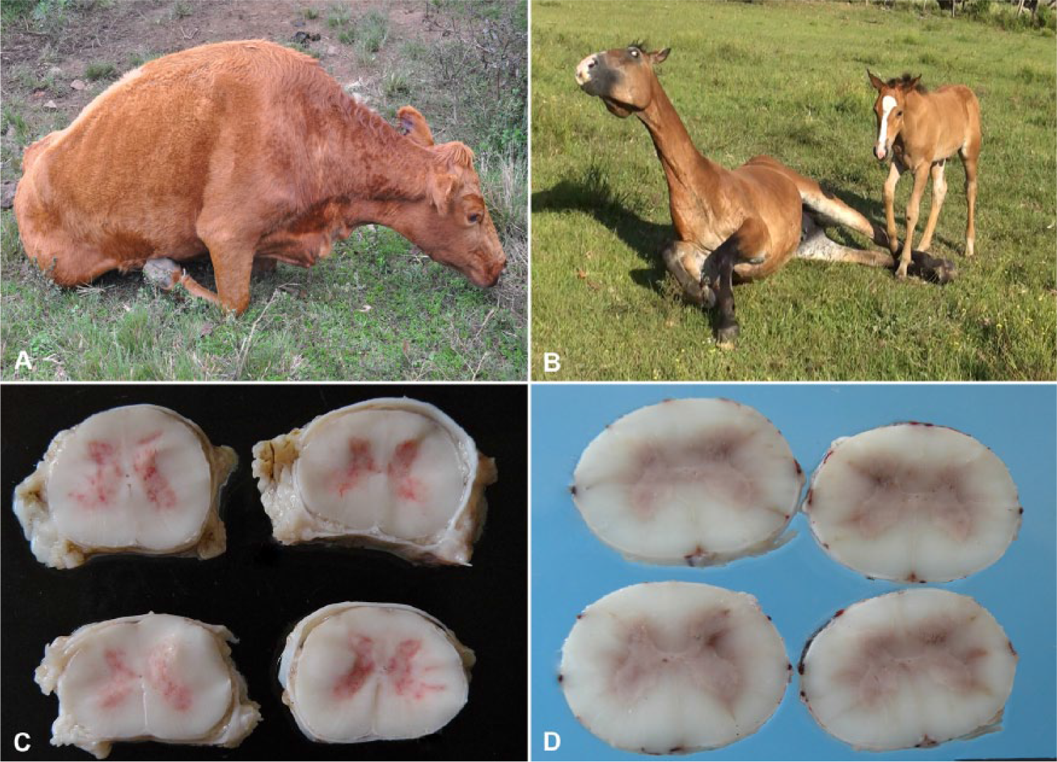
Clinicopathologic characterization of rabies in cattle and horses.
At autopsy, marked bladder distension and rectal ampulla filled with dry feces were observed in most of the animals examined. Additionally, relevant gross findings included hyperemia of leptomeningeal vessels, and multifocal areas of hemorrhage distributed especially through the gray matter of the spinal cord in horses (Fig. 1D); in cattle, these lesions were also present, but were fewer in number and intensity (Fig. 1C).
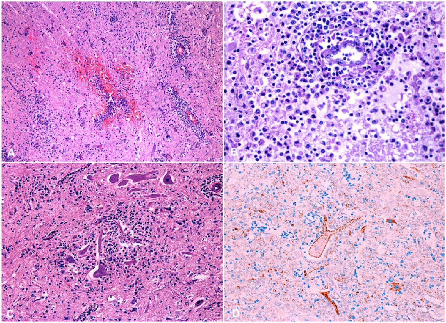
Only two horses exhibited lesions in the brain, which were located mainly in segments of the OB and CER. Lesions were characterized by mild to marked multifocal areas of inflammatory infiltrate around vessels (perivascular cuffs) containing mostly lymphocytes, and low numbers of plasma cells and macrophages. Additionally, moderate multifocal microgliosis, with mild neuronophagia, and lymphoplasmacytic leptomeningitis were also present. Inclusion bodies were not observed in any region of the brains of horses. Lesions of various degrees of severity were observed in the brain of the 25 cattle studied (25/26); sections of OB (23/26) and CER (21/26) were the most severely affected, followed by TH (11/26), HC (9/26), and CC (8/26). Leptomeningitis (73%) was observed predominantly in the cerebellar region. Furthermore, lymphoplasmacytic perivascular cuffs (92%), nodular microgliosis (92%) forming large glial nodules at times, and neuronophagia (42%) were also identified as important microscopic findings. Negri bodies were observed in 12 cases (46%).
The spinal cords of all cattle and horses exhibited histologic lesions. However, Negri bodies were observed in only 2 horses (28.5%) in L and LE segments, and in 19 cattle (73%). Additionally, all horses exhibited multifocal areas of hemorrhage in the spinal cord, which varied from mild to moderate (Fig. 3A).
Figure 2 depicts the inflammatory lesions observed in the spinal cords of horses, which were mild in the C and CE regions, moderate in the T, L, and S regions, and marked in the LE region. Similarly, the presence of malacia was mild in the C, CE, and T regions, moderate in the L, and S regions, and marked in the LE region (Fig. 3B). IHC staining intensity in spinal cord fragments varied from moderate to strong. During evaluation of the severity score index of bovine spinal cord lesions (Fig. 4), inflammatory lesions were found to be moderate in all spinal cord segments (Fig. 3C). Additionally, in contrast to that observed in the spinal cords of horses, presence of malacia was mild in all bovine spinal cord regions evaluated, except for the S region, where it was moderate. IHC staining intensity in bovine spinal cords was strong and homogeneous in all segments (Fig. 3D).
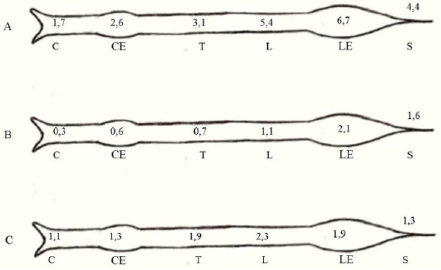
Severity score of horse spinal cord.
Characterization of histologic lesions and immunohistochemistry of spinal cords in cattle and horses with rabies.
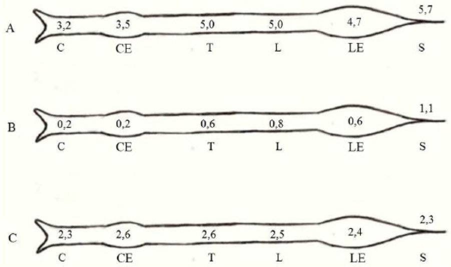
Severity score of bovine spinal cord.
The frequency of lesions per spinal cord segment was obtained through the sum of the animals that had lesions divided by the total of animals. In horses, lesions became more frequent from the T level on, independent of level of severity; however, IHC staining revealed only a mild variation between segments. Lesions in cattle exhibited variation only for malacia, which was more frequent in the L, LE, and S levels. All 33 cases exhibited positive immuno-staining as detected by IHC (100%); immunostaining was present in perikarya, axons, and dendrites of integer neurons or neurons undergoing neuronophagia, and was confined to granule aggregates or rounded structures associated with variable numbers of viral inclusion bodies. RABV was detected in 2 horses (28.5%) by DFAT. All cases that were negative for rabies as assayed by DFAT were also negative as assessed by the MIT. DFAT was positive for rabies in 20 (76.9%) of the 26 cattle studied. Samples that were negative for rabies as assessed by DFAT were subjected to MIT for validation of the results; this test indicated 5 samples were negative, and 1 sample was positive for RABV.
Fisher exact test was performed on the data using a level of significance of 5%, and the results showed that analysis of the spinal cord of horses allows for a 3.5-fold higher chance of detecting lesions caused by RABV as compared with analysis of brain samples alone. In cattle, this difference was not observed, as the frequency of lesions was similar in the brain and in the spinal cord.
Rabies usually raises some points to be discussed. Autopsy findings, generally, are not significant in these cases; however, possible findings include hyperemia of leptomeninges, and lesions that are secondary to neurologic impairment such as aspiration pneumonia, bladder and rectal distension caused by paralysis due to viral myelitis, and self-inflicted trauma. 10 Furthermore, hemorrhages can occasionally be observed macroscopically in the spinal cord of horses and cattle. 27 Previously published data corroborate the results found in our study; however, alterations such as those described herein are not always present in cases of rabies.10,28,34
The microscopic lesions observed in our study, which included nonsuppurative meningoencephalitis and meningomyelitis with foci of gliosis, neuronal necrosis, and neuronophagia with the presence of Negri bodies, were similar to those described by other authors.25-27 In the present study, spinal cord lesions were present in 100% of cattle and horses; brainstem and cerebellum were the next highest regions affected. In horses, lesions in the spinal cord were more frequent in the L and LE segments, and were characterized by moderate to marked inflammation, which included a dense perivascular lymphoplasmacytic infiltrate, nodular to diffuse microgliosis with neuronal loss, neuronophagia, and a prominent malacia. In some cases, an extension of the perivascular lymphoplasmacytic infiltrate was seen in the white matter of the spinal cord. A higher frequency of these lesions in lumbar sections of the spinal cord has also been described in a study of paralytic rabies in humans. 8 In the cattle studied by us, a homogeneous distribution of inflammatory lesions throughout all spinal cord segments was seen; on the other hand, malacic areas were more frequent in the L, LE, and S regions. These malacic lesions were predominantly mild, which is in contrast to those observed in the horses in our study. This lesion distribution found in horses and cattle can be explained by the centripetal course followed by the virus, which progressively travels through the spinal cord to the brain.9,16,42 Additionally, multifocal hemorrhagic areas were observed in the spinal cords of all horses, sometimes macroscopically, which corroborates findings of other authors.13,31 The exact mechanism by which RABV causes neuronal lesions and ultimately death is unknown. One hypothesis is marked viral-induced downregulation of genes in the brain. 45 An increase of nitric oxide has been observed in natural and experimental RABV-infected brains, suggesting nitric oxide neurotoxicity.21,38,43 The nitric oxide may play a role in blood-brain barrier leakage and may contribute to tissue damage due to the generation of peroxynitrite.2,15 Besides that, another possible mechanism of neuronal injury and lesions is virus-induced apoptosis, which was demonstrated in mouse models of rabies infection. 45
Even though Negri bodies are a characteristic finding of RABV infection, they are not evident in all cases. 19 In the present study, Negri bodies were detected at a higher frequency in spinal cord sections than in brain sections in 73% of cattle and in 28.5% of horses. The presence of Negri bodies is related to stage and clinical course of the disease, in addition to virus strain and viral concentration.10,27
Detection of RABV in horses can be unsuccessful if the search involves only the brain and cerebellum. 39 Statistical and comparative analysis of data regarding examination of the spinal cord, brain, and cerebellum showed that correct sample collection for the diagnosis of rabies in horses can result in a 3.5-fold higher chance of correct diagnosis, as only 2 horses exhibited brain lesions in the present study.
Sensitivity of the diagnosis of rabies using DFAT and IHC is ~98%. 46 IHC, in the present study, revealed the presence of viral antigen in all 33 rabies cases; this is in agreement with another work 40 in which the viral antigen was detected in all (39) of the cases analyzed. Additionally, 22 of the cases analyzed in our study (66.6%) were positive for rabies as detected by DFAT; this frequency is similar to that found in another study 31 in which 55% of the samples from horses diagnosed with rabies were positive as detected by DFAT. These data show that complete agreement between tests may not happen for all samples. 46 Divergence can be attributed to sample integrity; additionally, individual reaction to disease, incubation period, and clinical evolution can be influencing factors. 11 These results show the importance of IHC as an auxiliary diagnostic tool for RABV infection, especially in cases for which DFAT results are negative. IHC has become very popular in the past 2 decades owing to its ability to detect antigen in fixed tissue.17,23,36,37 Some studies show that the sensitivity of IHC is similar to that of DFAT when used for the diagnosis of rabies30,44; however, IHC can be even more sensitive in early diagnosis, when traditional histopathology and DFAT are still not able to detect viral antigens or characteristic lesions.3,24,44
Equine rabies is poorly studied in Brazil. 25 This disease is clinically indistinguishable from other types of encephalitis.12,20 Studies have shown that diagnosis of rabies in horses requires sampling methods different from those used for other species; additionally, DFAT and detection of Negri bodies were significantly less sensitive for the diagnosis of this disease in this species compared to cattle. 32 With this work, we can suggest that spinal cord histology associated with IHC is the most reliable way to diagnose this disease, especially in horses. Thus, the spinal cord of horses with neurologic signs submitted for autopsy should be collected to perform examination by histopathology and histochemistry.
Footnotes
Authors’ contributions
DM Bassuino contributed to conception and design of the study; contributed to acquisition, analysis, and interpretation of data; and drafted the manuscript. G Konradt, GS Silva, SP Pavarini, and D Driemeier contributed to conception of the study, and contributed to acquisition, analysis, and interpretation of data. RAS Cruz and DC Gomes contributed to conception of the study, and contributed to acquisition of data. All authors critically revised the manuscript; gave final approval; and agreed to be accountable for all aspects of the work in ensuring that questions relating to the accuracy or integrity of any part of the work are appropriately investigated and resolved.
a.
Rabies polyclonal DFA (no. 5199), Chemicon International Inc., Temecula, CA.
b.
AEC, Dako North America, Carpinteria, CA.
c.
NovaRED, Vector Laboratories Inc., Burlingame, CA.
d.
Excel 2010, Microsoft Corp., Redmond, WA.
e.
SPSS Statistics 18, IBM Corp., Armonk, NY.
Declaration of conflicting interests
The author(s) declared no potential conflicts of interest with respect to the research, authorship, and/or publication of this article.
Funding
The author(s) received no financial support for the research, authorship, and/or publication of this article.