
Abstract
Polymerase chain reaction (PCR) has gained increasing importance as a tool for directly demonstrating the presence of Chlamydophila in the placentas of aborted sheep and goats. However, because of the zoonotic potential of the disease, it is advisable to use fixed materials. To evaluate 4 different DNA extraction protocols in paraffin-embedded sections for PCR, previously immunohistochemically diagnosed placental samples from outbreaks of abortions in goats and sheep were used. The samples were also used to evaluate the effect of the duration of fixation in formalin on PCR. A protocol that uses Tris-HCl pH 8.5 with EDTA and subsequent digestion with proteinase K was found to be an easy protocol for obtaining excellent PCR products for Chlamydophila abortus diagnosis from formalin-fixed and paraffin-embedded specimens. It was also found that if samples are fixed in formalin for more than 2 weeks, the PCR technique is affected more adversely than immunohistochemical methods.
Chlamydophila abortus is an intracellular bacterium and the etiological agent of ovine enzootic abortion (OEA). This disease has spread worldwide and causes abortion in small ruminants during the last third of gestation. 15 Besides being the cause of important economic losses in agricultural industries, it also represents a zoonotic risk for pregnant women. 7
Adult ewes are infected as a result of contamination through the ingestion of organisms shed in vaginal fluids and placental membranes at the time of abortion or lambing or through inhalation of aerosol from the environment. In nonpregnant animals, few or no clinical signs are observed until the subsequent pregnancy, when the animals abort. 16 Abortion always appears in the last weeks of gestation regardless of the moment of infection 4 as a consequence of a suppurative necrotic placentitis. Experimental infections have shown that pathological lesions appear from 120 days of gestation, with the trophoblast cells being the main cell population affected. 14
Diagnosis of chlamydiosis is usually established by direct bacteriological examination of the specimens (isolation of the bacteria in cell culture or polymerase chain reaction [PCR]) or by indirect serological test (enzyme-linked immunosorbent assay, complement fixation test, or immunofluorescence). The disadvantage of indirect serological diagnosis is that it indicates that the animal has been exposed to Chlamydophila rather than detecting the organism. Since one of the most important strategies for prevention of OEA is the vaccination of flocks, serological analyses could lead to a false-positive diagnosis. However, among the direct diagnostic techniques, PCR has become a useful tool for the detection of Chlamydophila in biological samples. 8 PCR makes it possible to process a large number of specimens, is easy to use, and provides rapid results, besides being safer than culturing the microorganism in cell substrates. Polymerase chain reaction analysis using fresh or frozen samples is straightforward. Furthermore, PCR can be adapted for use on formalin-fixed and paraffin-embedded samples. The method permits samples to be kept for retrospective diagnosis from archival material and avoids the zoonotic risk of Chlamydophila abortus.
The aim of this study was to compare 4 different previously reported DNA extraction protocols using formalin-fixed, paraffin-embedded placental tissue from aborted sheep and goats and to study how the duration of fixation affects the usefulness of the PCR samples.
In the present study, 12 samples from different abortion outbreaks were used. Samples were fixed in 10% formaldehyde in phosphate-buffered saline and embedded in paraffin. Eleven of these samples were diagnosed as Chlamydophila abortus by histopathology and immunohistochemistry (IHC). The time of sample fixation varied between 3 and 20 days since the local veterinary services did not always send samples immediately.
For histopathological analyses, sections (5 μm) from formalin-fixed and paraffin-embedded tissue samples were cut, stained with hematoxylin and eosin, and examined for microscopic lesions. To demonstrate chlamydial antigen by IHC, tissue sections (5 μm) were stained with an antichlamydial-lipopolysacharide-specific biotinylated mouse monoclonal antibody, as previously described, 2 using the avidin-biotin-peroxidase complex method, according to the instructions of the manufacturer. a A positive reaction was demonstrated by the precipitation of diaminobenzidine tetrahydrochloride. Sections were counterstained with hematoxylin.
Eleven placenta sections were diagnosed positive and 1 was diagnosed negative by IHC staining. Positive placentas showed a suppurative placentitis associated with the presence of variable amounts of C. abortus antigen in the trophoblast cells of the periplacentomal chorioallantoic membrane, the base of the chorionic villi, and peripheral areas of the placentome. In some areas, many trophoblast cells were necrotic, and the choriallantonic membranes were ulcerated and covered with cell debris. The underlying mesenchyme showed significant neutrophil infiltration with areas of leukocyte degeneration and a large amount of both intracellular and extracellular C. abortus antigen.
The 12 formalin-fixed and paraffin-embedded tissue samples were used to compare 4 different previously described DNA extraction protocols. In protocol A, based on Fiallo et al., 5 six 10-μm sections were deparaffinized twice by adding xylene, which was removed from the samples with absolute ethanol. The air-dried pellets were then resuspended in a lysis buffer (100 mM Tris-HCl, 150 mM NaCl, 10 mM EDTA, 1 mg/ml of proteinase K) and incubated for 2 hours. To extract DNA, the samples were heated for 10 minutes and then cooled. The DNA extract was stored at −20°C until DNA amplification.
In protocol B, based on Marchetti et al., 11 three 2-μm sections were deparaffinized twice by adding xylene, which was removed from the samples with absolute ethanol. The resulting pellets were air dried overnight and then, instead of following the phenol-chloroform method to extract the DNA, a DNA extraction kit b was used following the manufacturer's instructions. The extracted DNA was stored at − 20°C until DNA amplification.
In protocol C, based on Müller et al., 13 one 4-μm section was deparaffinized by adding a digestion buffer (50 mM Tris-HCl, pH 8.5; 1 mM EDTA) and incubating the samples with proteinase K before being inactivated at 95°C. Finally, the supernatant containing the extracted DNA was stored at − 20°C until DNA amplification.
Finally, in protocol D, based on Shi et al., 18 two 10-μm sections were deparaffinized by adding NaOH and heating. A slight modification was introduced, again using a commercial DNA extraction kit, b instead of following the phenol-chloroform DNA extraction. The DNA extracts were stored at − 20°C until DNA amplification.
Each DNA extract was used as a template for PCR amplification using primers CpsiA (5'-ATG AAA CAT CCA GTC TAC TGG-3') and CpsiB (5'- TTG TGT AGT AAT ATT ATC AAA-3'), which permit the very sensitive amplification of C. abortus DNA 6 and were designed from the 4 available C. abortus pmp gene sequences. 9 The amplified products of target sequences (pmp91A, pmp90A, pmp91B, and pmp90B genes) obtained with these primers were roughly 300 bp in length, as was expected. Polymerase chain reaction was performed on 2 μl of each DNA extract in a total volume of 25 μl. 8 The final reaction mixture contained 2 μM of each primer, 200 μM of each deoxynucleoside triphosphate, 3 mM MgCl2, 0.5 U Taq DNA polymerase, c and 5 μl10× Ex Taq Buffer. c Reactions were performed in an automated DNA thermal cycler, d in which the samples were subjected to 30 amplification cycles. Prior to this, DNA samples were denaturated at 95°C for 5 minutes. An amplification cycle consisted of denaturation at 94°C for 30 seconds, primer annealing to the template at 50°C for 1 minute, and primer extension at 72°C for 2 minutes. Finally, the products were incubated at 72°C for 10 minutes and then cooled to 4°C. The PCR products were examined by electrophoresis or were stored at 4°C until examination. To detect PCR products, 5 μl of each sample was examined by electrophoresis in 1.5% agar gel stained with 0.5 μg/ml ethidium bromide. The results were visualized under a transilluminator and photographed.
Two controls were included in each amplification. As a positive control, C. abortus DNA, extracted from the frozen placenta of an aborted sheep that tested positive for C. abortus by IHC analysis, was used. As a negative control, DNA extracted from a frozen sheep placenta, negative for C. abortus by IHC analysis, was used.
To evaluate the reproducibility of the experiments, all samples were subjected to the whole procedure, from DNA extraction to PCR amplification, 3 times. Furthermore, to minimize false-positive results, amplified products were physically separated from the starting material. Preparation of the reaction mixture and the set up of the amplification procedures were performed in a sterile area room with no circulating air and ultraviolet light.
Successful C. abortus DNA extraction from formalin-fixed and paraffin-embedded cotyledons was achieved with the all the extraction protocols used in this study. However, clear differences were observed between the 4 protocols. Samples processed following protocol A (Fig. 1A) showed a poor-quality PCR product with bands clearly visible only in samples 1, 8, and 9 and less clearly in samples 4, 5, 6, and 7. In fact, some samples that were positive by IHC (samples 2, 3, 11, and 12) did not show any band in the ethidium bromide-stained gels. By chance, these 4 samples were formalin fixed more than 2 weeks before being used for DNI extraction. Moreover, this DNA extraction protocol was quite long.
Protocol B provided poor-quality PCR products (Fig. 1B), with the bands being clearly visible only in samples 1, 6, 8, and 9. No bands were detected in samples 2, 3, 4, 5, 7, 10, 11, and 12. This protocol had the lowest number of positive samples of the 4 protocols used in this study.
Protocol C was the fastest. Moreover, samples processed by this protocol showed good-quality PCR products and the most clearly visible bands (Fig. 1C). However, samples 2, 3, and 12, which had been formalin fixed more than 2 weeks previously, were again negative.
The samples processed following protocol D (Fig. 1D) showed similar results as protocol C. Moreover, as with protocols A, B, and C, there were still negative samples that were positive by IHC (samples 2, 3, and 12). Protocol D was easy to carry out but needed a DNA extraction kit, which resulted in extra time and extra expenditure. The use of a DNA extraction kit considerably improved the PCR products compared with those obtained using phenol-chloroform DNA extraction (data not shown).
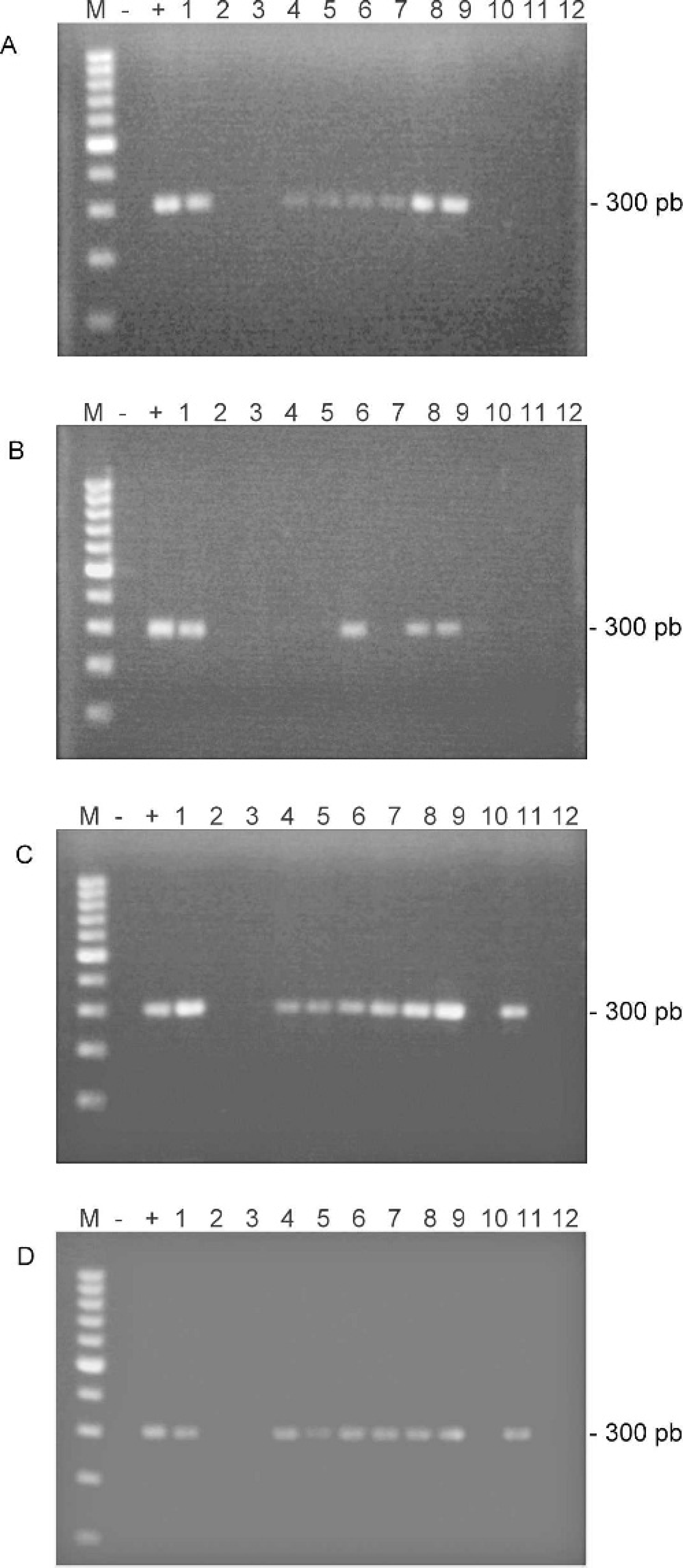
Gel electrophoresis of PCR analyses.
PCR products were sequenced in collaboration with the sequencing service of the University of Murcia (SACE-CAID), with an ABI Prism 3130 sequencer. e The obtained sequences were compared with the sequences available in the GenBank by using the BLAST server from the National Center for Biotechnology Information (http://www.ncbi.nlm.nih.gov/BLAST). All the PCR products were 98% to 99.4% identical to C. abortus strain S26/3.
To ascertain whether negative PCR results could be due to a loss of sensitivity associated with the duration of formalin fixation, 2 new IHC-positive C. abortus samples from an abortion (samples 1' and 2') were maintained in formalin for 1, 2, 3, and 6 weeks before being used for DNA extraction (following protocol C) and PCR. It was determined that samples fixed in formalin for more than 2 weeks before being paraffin embedded showed a loss of sensitivity in the PCR technique (Fig. 2), which could explain why some of the samples showed false-negative PCR results.
A common problem that arises in the diagnosis of abortion in small ruminants is the large number of samples that need to be analyzed at certain periods of the year. Therefore, most laboratories store samples before performing diagnostic testing. Although frozen samples are frequently used, fixed and paraffin-embedded samples permit IHC and PCR to be used for diagnostic purposes.
In contrast to the widely described use of fresh tissues for the diagnosis of Chlamydophila spp. by PCR, 1,10,12,17 the use of formalin-fixed and paraffin-embedded samples collected from small ruminant abortions for C. abortus diagnosis by PCR has not been optimized for sensitivity. Even though fixing in formalin has been demonstrated to reduce PCR effectiveness 3 and to contribute to reducing immunostaining efficiency, 19 it is still used by a large number of pathologists since fixation permits retrospective analysis and provides a safe way to process potentially hazardous abortion specimens that may contain not only C. abortus but also other potential zoonotic agents such as Coxiella burnetii, Listeria monocytogenes, and Brucella melitensis. An additional advantage of using formalin-fixed and paraffin-embedded tissue sections is that it permits parallel studies to be carried out using immunohistopathological analysis to show the necrotic purulent placentitis associated with chlamydial abortion and consequently to obtain an accurate PCR diagnosis. Furthermore, the PCR technique permits the rapid and objective analysis of a large number of samples, avoiding the interpretation or the background problems that could arise in IHC.
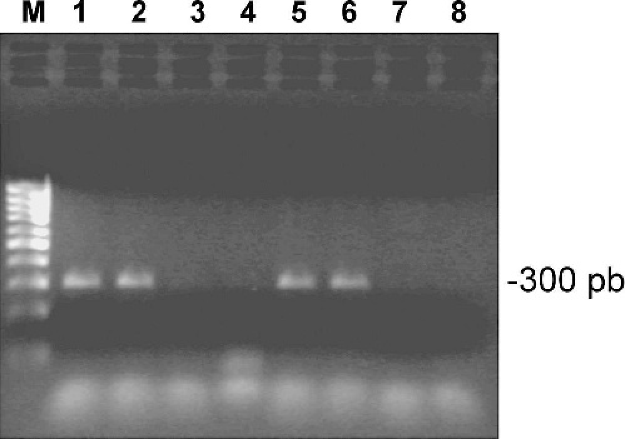
Gel electrophoresis of PCR analyses of 2 C. abortus placental tissues maintained in formalin for different times. M = size marker.
When 4 DNA extraction protocols were compared for use with formalin-fixed and paraffin-embedded cotyledons, protocols B and D needed to be modified by adding the extra step involved in using a DNA extraction kit because the classical phenol-chloroform DNA extraction procedure is risky for operators, prone to mistakes because of a cumbersome procedure, and also yielded a lower percentage of positives in relation to the use of a DNA extraction kit (data not shown). Protocols A and C were followed as described by the authors, without using a DNA extraction kit. This reduced not only the cost, an important aspect to be taken into account in small ruminant diagnosis, but also the time necessary to carry out the analysis. At the same time, it reduced possible manipulation mistakes in the DNA extraction step. Furthermore, the best quality PCR products were observed using these protocols, especially protocol C. 13
A recent study of chlamydial abortions in cattle using fixed and paraffin-embedded samples 1 has shown that the results of IHC diagnosis can be improved by PCR. Borel et al. 1 used a DNA extraction kit, while the present study shows that a simpler and cheaper protocol can be used before PCR amplification for the differential diagnosis of abortion in small ruminants. Moreover, it can be used alone or combined with IHC diagnosis.
An important conclusion of this study is that fixation affected the PCR results to a greater extent than those of IHC analyses since a loss of sensitivity was detected in samples that had been fixed in formalin more that 2 weeks prior to using the PCR technique. Perhaps DNA integrity is reduced during the time samples are fixed in formalin, reducing the suitability of such samples for PCR. Special care must therefore be observed with regard to the length of time that samples remain in formalin.
In conclusion, a standardized, PCR-based diagnosis of C. abortus infection in formalin-fixed specimens is feasible. Although the technique is more adversely affected than IHC techniques by the duration of fixation, PCR represents a reliable and sensitive diagnostic tool, which, together with other diagnostic methods, can be used in samples that have been fixed in formalin (rather than frozen) before being sent to diagnostic laboratories. The protocol described by Müller et al. 13 permits good-quality PCR products to be obtained for C. abortus diagnosis.
Acknowledgements. This study was supported by a Fundación SENECA grant from Región de Murcia, code 0055-0/PI/04. We are grateful to Dr. Pilar de la Rua and all her technical staff of Departamento de Biología Animal, University of Murcia, for all of their advice. N. Ortega and C. M. Martinez are the recipients of a postdoctoral grant from the University of Murcia and a predoctoral grant from the Spanish Ministry of Science and Technology, respectively.
Footnotes
a.
Vector Laboratories, Burlingame, CA.
b.
DNeasy Tissue Kit, Qiagen, Hilden, Germany.
c.
Takara Ex Taq, Takara Bio Inc., Shiga, Japan.
d.
Eppendorf Mastercycler Personal, Hamburg, Germany.
e.
Applied Biosystems, Foster City, CA.