
Abstract
An in situ hybridization (ISH) procedure with a digoxigenin-labeled oligonucleotide probe for detection of Lawsonia intracellularis in paraffin-embedded tissue is described. This technique recognized 71% of PCR-positive cases and was thus superior to Warthin-Starry silver stain, which only detected 41%. The presented ISH is of comparable sensitivity to previously published immunohistochemical assays and is recommended for laboratories wishing to diagnose L. intracellularis infections in tissue sections but without access to antibodies.
Lawsonia intracellularis is the causal agent of porcine proliferative enteropathy (PPE), a transmissible enteric disease of growing pigs. The disease is characterized by adenomatous proliferation of immature intestinal epithelial cells in small and large intestine, but particularly in the ileum. 10 Porcine proliferative enteropathy is present in all countries with intensive pig production and has a significant economic impact. 13 Reliable and timely diagnosis of the disease is thus a prerequisite for efficient control measures. Because the bacterium does not grow in conventional bacterial culture media, other means of diagnosis are required. In advanced cases, the macroscopic and histological lesions are pathognomonic. 14 Frequently, however, additional diagnostic methods must be used for unequivocal diagnosis. Currently, these methods are performed on fecal samples, intestinal scrapings, and frozen or paraffin sections. The methods applied include simple staining procedures (e.g., modified Ziehl-Neelsen stain) 3 or the more sophisticated Warthin-Starry silver stain, 5 , 6 both of which are nonspecific for L. intracellularis. Specific methods targeting antigens or genome of the agent are also available, e.g., several PCR protocols, 8 , 9 , 12 fluorescent antibody techniques, 6 immunohistochemistry (IHC), 7 , 11 and certain in situ hybridization (ISH) protocols. 2 , 4 Although these methods have the advantage of being specific and sufficiently sensitive, most of them also have disadvantages: PCR simply indicates the presence of bacterial genome, without localizing the bacteria within lesions. The localization of bacteria within the altered epithelial cells is achieved by combined morphological and antigen/genome detection techniques, which for best preservation of morphology should be performed on paraffin-embedded tissue. This is perfectly achieved by IHC. Although it provides excellent results with the highest specificity and sensitivity, including morphological localization of the bacteria, this method is not accessible to the entire scientific community and all diagnostic laboratories because antibodies are not commercially available. Alternatively, ISH might enable laboratories without access to antibodies to employ a method with advantages similar to IHC. The ISH procedures published so far, however, have certain disadvantages. One technique requires frozen sections with the inherent flaw of poor detail resolution. 4 Another protocol works with fluorescent probes, 2 resulting in only a temporary signal and requiring special microscopic equipment. Also, simultaneous evaluation of histopathological changes is more difficult than with light microscopy. The goal of this study was to establish an ISH protocol generating a permanent reaction product in paraffin sections that is visible with a conventional light microscope.
The DNA oligonucleotide probe used targeted a portion of the 16S ribosomal RNA (rRNA) of L. intracellularis and was designed based on a previously published sequence. 2 Whereas the 3′ end was left identical, 15 additional nucleotides were added to the 5′ end, because according to the authors' experience, oligonucleotide probes with a length between 30 and 40 nucleotides give the best signal/background ratio. The probe sequence was: 3′-AACCGGAG CAGTCTCTCTAGAGTGCCCAACTTTAC-5′, which is complementary to nucleotides 1142–1177 of rRNA of L. intracellularis (GenBank accession number L15739). The probe was labeled with digoxigenin at the 3′ end. a The staining protocol was established using samples of paraffin-embedded ileum from 3 pigs with classical PPE that were also positive by IHC (using the antibody Law1-DK, which detects a protease resistant molecule of 21 kDa, presumably lipopolysaccharide) 1 and PCR (according to Jones et al. 8 )as positive controls. Best results (crisp dark-blue specific signal with minimal background) were achieved using the following protocol: After dewaxing in Neoclear b and rehydration in graded ethanols, proteolytic treatment was done with proteinase K, c 5 μg/ml at 37°C for 30 minutes. Then the slides were washed and covered by the hybridization mixture, 100 μl of which contained 50 μl deionized formamide, d 20 μl 20x standard sodium citrate (SSC), 10 μl dextrane sulfate b (50%w/v), 2 μl 50x Denhardt solution, e 5 μl boiled herring sperm DNA f (50 mg/ml), 11 μl distilled water, and 2 μl probe with a final concentration of the probe of 200 ng/ml. Then the slides were heated at 95°C for 6 minutes. After cooling on crushed ice, the slides were hybridized for 16 hours in a humid chamber at 40°C. Thereafter, they were washed in 2x SSC, 1x SSC, and 0.1x SSC (10 minutes each) at room temperature (approximately 22°C). For detection of the digoxigenin-labeled hybrids, the slides were incubated for 60 minutes with anti-digoxigeninAP Fab fragments c (dilution 1 in 100) at room temperature. The slides were rinsed twice and the signal was visualized with 5-bromo-4-chloro-3-indolyl phosphate (BCIP c ) and 4-nitro blue tetrazolium chloride (NBT c ) for 16 hours at room temperature in the dark. Then the color development was stopped with TE buffer (pH 8.0), and the slides were counterstained with haemalum, b dehydrated, and mounted under coverslips with Aquatex. b
Comparison of 3 methods for detection of Lawsonia intracellularis in 49 ileal samples.
Negative controls consisted of tissue sections containing other bacteria (Nocardia asteroides, Brachyspira hyodysenteriae, Helicobacter pylori) and application of an irrelevant oligonucleotide probe (to porcine circovirus type 2) to positive control sections.
The Lawsonia-specific ISH produced prominent blue-black rims at the luminal border of the hypertrophic enterocytes consistent with the localization of the bacteria. Individual bacteria were not discernible. In addition, signals were also present in the cytoplasm of histiocytic cells in the propria and submucosa. Negative controls did not show any specific signals.
To evaluate the sensitivity and specificity of this new technique on diagnostic material, it was applied on ileum sections of 49 pigs and compared to 2 other techniques frequently used for the diagnosis of PPE: PCR (according to Jones et al. 8 ) and Warthin-Starry silver stain (Table 1). Twelve samples (71% of the 17 PCR positives, PCR results not shown) were also positive with ISH. In 5 PCR-positive cases no signal could be found in the examined sections. However, Warthin-Starry silver stain only detected 7 positive samples (41% of the PCR positives), all of which were found positive by ISH. The ISH-positive cases showed variable staining patterns. First, there were cases with abundant diffuse staining and abundant focal staining with clear histological evidence of PPE (7 cases) (Fig. 1). These cases were also identified by Warthin-Starry stain. Second, there were cases with scarce signals in single crypts or groups of crypts with no obvious histological alterations (2 cases) (Fig. 2). Third, there were a few cases with signals exclusively present in the propria and submucosa without histological lesions consistent with PPE (3 cases) (Fig. 3). The latter two patterns were consistently not detected by Warthin-Starry stain. The 5 cases positive by PCR but not yielding signals by ISH also did not show any histological indication of PPE.
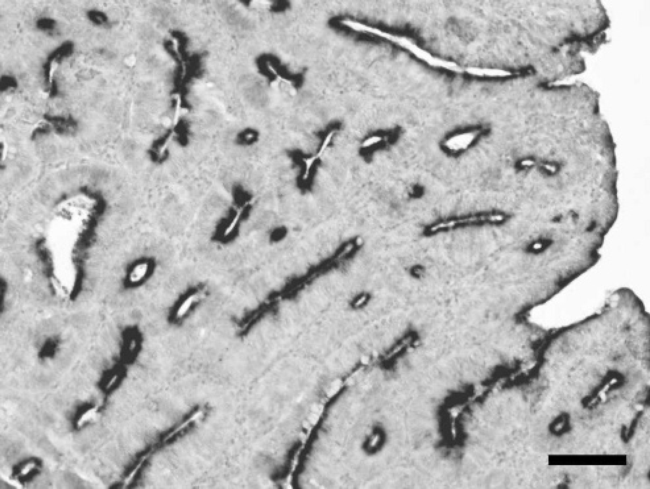
Lawsonia-specific ISH produces distinct blue-black rims consistent with the localization of bacteria at the luminal border of the majority of hypertrophic enterocytes. Abundant diffuse staining reaction in a case with histological lesions characteristic of PPE. ISH. Bar = 100 μm.
Taken together, the data of this investigation are clearly in line with results from other comparative studies. It has been repeatedly shown that the sensitivity of IHC is superior to the Warthin-Starry technique and that PCR tends to give positive results in cases without clinical or pathological suspicion. 5–7 Whereas diagnosis of classical PPE is usually unproblematic and there is good correlation between macroscopic lesions, histological enterocyte hyperplasia, and detection of Lawsonia within enterocytes by Warthin-Starry stain and IHC or ISH, cases of L. intracellularis infection without prominent enterocyte hyperplasia are diagnostically more challenging. Such cases are frequently macroscopically or histologically missed, and the bacteria are usually not found by Warthin-Starry stain 7 because the pathogen is present only in scattered groups of epithelial cells and/or macrophages of the propria and submucosa. Immunohistochemistry and ISH, however, easily enable one to specifically localize the bacteria in these cases, 5 , 7 which are considered to indicate recovery from PPE. In this study, PCR of mucosal scraping identified a few more positives than ISH. Similar results have been found when comparing PCR with IHC data. 7 Possible explanations for these differences could be the focal presence of bacteria, which is more easily missed by IHC or ISH examination of 1 transversal 3-μm section of ileum than by scraping a more extended area for PCR examination, or the presence of very small numbers of predominantly extracellular bacteria that escape detection by IHC or ISH. From a clinical point of view the detection of such low amounts of bacteria, most likely representing the final stage of recovery from infection, 7 is probably not very relevant. Thus a combined morphological and pathogen detection method, like IHC or ISH, is considered the most powerful diagnostic technique, because it is able to distinguish between different stages of the infection. The application of this technique at herd level, examining more than 1 suspicious animal per group, will also increase the likelihood of discovering resolving cases or very early stages of disease, both with low amounts of bacteria.
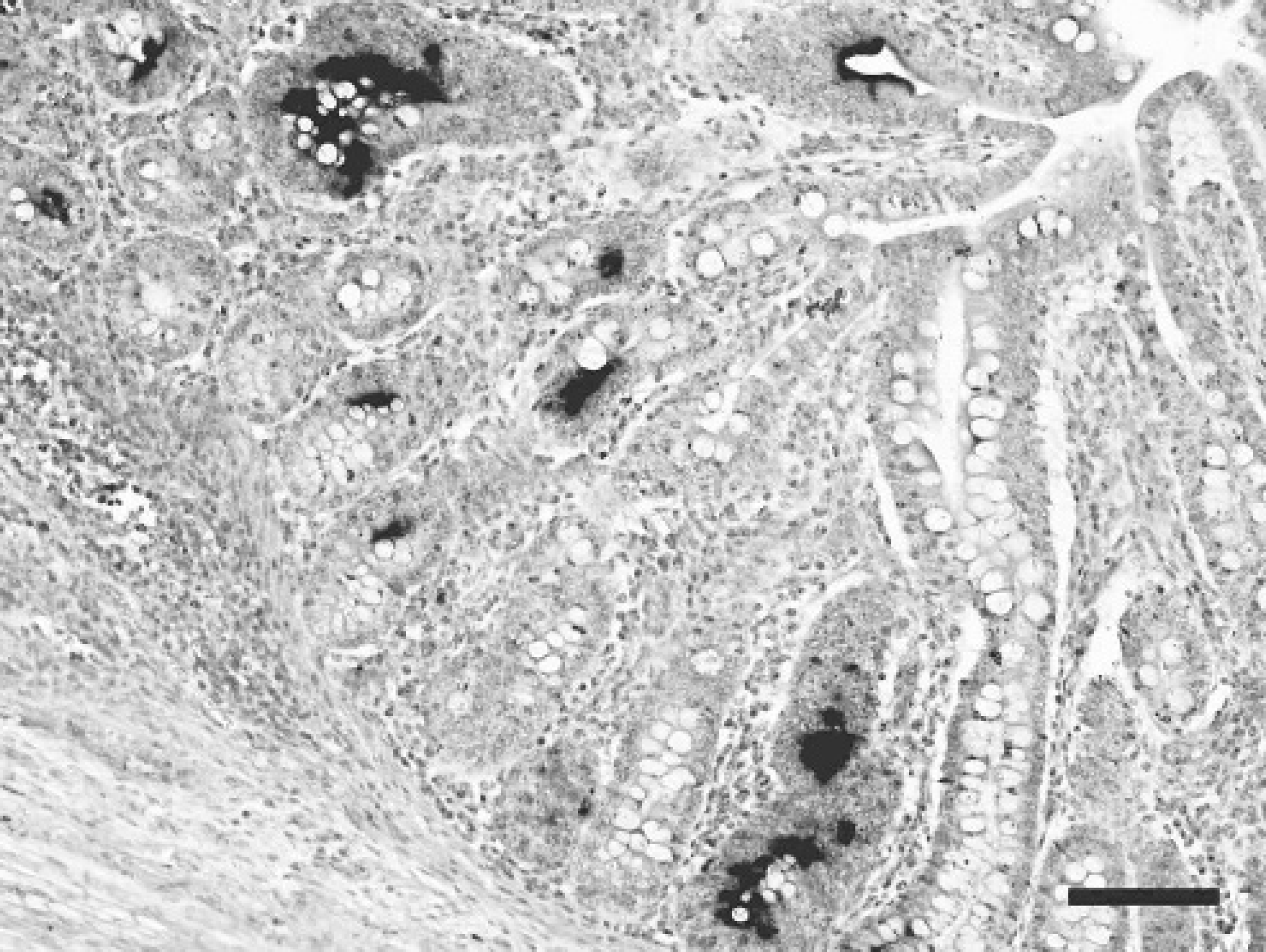
Case with focal moderate staining reaction. Signal is present within a few hypertrophic enterocytes. This case was negative with Warthin-Starry stain and showed no histological lesions indicative of PPE. ISH. Bar = 100 μm.
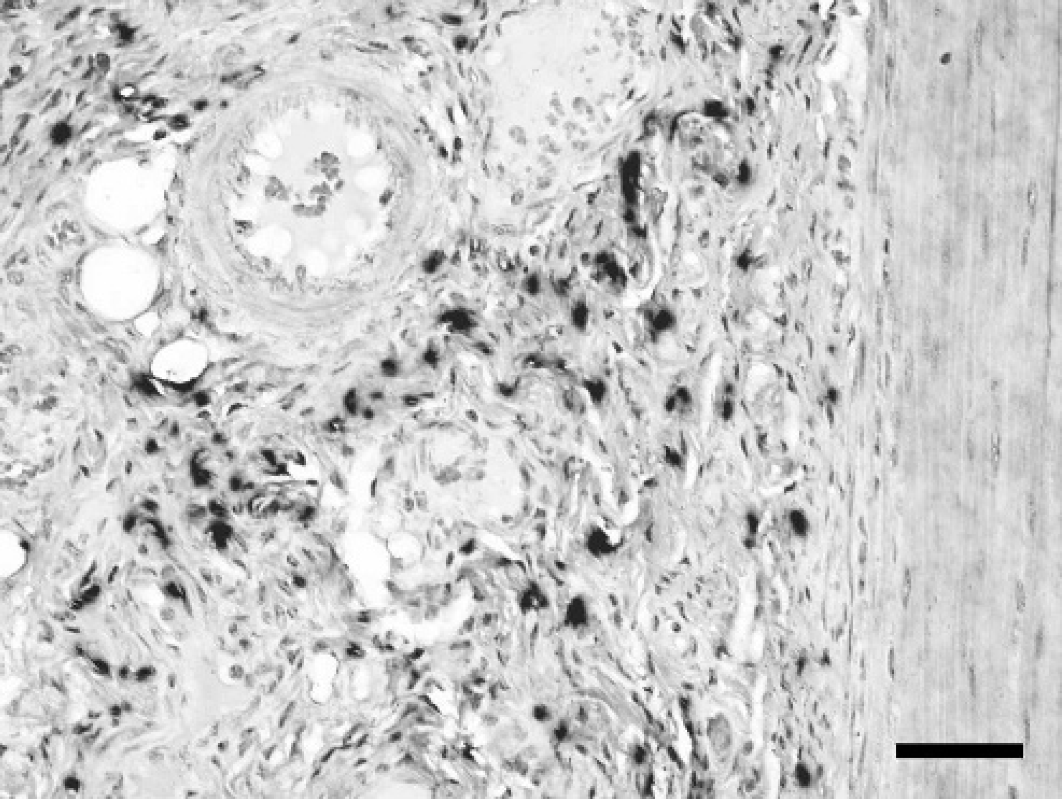
Case with specific staining in the submucosa. In this case, neither staining of enterocytes by ISH nor histological lesions characteristic for PPE were present. Warthin-Starry stain was negative. ISH. Bar = 50 μm.
The sensitivity of ISH seems to be comparable to IHC and its slightly more laborious protocol is counterbalanced by the easy availability of customized modified oligonucleotide probes. The present technique provides several advantages as compared to previously described ISH protocols. The method described by Gebhart et al 4 was developed to confirm whether cloned DNA probes generated from DNA of the bacterium (then still not fully characterized) were able to hybridize with the intracellular organisms that were suspected as the etiologic agent. The probe was generated from an unknown region of genomic DNA and seemed to have generated a hybridization signal only on frozen sections. From the diagnostic point of view, both the complicated probe synthesis and the need for frozen sections make this technique less attractive. A protocol later published by Boye et al. 2 meets diagnostic needs more adequately. It takes advantage of easily accessible oligonucleotide probes targeting the abundant bacterial rRNA. The fluorescent label makes it an excellent tool for a rapid diagnostic assay, because the entire protocol requires only a few steps. The method introduced here uses a DNA oligonucleotide probe complementary to a region of rRNA, providing the advantages of an abundant presence of target nucleic acid in the samples and easy probe availability, because numerous biotechnology companies offer rapid synthesis of oligonucleotide probes according to customers' requirements. The disadvantages of the method of Boye et al. (1998) (a temporary signal, limited correlation of the signal with the lesion in dark field microscopy, and the need for fluorescence microscopic equipment) are eliminated by using a digoxigenin-labeled probe followed by immunological detection by an alkaline phosphatase-antibody conjugate and the permanent color substrate NBT/BCIP. It is suggested that this method is a suitable alternative to IHC for those who wish to demonstrate L. intracellularis in paraffin-embedded tissue but have no access to primary antibodies.
Acknowledgements. We thank Henriette Toft Boesen, Danish Institute for Food and Veterinary Research, for the generous gift of the antibody LAW1-DK. The help of Klaus Bittermann with the digital images is gratefully acknowledged.
Footnotes
a.
MWG, Ebersberg, Germany.
b.
VWR, Vienna, Austria.
c.
Roche, Basel, Switzerland.
d.
Fluka, Buchs, Switzerland.
e.
Sigma, St. Louis, MO.
f.
Invitrogen, Carlsbad, CA.