
Abstract
In this retrospective study, the authors describe the gross and histologic changes associated with rupture of an abdominal artery aneurysm in 33 mature female Holstein cattle between January 1980 and June 2005 from 29 farms in upstate New York and northern Pennsylvania. Over this period, there was an increase in the number of cases submitted for necropsy per year, and a seasonal trend did not exist. Affected animals ranged from 2.5 to 5.5 years of age. Grossly affected cattle exhibited marked hemoabdomen. There was marked dilation and rupture of the abdominal aorta or one of its branches, including the mesenteric, left gastric, celiac artery, right ruminal artery, or left ruminal artery. Histologically, the tunica media of the affected arteries was often thin and irregular in width with disrupted, fragmented, and coiled elastin. Granulation tissue and hemorrhage was localized at the site of rupture. There was mild-to-marked hyperplasia of the tunica intima that was irregular and disorderly with adjacent smooth-muscle hyperplasia within the tunica media. In addition, in a fraction of cases, there was mild-to-moderate mucinous change (48%) and mineralization (30%) within the tunica media. To the authors' knowledge, this is the first description of the gross and histologic changes in Holstein cows with abdominal artery aneurysm and rupture.
Introduction
Rupture of abdominal arteries in cattle has been recognized by New York practitioners for at least 20 years (personal communication with New York veterinarians). There have been 2 published reports of individual cows affected with abdominal arterial aneurysm and rupture (1 in Minnesota and 1 in the Netherlands), and the etiology was not determined. 2 , 10 These cases were both 4- to 5-year-old, female Holstein dairy cattle. In these 2 cases, death was attributed to rupture of the cranial mesenteric artery with subsequent exsanguination. In the first case, fibrin and hemorrhage were present histologically at the site of arterial rupture. Thickening and irregularity of the vessel wall were also described. 2 In the second case, rupture of the internal elastic lamina with chronic proliferation of the intima was observed histologically within the ruptured artery wall. 10
Ruptures of abdominal arteries have also been reported in particular genetic lines of cattle and have been traced back to a single bull. 9 Ruptures of the thoracic aorta in cattle have been linked to an inherited defect related to decreased fibrillin production and has been termed bovine Marfan syndrome, analogous to Marfan syndrome seen in humans. 8
In this study, information was gathered from several affected cattle that were believed to be unrelated and located on several different farms throughout upstate New York and northern Pennsylvania. To the authors' knowledge, the signalment, gross necropsy findings, and histologic changes in cattle with abdominal artery aneurysm and rupture have not been previously described on such a large scale.
Materials and methods
Study population. The Cornell University Anatomic Pathology computer data base was searched for any domestic bovid with abdominal artery rupture submitted through the surgical or necropsy service from January 1980 to June 2005. Additional cases fitting the same criteria submitted through the Anatomic Pathology Service by the Ambulatory Clinic at Cornell University between January 2004 and June 2005 were also included. The cases from the Ambulatory Clinic were collected in the field and were brought to the attention of the authors for inclusion in the study. For an animal to be included in the study, it had to be a domestic bovid (Bos taurus) of any age with rupture of an abdominal artery and subsequent hemoabdomen confirmed on postmortem examination.
Data. Data obtained for each case included age, sex, breed, month of submission, year of submission, and the anatomical location of the arterial rupture. The necropsy reports were obtained and reviewed for cases meeting the above criteria. Available histologic slides of the arteries stained with hematoxylin and eosin as well as Verhoeff elastin stain were also reviewed. Selected sections were stained with Von Kossa for mineral. Samples of the major abdominal arteries were collected from 6 adult female Holstein dairy cows for which the cause of death was not directly related to the cardiovascular system. These samples were routinely processed and stained with hematoxylin and eosin as well as Verhoeff elastin stain for comparison with affected animals.
Age distribution of cattle with rupture of abdominal arteries submitted through the necropsy or surgical service between 1980 and 2004. Affected cattle were between the age of 2.5 and 5.5 years.
Descriptive statistics were calculated for all available numerical data. Cases from 2005 and from the ambulatory service were not included in the statistical risk-assessment analysis because of an inability to calculate a denominator properly. For statistical analysis, the ages were broken into 3 groups (<2.5 yr, 2.5–5.5 yr, and >5.5 yr). For analysis of breed affected, the number of Holsteins was compared to the total number of cattle for all other breeds. For analysis of number of cases per year, the numbers of affected and unaffected cattle were grouped in 5 consecutive year periods. For analysis of season, the numbers of affected and unaffected cattle were grouped based on 4 periods (winter = Dec-Feb, spring = Mar-May, summer = Jun-Aug, fall = Sep-Nov).
The total number of domestic bovids submitted through the Anatomic Pathology service from January 1980 to December 2004 was calculated based on age, sex, breed, month of submission, and year of submission. Information from these submissions was used as the denominator in chisquare (χ2) analyses to determine if age, sex, breed, season of submission, and year of submission were risk factors. Graphs and regression lines were created using Excel. a Statistical analysis was performed using Statistix 8. b
Results
Signalment. Between January 1980 and June 2005, a total of 33 cases from 29 different farms were submitted. Twenty-nine of these cases were submitted through the Pathology Service, and 4 cases were submitted through the Ambulatory Clinic. Submitting farms were located throughout upstate New York as well as northern Pennsylvania. In 10 of these cases, only gross necropsy evaluation was performed. Histologic sections of abdominal arteries were available for review in 23 cases.
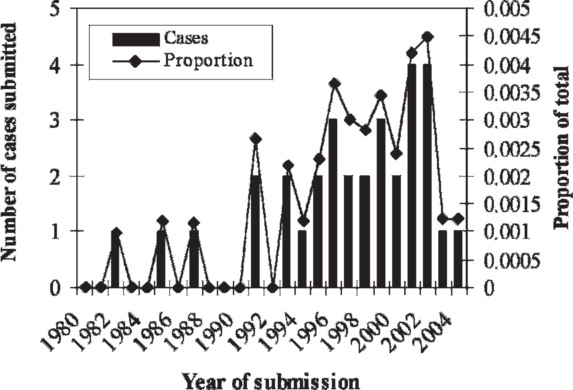
Secular trend of rupture of abdominal arteries in cattle submitted through the necropsy or surgical service between 1980 and 2004. The proportion of total cattle submitted to the necropsy or surgical service with aneurysm and rupture over this period is provided (P = 0.0004, R 2 = 0.5021, χ2 = 20.49).
The age range of the affected animals was 2.5 to 5.5 years, with a median of 4 years (Fig. 1). Eight cases were recorded only as “adult,” and for 2 cases, age was not provided. Age appeared to be a risk factor for the development of abdominal artery aneurysm and rupture (χ2 = 45.64, df = 2, P < 0.0001).
Thirty affected animals were Holstein dairy cows. Information on breed was not provided for 3 cases. Holsteins were at higher risk for developing rupture of an abdominal artery than other breeds (χ2 = 9.45, df = 1, P = 0.0021). Thirty of the affected animals were female. Sex was not listed in the report for 3 cases. Analysis of sex was not done because there were too few male submissions.
Annual trends and seasonality. The number of cases submitted for necropsy per year increased significantly from 1980 to 2005 (Fig. 2, chi-square test for heterogeneity of independence comparing 5-year periods: χ2 = 20.49, df = 4, P = 0.0004). There was not a seasonal pattern (Fig. 3, chi-square test for heterogeneity of independence comparing seasons in 3-month periods: χ2 = 5.74, df = 3, P = 0.13).
Pathology. Severe hemoabdomen and variably sized blood clots were present within the abdomen of all animals. These clots were intimately associated within the right lateral serosal surfaces of the forestomachs (Fig. 4). There was variable dilation and rupture of one of the major abdominal arteries in each of the cases examined (Fig. 5). The mesenteric artery was affected in 10 cases, the left gastric artery in 9 cases, the celiac artery in 4 cases, the abdominal aorta in 2 cases, the left ruminal artery in 1 case, and the right ruminal artery in 1 case. The location of arterial rupture was not provided in the report of 6 cases. Large hemotomas often covered the site of rupture and occasionally extended through the adjacent connective tissue.
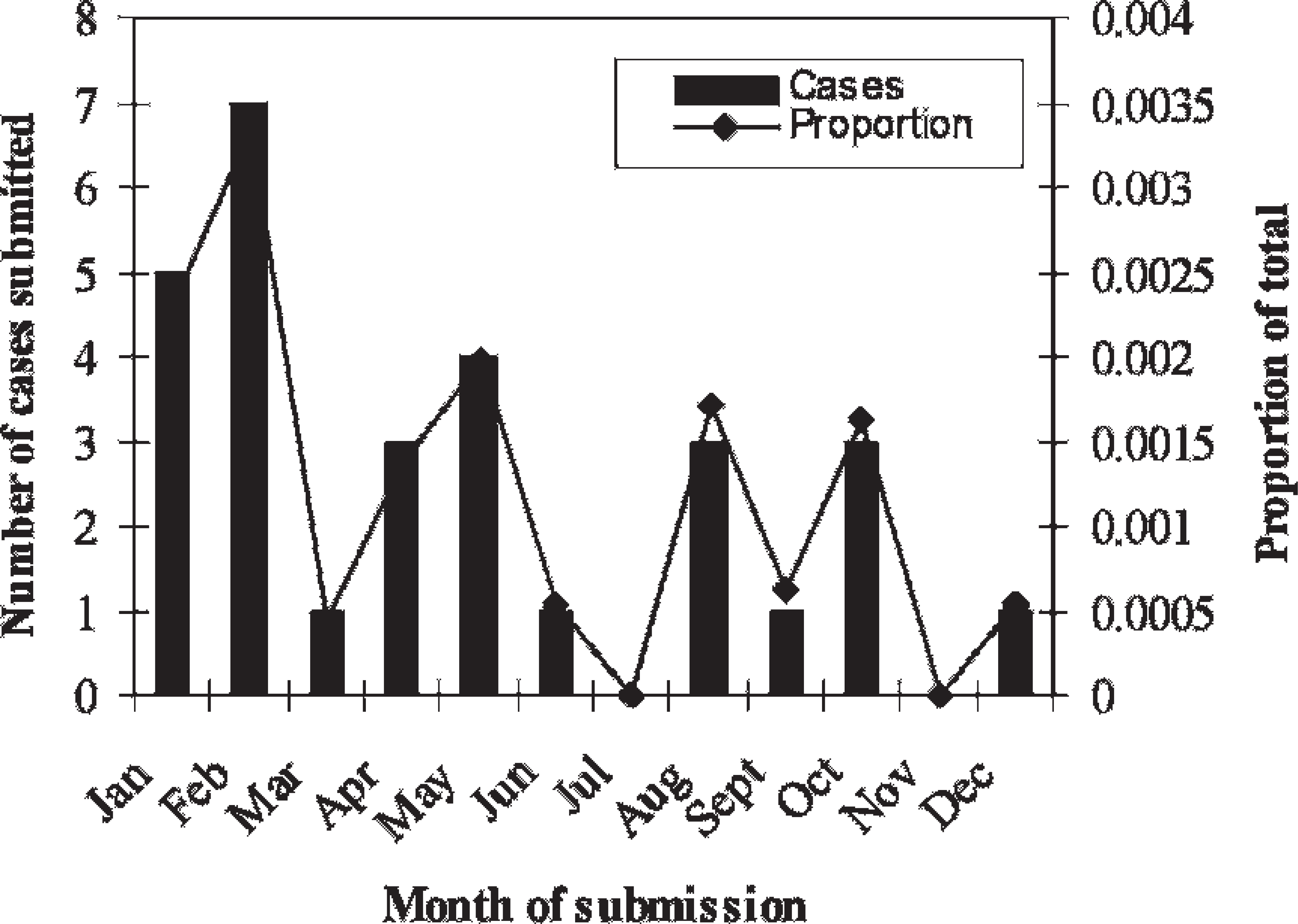
Monthly distribution of cattle with abdominal arterial rupture submitted through the necropsy and surgical services. The proportion of the total cattle submitted for necropsy with aneurysm and rupture by month is included. There was no seasonal trend (P = 0.13, df = 3, χ2 = 5.74).
Striking histologic changes were present within the walls of the arteries at the site of arterial rupture. The wall of ruptured arteries was markedly thin and irregular in width. The tunica intima and portions of the tunica media were often absent in the more affected areas. Aggregates of neutrophils and fibrin were clumped along the luminal surface within the attenuated areas, occasionally obscuring the adjacent architecture (Fig. 6). Hemorrhage consistently surrounded the site of rupture, and the amount of hemorrhage varied from mild to severe. At the site of rupture, there was a variable amount of granulation tissue, which extended along the serosal surface and occasionally along the luminal surface (Fig. 7). Within areas of the most pronounced granulation tissue, there were moderate numbers of infiltrating neutrophils with fewer numbers of macrophages, lymphocytes, and plasma cells.
Within all of the abdominal arteries examined, there were profound changes in the elastin. The elastin within the tunica media was affected at the sites of rupture, with minimal elastin evident in the more affected areas. The remaining elastin within the tunica media lost the typical parallel arrangement and was disorganized, fragmented, and had a slightly frayed appearance. The amount of elastin within the tunica media proximal and distal to these areas was also often decreased. These changes were not noted within the thoracic aorta or within the larger arterioles of the major abdominal or thoracic organs.
The internal elastic lamina was also variably disrupted and occasionally completely absent at the site of vessel rupture in 87% of cases. Fragments of coiled internal elastic lamina were embedded in the more affected areas in 22% of the cases (Fig. 8). Within the arteries proximal and distal to the site of rupture, the internal elastic lamina was occasionally disrupted and slightly coiled, with portions often absent. Areas of internal elastic lamina loss were characterized by intimal thickening and smooth-muscle hyperplasia within the adjacent tunica media. Adjacent to the site of rupture, there was moderate-to-marked multifocal expansion of the tunica intima with smooth-muscle hypertrophy and hyperplasia within the underlying tunica media in 61% of the cases (Fig. 9). In a single affected animal, there was mineralization of the internal elastic lamina and the elastin within the tunica media multifocally.
In 48% of cases, there were multifocal areas of mucinous change within the tunica media, expanding and separating the normal architecture. Similarly, there were areas of mineralization within the tunica media that completely replaced the normal architecture in 30% of the cases. There was variable intimal thickening, mucinous change, and mineralization within the arteries proximal and distal to the sites of rupture.
Compared to 6 control Holstein cows, the changes in the arteries of affected animals were quite pronounced. In 3 of 6 of the control animals, there was only very mild, multifocal, patchy thickening of the tunica intima in arteries. One control had small areas of mineralization within the tunica media. No other changes were noted in sections from control animals.
Discussion
This report is the first to describe the demographics, gross findings, and histologic changes associated with the rupture of abdominal artery aneurysms in a group of affected, unrelated cattle from different farms. This disease appears to affect only 2.5- to 5.5-year-old Holstein cattle, which is consistent with 2 previous case reports. 2 , 10
Age appears to be a factor in the development of disease, with affected cattle ranging in age from 2.5 to 5.5 years. It is possible that the pathogenesis of the disease is over a long course of time and that the changes within the vessel occur slowly, until eventually, the artery ruptures and exsanguination occurs. The chronicity of the lesion is supported by the presence of smooth-muscle hypertrophy and granulation tissue within the vessel wall at the site, both of which require several weeks to form. Similar histologic changes have also been reported in humans with long-term microdamage to the arterial wall. 5

Abdomen of a cow with abdominal arterial rupture. A large amount of unclotted blood from the abdomen has pooled on the ground. A large blood clot is intimately associated with the serosal surface of the omasum (arrow).

Arterial branch from a cow. There is an 8-cm-diameter dilation of the left gastric artery with rupture and adherent blood clot at the site (arrow).

Abdominal artery from a cow with arterial rupture. A thick aggregate of neutrophils and fibrin is adhered to the luminal surface. Hematoxylin and eosin 1.25X.
Breed also appears to be a risk factor for the disease. It is possible that sudden death in Holsteins is more likely to be followed up through necropsy. It is also possible that the intense management and high production in Holsteins are related to development of this disease. Furthermore, there may be a breed-related predisposition for development of abdominal artery rupture, similar to bovine Marfan syndrome. 8
All affected cattle in this study were female. It is the impression of local practitioners that this is a disease of female dairy cows, and it has not been seen in male dairy cattle or in beef herds (personal communication with New York veterinarians). This clinical impression could be a result of several different factors. It is possible that female dairy cattle so outnumber males that the chances of recognizing a relatively uncommon disease in a bull are extremely small. It is also possible that the intense management, diet, and/or high milk production in dairy cattle play a role in the pathogenesis of the disease.
Season does not appear to play a role in the development of aneurysm with rupture in the abdominal arteries of cattle. However, there does appear to be an increasing trend of cases submitted over time, from 0 cases in 1980 to 4 cases in 2002. This increase in submissions might be due to several possible factors. Perhaps submitting dairies in the region are becoming larger, increasing the chances of the disease being observed within a herd. It might also be due to increased awareness of the disease and subsequent increased submission for necropsy of sudden death animals. Finally, it is possible that changes in management practices in dairies are playing a role in the increased development of vascular degeneration resulting in eventual dilation and rupture.
Rupture of abdominal arteries in cattle appears to be sporadic. The common presentation is sudden death of a single animal. However, it has been noted by local practitioners that the disease occasionally occurs in clusters, with 2 to 4 animals dying suddenly within a few days on a single farm. The reason for the variation in the number affected at a single time is not clear in this study.
The pathogenesis of the development of the histologic changes is unknown. The changes suggest a period of weakening within the vessel wall followed by attempts at repair before final rupture and exsanguination. The granulation-tissue formation, smooth-muscle hypertrophy, and intimal thickening all appear to be attempts at repair and stabilization of the vessel wall. It is difficult to know if the changes in the internal elastic lamina and the elastin within the tunica media are the cause or the consequence of vessel damage. In pigs and turkeys, defects in lysyl oxidase secondary to copper deficiency result in decreased and abnormal layering of elastin, resulting in subsequent arterial rupture. 3 , 4 , 11 The elastin within the vessels in cattle with Marfan syndrome is similarly abnormal, resulting in vascular rupture. 8
In humans with abdominal artery rupture, direct damage to the vessel wall or damage secondary to deposition of antigen-antibody complexes can result in fragmentation and break down of elastin. 5 , 6 This damage to the arterial wall integrity would result in secondary changes, such as an increase in smooth-muscle cells, as is seen in this study and which has also been reported in rats. 1 Furthermore, a primary defect in collagen, such as is seen with Ehler's Danlos syndrome in humans, could cause weakening of the vessel wall with secondary fracturing of the elastin. 5 , 7 It is likely that abdominal artery rupture in cattle is multifactoral, as it is in humans. The pathogenesis and etiology of the histologic changes in the cases presented were beyond the scope of this study. Preliminary data suggest that liver iron levels less than 60 mg/kg are associated with the development of aneurysm and rupture of the major abdominal arteries in dairy cattle (Lamm et al., unpublished data).
In summary, aneurysm with rupture of the abdominal arteries in cattle is primarily seen in mature female Holsteins. There is no seasonal trend, and there have been increasing numbers of submissions over the past 25 years. Grossly, there is dilation and rupture of a single vessel with hemoabdomen. Histologically, there is marked disruption of the tunica media with an overall decrease in the amount of elastin and histological evidence of attempts at repair. In aggregate, these lesions suggest chronic damage to the vessel wall with eventual acute rupture and exsanguination.

Abdominal artery from a cow with arterial rupture. A large amount of granulation tissue is present along the serosal surface (asterisk). Hemorrhage dissects through the arterial wall (arrow). Hematoxylin and eosin 1.25X.

Abdominal artery from a cow with arterial rupture. The internal elastic lamina has ruptured and coiled in upon itself (arrow). Hematoxylin and eosin 10X.

Abdominal artery from a cow with aneurysm and rupture. There is smooth-muscle hypertrophy and hyperplasia within the tunica media (large asterisk). There is a focal area of mineralization (arrow). Hematoxylin and eosin 4X.
Footnotes
a.
Microsoft Excel, Redmond, WA.
b.
Statistix 8, Tallahassee, FL.