
Abstract
Reported clinical signs of coccidiosis in South American camelids include anorexia of a few days duration, sudden death, and diarrhea. Antemortem diagnosis of clinical coccidiosis is usually based on clinical signs and supported by detection of coccidial oocysts in feces. This report describes 2 atypical cases of coccidiosis in South American camelids that had no coccidial oocysts detected on antemortem fecal flotation, prolonged weight loss, and normal fecal consistency.
Keywords
Several species of coccidia have been reported in South American camelids (Eimeria lamae, Eimeria alpacae, Eimeria macusaniensis, Eimeria punoensis, and Eimeria peruviana, Eimeria ivitaensis). 5,6,12,14 No cross-transmission studies between South American camelids and other domestic animals have been reported, but cross-transmission is believed to occur among the guanaco, alpaca, and llama. 16 Eimeria lamae and E. macusaniensis are considered highly pathogenic. 5 Reported clinical signs of coccidiosis in South American camelids include anorexia of a few days duration, 16 sudden death, 10,15 and diarrhea. 12,15 The diagnosis of clinical coccidiosis is usually based on clinical signs and supported by the detection of large numbers of coccidial oocysts in feces. 13 However, oocysts detection is dependent on detection technique, 3,5,8,16 and the output of oocysts can decline rapidly in acute infections resulting in clinically affected animals having low oocyst counts. 13 Hence, fecal examination may not always aid in diagnosis. This report describes 2 atypical cases of coccidiosis in South American camelids that had no coccidial oocysts detected on antemortem fecal flotation, prolonged weight loss, and normal fecal consistency.
The first case involved a 10-year-old, 61-kg, female alpaca that presented with appetite loss and chronic weight loss of 5-week duration. The alpaca was presented to the hospital in winter (December). The alpaca had been housed at a breeding facility 2 weeks before presentation. The alpaca's diet consisted of free choice alfalfa hay and a commercial concentrate diet a formulated for llamas and alpacas. The alpaca was nursing a 6-week-old healthy cria. The alpaca was vaccinated yearly with Clostridium perfringens types C and D and Clostridium tetani and dewormed monthly with injectable ivermectin. 13
Physical examination revealed poor body condition (body condition score 2 of 10). 5,9 Rectal temperature was 38.3°C (reference range, 37.5°C-38.9°C 5 ), heart rate was 112 beats/minute (reference range, 60–90 beats/minute 5 ), and respiratory rate was 28 breaths/minute (reference range, 10–30 beats/minute 5 ). The initial problem list based on history and physical examination included tachycardia, anorexia, and chronic weight loss. Tachycardia was most likely caused by stress at the time of admission. Inappetence was considered secondary to an underlying primary disease. Differential diagnoses for weight loss included insufficient caloric intake attributable to anorexia, intestinal parasitism, Mycobacterium avium ssp. paratuberculosis (MAP), neoplasia, renal disease, liver disease, third stomach compartment ulcers, internal abscessation, tuberculosis, coccidioidomycosis, 17 bovine viral diarrhea (BVDV), 2,11 and immunodeficiency syndrome. 1 The initial diagnostic workup included a complete blood count (CBC), serum biochemistries, urinalysis, and fecal flotation.
The CBC was unremarkable. Serum biochemical abnormalities included hypoproteinemia (4.2 g/dl; reference range, 4.7-7.3 g/dl 5 ) characterized by hypoalbuminemia (1.6 g/dl; reference range, 2.9-5 g/dl 5 ) and an elevated aspartate aminotransferase (AST) activity (634 U/L; reference range, 128–450 U/L 5 ). Urinalysis revealed ketonuria. Fecal flotation for the detection of parasite ova or coccidial oocysts was performed using a saturated sucrose solution with a measured specific gravity of 1.27. 3 No trichostrongylid ova or coccidial oocysts were observed. Hypoalbuminemia can result from the loss of albumin through the gastrointestinal tract or kidney, decreased synthesis by the liver, loss into body cavities, or hemorrhage. Renal disease was unlikely as there was no evidence of renal dysfunction on either the serum biochemical analysis or urinalysis. Elevated AST activity can indicate myonecrosis or hepatocellular damage. Creatine phosphokinase, a specific enzyme indicating muscle damage, was within reference ranges; hence, the elevation in AST activity was considered most likely because of hepatocellular injury.
Further testing included an abdominal ultrasound to evaluate the liver, and percutaneous liver biopsies were obtained to help determine the cause of the elevated AST activity. Inguinal lymph node biopsies were obtained to rule out immunodeficiency syndrome, and serum was submitted for detection of antibodies against MAP. Ultrasonographic evaluation of the liver was unremarkable, percutaneous liver biopsies and lymph node biopsies revealed no abnormalities, and agar-gel-immunodiffusion for MAP was negative.
Minimal improvement was noted after 6 days of symptomatic and supportive treatment that included intravenous (IV) fluids and electrolytes and ceftiofur sodium c (2 mg/kg IV). Seven days after presentation the alpaca exhibited signs of abdominal discomfort, which included frequent lying in lateral recumbency and kicking at the abdomen. A follow-up serum biochemical evaluation at that time revealed hypoproteinemia (4.4 g/dl) characterized by hypoalbuminemia (2.1 g/dl). Minimal improvement was noted after treatment with flunixin meglumine d (2 mg/kg BID) and an exploratory abdominal laparatomy was performed. The first (C1) and second (C2) stomach compartments were relatively empty. The third stomach compartment was slightly distended, but no other gross abnormalities were noted. Biopsies of the liver, kidneys, pylorus, jejunum, and ileum were performed, and sections of each were stained with hemotoxylin and eosin and examined. Sections of pylorus were obtained to rule out a possible pyloric outflow disorder. Examination of liver sections revealed swollen hepatocytes with moderate to abundant bile consistent with cholestasis. Sections of each kidney and pylorus were unremarkable. Sections of ileum revealed numerous coccidial organisms in the lamina propria in various stages of development along with large numbers of eosinophils.
Supportive therapy was continued after surgery, and treatment with sulfadimethoxine e (55 mg/kg on the first day and 27.5 mg/kg thereafter for 4 days) was initiated to treat the coccidiosis. Minimal improvement was noted after 5 days of treatment with sulfadimethoxine. Because of the animal's poor appetite and lack of response to therapy the owner elected to euthanize the animal 20 days after admission to the hospital.
Gross postmortem examination indicated focal serosal hemorrhage at the pylorus, ileum, and jejunum, most likely from the biopsies obtained during surgery. Histopathology revealed numerous coccidia at various stages of development in the lamina propria of the jejunum and ileum (Fig. 1). The coccidial organism was identified as E. macusaniensis based on the thickness of the oocyst wall and large size of the oocysts (90 mm × 64 μm). 7 Hepatocytes in all zones contained moderate to abundant bile pigment consistent with cholestasis. Fecal culture for MAP was negative. No significant growth of enteric pathogens was obtained from a culture of intestinal samples. Histopathology of other organ systems was unremarkable. Isolation of BVDV on necropsy was not performed, but there were no histologic findings consistent with BVDV infection. In the absence of other significant lesions, a diagnosis of intestinal coccidiosis with cholestasis was made based on necropsy and antemortem biopsy results.
The second case involved a 2-year-old, 144-kg, intact male llama that was examined for weight loss of 17-day's duration. The alpaca was presented to the hospital in the spring (April) and had a good appetite. The llama's diet consisted of 1 kg of a commercial concentrate a recommended for llamas and alpacas twice daily and free choice fescue grass hay. The llama was vaccinated yearly with C. perfringens types C and D and C. tetani and dewormed 3 times a year with injectable ivermectin. 13
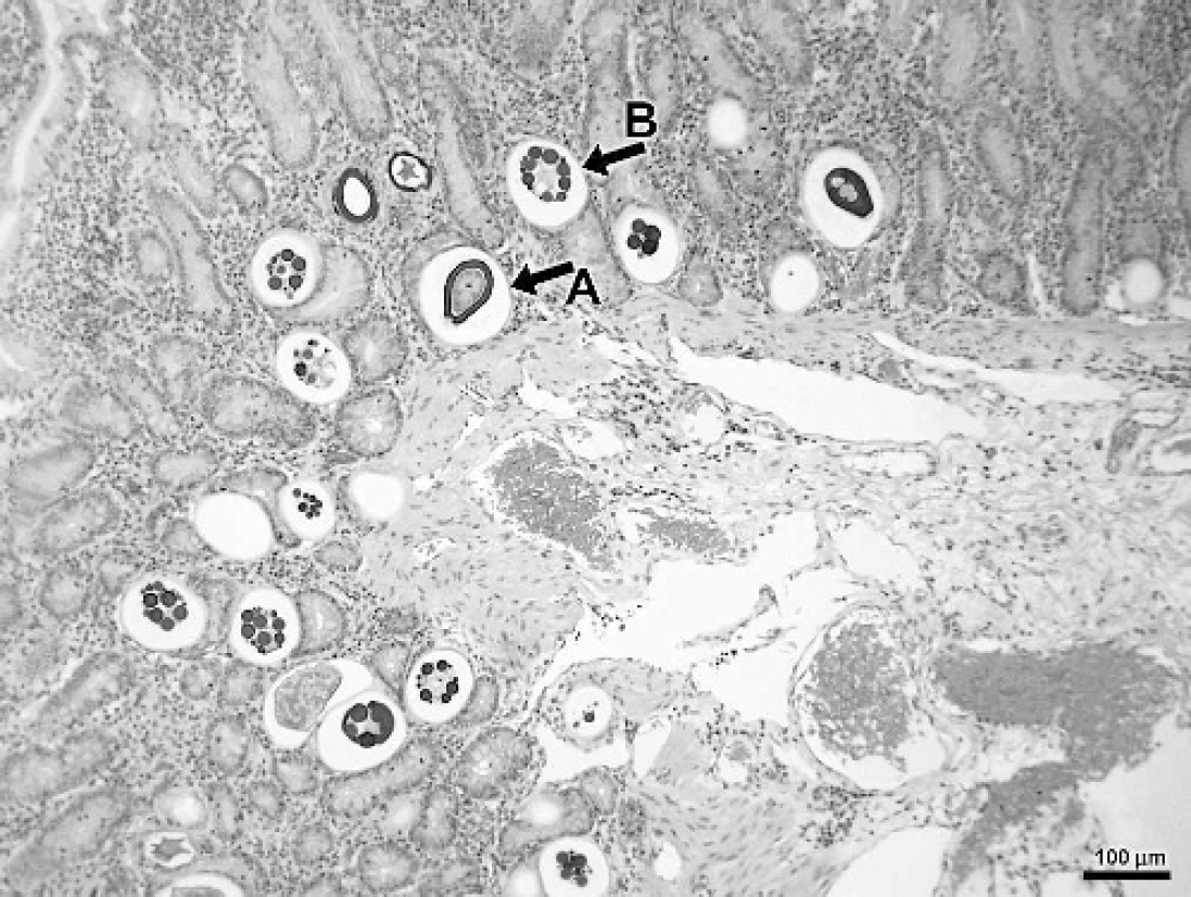
Hemotoxylin and eosin-stained sections of ileum showing an unsporulated oocyst,
On physical examination the llama had a moderate body condition (body condition score 5 of 10). 5,9 Rectal temperature was 37.9°C, the heart rate was 92 beats/minute, and respiratory rate was 32 breaths/minute. The initial problem list based on history and physical examination included mild tachycardia and tachypnea and weight loss. Tachycardia and tachypnea were attributed to stress and excitement during examination. Differential diagnoses considered for weight loss were the same as those for the case described above. Insufficient caloric intake was considered unlikely because the llama had a good appetite and the ration appeared adequate. The initial diagnostic workup included CBC, serum biochemistries, fecal flotation, and urinalysis.
The CBC and urinalysis were within reference ranges. The only serum biochemical abnormality was hypoalbuminemia (2.2 g/dl). Fecal flotation performed as described in the first case revealed a few trichostrongylid ova. The causes of hypoalbuminemia that were considered were the same as those described above. Given that no abnormalities consistent with liver or renal disease were detected on the serum biochemical profile and urinalysis, gastrointestinal loss of albumin was considered likely.
The llama was hospitalized and monitored for signs of reduction in feed intake. No treatments were done while the llama was hospitalized. Repeat serum biochemistries performed 2 days after admission were unremarkable. The owner elected to monitor the llama at home, and the animal was discharged 4 days after presentation. Twelve days after being discharged from the clinic the owner reported that the llama had developed diarrhea of 2-day's duration and subsequently died. The llama was submitted for necropsy.
Gross postmortem examination revealed multiple petechial and ecchymotic hemorrhages in the mesentery and omentum. The liver was diffusively yellow. The jejunum and ileum had segmental areas filled with blood. Fecal flotation performed at the time of the necropsy revealed moderate numbers of trichostrongylid ova and a few Nematodirus spp. and Trichuris spp. ova, but no coccidial oocysts.
Histopathology revealed numerous Eimeria oocysts at various life-cycle stages in the mucosa and lamina propria of the jejunum and ileum. Tissue sections from the second case were no longer available for retrospective speciation of the Eimeria spp. reported in the original postmortem report. Hemorrhages were present in the mesentery and omentum. Hepatocytes were swollen and had vacuolated cytoplasm consistent with hepatic lipidosis. Histopathology of other organs was unremarkable. Bacteriological cultures of tissues from the intestine, liver, and lungs were negative. The hemorrhages observed in the mesentery and omentum were considered an extension of the inflammation in the ileum and jejunum caused by the coccidia. A diagnosis of intestinal coccidiosis and hepatic lipidosis was made based on necropsy results. Antemortem diagnostic workup in this patient was minimal because of the unremarkable physical examination and laboratory findings.
The described cases were atypical in that they were presented for ill-thrift and weight loss of more than 2 weeks duration and had formed feces on initial presentation. In contrast, anorexia of a few days duration, 16 diarrhea, 12,15 and sudden death 10,15 have been reported as presenting clinical signs with coccidiosis in South American camelids. Whereas the second case had acute diarrhea before death, the first case never developed diarrhea. Hepatic lipidosis in South American camelids is associated with anorexia, weight loss, and physiologic conditions with a high demand for energy such as pregnancy and lactation. 18,19 In the second case presented here, the hepatic lipidosis was consistent with the history of weight loss. Colic observed in the first case may have been caused by the severe coccidial enteritis observed during postmortem examination. 17
The primary site for coccidiosis in South American camelids is the small intestine. 5 The absence of coccidial oocysts on routine fecal flotation examination can be because of an incomplete prepatent period or the use of an insensitive fecal flotation technique. The prepatent period of Eimeria spp. in South American camelids ranges from 10–34 days, with Eimeria macusaniensis having the longest prepatent period of 33–34 days. 3,4 The first case in this report demonstrated ill-thrift for at least as long as the average prepatent period of E. macusaniensis, and hence, fecal flotation should have revealed coccidial oocysts. Consequently, a negative fecal examination is possible in South American camelids with coccidiosis, and a negative fecal examination cannot be used to rule out ill-thrift secondary to coccidial infiltration of the small bowel. Jarvinen 8 reported that fecal flotation solutions with a specific gravity ≤1.20 may fail to detect E. macusaniensis, especially at low oocyst concentrations, whereas a saturated sugar solution with a specific gravity ≥1.28 gave comparable results to fecal sedimentation. Fecal sedimentation to detect oocyst of E. macusaniensis has been recommended because of their large size and higher specific gravity. 7,16 The fecal flotation technique used for the 2 cases reported here was a saturated sucrose solution with a specific gravity of 1.27. Hence, the technique should have detected oocysts in feces of the described cases.
Ruminants develop an immune response to infection with coccidia and are therefore less likely to develop clinical disease in adulthood. It is unknown whether this occurs in South American camelids. 5 Experimental infections with Eimeria alpacae and Eimeria punoensis did not produce clinical signs in healthy animals older than 1 year. 4 The prevalence of E. macusaniensis has been reported to be significantly lower in adults than in animals <1 year of age. 8 However, E. macusaniensis with secondary necrotizing bacterial enteritis and sudden death has been reported in a 10-year-old alpaca. 10 In the present report, both animals were older than 1 year, and hence, coccidiosis must be considered as a differential diagnosis in adult South American camelids with inappetence, weight loss, normal fecal consistency, and a negative fecal flotation for coccidial oocysts.
Footnotes
a.
Mazuri Alpaca/Llama Growth and Repro diet, Brentwood, MO.
b.
Ivomec, Merial, Iselin, NJ.
c.
Naxcel, Pfizer Animal Health, Kalamazoo, MI.
d.
Banamine, Schering-Plough Animal Health Corp., Union, NJ.
e.
Albon, Pfizer Animal Health, Exton, PA.