
Abstract
Clostridium difficile infection in swine has most often been described in suckling pigs, where it has been associated with mesocolonic edema and typhlocolitis. This prospective study was designed to assess the correlation between the presence of C. difficile toxins (TCd) in the colon contents of neonatal pigs and a number of parameters, including gross evidence of diarrhea, mesocoloninc edema, typhlitis, and colitis. C. difficile was isolated from 51% (66/129) of large intestines and TCd was detected in the colon contents of 50% (65/129) of the piglets. Fifty-eight percent (38/65) of TCd-positive piglets had normal to pelleted colon and rectal contents, whereas 75% (48/64) of TCd-negative pigs had gross evidence of diarrhea. Clostridium difficile toxin-positive animals were significantly more likely to have normal to pelleted feces. Edema of the mesocolon was observed in 38/65 (59%) of TCd-positive piglets. Because a high number of TCd-positive piglets (41%) lacked edema of the mesocolon and a high number of TCd-negative pigs had mesocolonic edema (51%), a statistically significant association between TCd and mesocolonic edema was not identified. Seventy-five percent (49/65) of TCd-positive piglets had colitis and 47/65 (72%) had typhlitis. The association between TCd and both colitis and typhlitis was statistically significant. Apparently healthy piglets were obtained from 5 separate sites. Because TCd was detected in the colon contents of 23/29 (79%) apparently healthy piglets obtained from 5 separate sites, and 70% of TCd-positive control pigs had colitis, C. difficile may represent an important subclinical issue in neonatal swine.
Introduction
Clostridium difficile is an anaerobic, gram-positive, spore-forming bacillus that has been reported to cause enteric disease in several animal species, including cats, dogs, guinea pigs, hamsters, horses, ostriches, pigs, and rabbits. 10,12,14,18–20,26–28,30–32,35 Clostridium difficile is also an important cause of nosocomial diarrhea in humans. 22 The mature colonic bacterial flora of healthy adults resists C. difficile colonization, and C. difficile associated disease (CDAD) in humans is generally linked to procedures that alter the normal enteric flora, such as antibiotic treatment or antineoplastic chemotherapy. 3,4,7,22,33 Clinical disease can be precipitated by antibiotic treatment in other species as well, including horses, guinea pigs, and hamsters. 2,15,19,21,26,27
A number of virulence factors, including flagella and hydrolytic enzymes produced by the organism, have been associated with the development of disease. 17,25,29 The best characterized and most important virulence factors are the C. difficile exotoxins, toxins A and B. 6,8,17,23 Both of these toxins are cytotoxic for a number of different cell types, and cause increased vascular permeability. 6,16,17,23,24 Both toxins also induce the production of a variety of inflammatory mediators, including tumor necrosis factor-alpha and interleukins, which contribute to the associated inflammatory response and pseudomembrane development in humans. 23 Toxin A is thought to play a more critical role than toxin B in the pathogenesis of C. difficile diarrhea, because only toxin A is associated with extensive tissue damage and fluid accumulation in experimental animal models. 6,23
Clostridium difficile has been reported to cause diarrhea, mesocolonic edema, and colitis in swine. 28,30,35 Unlike other species, disease in swine has most often been described in suckling pigs, and is not necessarily associated with antibiotic usage. 28,30,35 The common occurrence of mesocolonic edema in young pigs submitted to the Iowa State University Veterinary Diagnostic Laboratory (ISU VDL) with a history of diarrhea, lead to an initial prospective study to assess the frequency of C. difficile toxin detection in piglets with diarrhea. Clostridium difficile toxins (TCd) were detected in at least 1 animal in 55% of neonatal diarrhea submissions and in 29% of assessed piglets less than a week of age. 35 The positive predictive value of mesocolonic edema for TCd was low (42%). 35 However, mesocolonic edema appeared to represent a reasonable screening tool as TCd was not detected in any piglet without mesocolonic edema. 35 Colitis was a much better predictor of TCd as 84% of piglets with colitis were positive for C. difficile toxins. 35
Continued evaluation of field cases of C. difficile colitis submitted to the ISU VDL raised concerns about the association between C. difficile toxins, clinical signs and lesions. This report details the findings of a second prospective study designed to assess the correlation between the detection of TCd and a number of parameters, including gross evidence for diarrhea, mesocoloninc edema, typhlitis, and colitis in piglets less than a week of age.
Materials and methods
The study population consisted of 100 live piglets, 1–7 days of age, submitted to the ISU VDL with the clinical complaint of diarrhea, and 29 apparently healthy control pigs purchased from local farms. The 100 piglets represented 36 cases from 34 different sites. The 29 control pigs were purchased from 5 separate farms. These animals were from litters that did not have a history of diarrhea, were apparently healthy, and had no visible evidence of diarrhea. All animals were evaluated between Jan 1, 2001 and October, 2001.
Necropsy
The following gross observations were made at necropsy: 1) body condition (normal, thin, emaciated), 2) hydration status (normal, mild, moderate, severe dehydration), 3) fecal staining of the perineum (none, mild, moderate, severe), 4) consistency of the contents of the distal small intestine, cecum, colon, and rectum (−1 = firm/pelleted, 0 = normal, 1 = pudding-like, 2 = watery), 5) the presence of necrotizing lesions in the small or large intestine, 6) mesocolonic edema (mild = 1 mm separation between loops, moderate = 2–3 mm separation between loops, severe >3 mm separation between loops), and 7) additional gross lesions.
Diarrhea scores
A scoring system was devised to assess for gross evidence of diarrhea at necropsy. An animal received a diarrhea score of −1 if there was no fecal staining of the perineum, it did not appear dehydrated, and the contents of the colon and rectum were dry or pelleted. An animal received a diarrhea score of 0 if there was no fecal staining of the perineum, it did not appear dehydrated, and the contents of the colon and rectum were of normal consistency. A diarrhea score of 1 was given if a piglet fulfilled two of the following three criteria: 1) mild fecal staining of the perineum, 2) mild dehydration, and 3) the consistency of colon and rectal contents were pudding-like. The animal received a diarrhea score of 2 if the contents of the cecum, colon, and rectum were watery, mild to marked fecal staining of the perineum was observed, and mild to severe dehydration was identified.
Histopathology
Thymus, lung, heart, liver, spleen, kidney, mid-jejunum, distal jejunum, ileum, cecum, and a cross section through the spiral colon containing at least 3 separate loops, were harvested from each pig, placed in 10% neutral-buffered formalin for 24 hrs, and routinely processed. All tissues were evaluated blindly, without knowledge of TCd levels. Purchased, apparently healthy pigs with normal feces that were TCd-negative and in which enteric pathogens were not detected were used as histologic controls. In these control piglets, epithelial cells lining glands in the cecum and colon were composed of 25–80% goblet cell and 0−1 mitotic figures were observed in every 3 crypts. When there were fewer than 25% goblet cells in 2 or more adjacent glands, this was considered to be mild loss of goblet cells. When 30–50% of glands had less than 25% goblet cells, this was considered to be a moderate change. When >50% of glands had <25% goblet cells, this was considered to be severe loss of goblet cells. Aggregates of 3–20 neutrophils (PMNs) in the superficial lamina propria received a score of 1. Accumulations of >20 PMNs were given a score of 2. Rare, superficial erosions were given a score of 1. A moderate number of erosive lesions was given a score of 2.
Laboratory tests
The following laboratory tests were undertaken on specimens from each piglet: 1) cecal contents were assayed for rotavirus with a commercially available ELISA a that detects type A rotavirus; 2) immunohistochemistry (IHC) for rotavirus and TGE on sections of small intestine; 3) PRRSV IHC on a single slide containing lung, liver, spleen, and thymus as previously described; 34 4) sections of mid-jejunum, ileum, and 2 sections of spiral colon were cultured for Escherichia coli, Clostridium perfingens, Enterococcus durans, C. difficile, and Salmonella sp.; 5) contents of the jejunum, ileum, and proximal and distal colon were assessed for C. difficile toxins with a Tox A/B ELISA kit b using the colorometric chart provided to subjectively grade toxin levels on a scale from 0 through +4.
Diagnostic criteria
The following criteria were used to establish diagnoses. A piglet was considered to have colibacillosis when a heavy and relatively pure growth of one colony type of E. coli was isolated from intestine and evidence for villus colonization was observed. Escherichia coli genotyping was not undertaken. Clostridium perfringens type C enteritis was diagnosed when the animal had a necrotic enteritis, a heavy growth of C. perfringens was isolated, and this isolate was positive for alpha and beta toxin genes (±beta-2 toxin). 11
An animal was considered to have E. durans enteritis when a heavy growth of the organism was isolated from small intestine and microscopic examination of the intestine revealed carpeting of villi by coccoid bacteria. The identification of rotavirus, TGE, or PRRSV antigen was sufficient to constitute a diagnosis of each of these agents. For the purpose of this study, a piglet was considered to have C. perfringens type A enteritis when a moderate to heavy growth of C. perfringens was isolated from mid jejunum, sections of small intestine had numerous large intraluminal bacilli, and the isolate was positive for alpha-and beta-2 toxin genes. 11
Clostridium difficile isolation
Contents from the jejunum, ileum, proximal and distal colon from each pig were cultured for C. difficile. Approximately 1 ml of intestinal contents was placed into a 5 ml sterile, plastic snap cap tube. One milliliter of absolute ethanol was added and the mixture was homogenized on a vortex machine. The solution was incubated at room termperature for 30 min and 0.1 ml of the mixture was plated onto anaerobic blood agar, and C. difficile select agar (CDSA) and streaked for isolation. Plates were incubated anaerobically in a GasPak jar for 48 hr, and re-examined at 96 hr. Plates were examined for typical colonies (nonhemolytic, grey, raised, “ground glass” texture, with a filamentous edge). Plates with suspect colonies were placed under a woods lamp where C. difficile colonies exhibit chartreuse fluorescence. Gram stains of suspect colonies yielded gram-positive (destaining) large, long, thin, straight, rods with numerous oval subterminal spores. Two to 4 suspect colonies from each plate were subcultured and incubated anaerobically in a GasPak jar for 48 hr and re-examined at 96 hr to prove the isolates were a strict anaerobe. The identity was confirmed with a positive
Statistical analysis
Chi-square was used to determine the significance of differences between TCd-positive and TCd-negative groups. Positive predictive values were calculated for the prognostic value of mesocolonic edema and colitis as an indicator of the presence of C. difficile toxin. The positive predictive value of mesocolonic edema for TCd was calculated by comparing the total number of toxin-positive piglets with mesocolonic edema to the total number of piglets with mesocolonic edema. The positive predictive value of colitis for TCd was calculated by comparing the total number of toxin-positive piglets with colitis to the total number of piglets with colitis.
Results
Necropsy results
All piglets were evaluated for gross evidence of diarrhea. The vast majority (83%) of control piglets lacked gross evidence of diarrhea. Ten percent (3/29) had pelleted contents in the colon and rectum (−1), 72% (21/29) had normal contents (0), and 17% (5/29) had mild diarrhea (+1). Seventy percent (70/100) of the case piglets submitted with a clinical complaint of diarrhea did have gross evidence of diarrhea. Twenty-four percent (24/100) had mild diarrhea (+1) and 46% (46/100) had severe diarrhea (+2). Somewhat surprisingly, 30% of the piglets submitted with a history of diarrhea, did not have gross evidence of diarrhea. Twenty-two percent (22/100) had pelleted contents in the colon and rectum (−1) and 8% (8/100) were considered to have normal contents (0) in the distal intestinal track.
Mesocolonic edema was identified in 38% (11/29) of control pigs, and 72% (72/100) of scouring pigs. When both groups were combined, mesocolonic edema was observed in 64% (83/129) of the piglets. Additional gross lesions were not apparent in control piglets. Two of the scouring piglets had peritonitis associated with umbilical infections, 5 had necrotic enteritis, and 1 had an entrapped and strangulated segment of small intestine within an inguinal hernia.
Clostridium difficile isolation and TCd testing were undertaken on a segment of jejunum, ileum, proximal and distal colon from each pig. Clostridium difficile was isolated from the small intestine of 61/129 (47%) piglets and large intestine of 66/129 (51%) of the animals. The organism was isolated from both small and large intestine in 31% (40/129) of the pigs.
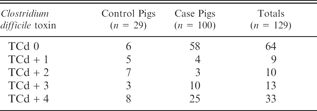
Clostridium difficile toxins were detected in the colon contents of 50% (65/129) of the study piglets (Table 1). This included 23 of 29 control piglets and 42 of 100 case pigs. Clostridium difficile toxins were only detected in 3 small intestinal samples, where 2 of the samples were +1 TCd and the third was +2 TCd. There was a statistically significant difference in the C. difficile isolation rate from small and large intestine compared to the rate at which toxin was detected in these 2 segments of bowel (P ≤ 0.001).
In 31% (40/129) of the piglets, C. difficile culture and TCd testing were both negative. In 35% (45/129) of the piglets both culture and TCd testing were positive. Clostridium difficile was isolated from 18% (23/129) of TCd-negative piglets, and TCd was detected in 21 of the culture negative piglets.
In the control group, 12/23 (52%) TCd-positive pigs had toxin levels ≤2, whereas in case piglets only 7/42 (17%) of TCd-positive piglets had toxin levels of ≤2 (Table 1). There was a statistically significant difference (P ≤ 0.001) in the proportion of control piglets with low levels of toxin (+1, +2) compared to the case piglets.
Agents identified in both the control and case pig populations are listed in Table 2. In total, TCd were detected in 65/129 (50%) piglets evaluated in this study. Clostridium difficile was the sole agent detected in 46 of the 129 pigs (36%) pigs. Clostridium difficile toxins, in combination with other agents/processes were identified in 19 (15%) of the piglets. Clostridium difficile toxins were most commonly detected in combination with C. perfringens type A (9) and rotavirus (4).
Subjective Clostridium difficile toxin (TCd) levels in control and case populations.
If it is assumed that each agent occurs independently, then multiplying the diagnostic frequency percentage of 2 agents would yield the proportion that would be predicted to occur together by chance. Calculations indicate that C. perfringens type A and TCd would occur together by chance in nine piglets, which was the actual number observed. By chance, rotavirus and TCd should occur together in 6 piglets, and the two were actually observed in combination in 4 animals. The rate at which combinations of pathogens were identified was not significantly different than would be expected by chance.
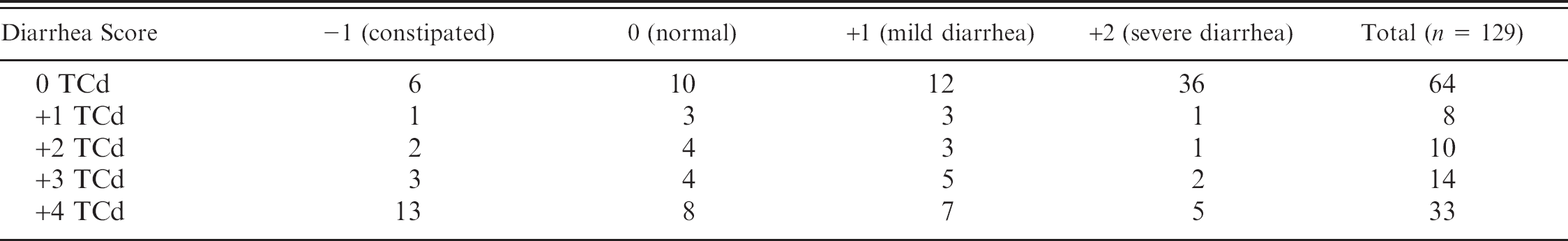
Gross evidence of diarrhea was assessed in each pig. Nineteen TCd-positive pigs were constipated (−1), 19 TCd-positive pigs were normal (0), 18 TCd-positive pigs had mild diarrhea (1), and 9 TCd-positive pigs had severe diarrhea (+2) (Table 3). When compared with the diarrhea scores of TCd-negative pigs, there was a significantly greater (P ≤ 0.001) likelihood that TCd-positive pigs would have normal or pelleted material in their colon and rectum. This difference was slightly more pronounced at high levels of TCd (+4), as 21/33 (64%) piglets with +4 TCd had diarrhea scours of −1 or 0.
The presence of additional enteric pathogens or inflammatory processes has the potential to influence gross lesions. Additional agents (Table 2) or inflammatory processes (peritonitis, strangulated inguinal hernia) were identified in 19 TCd-positive pigs. When these piglets were eliminated from the TCd-positive population, 14/46 TCd-positive pigs were constipated (−1), 13/46 TCd-positive pigs were normal (0), 14/46 TCd-positive pigs had mild diarrhea (+1), and 5/46 TCd-positive pigs had severe diarrhea (+2). Following the elimination of confounding agents, TCd-positive pigs were still significantly (P < 0.001) more likely to have normal or pelleted feces compared to the TCd-negative population. At the highest toxin level (+4), this association was significantly (P ≤ 0.025) more pronounced as 18/25 (72%) of these pigs had diarrhea scores of −1,0.
Survey of C. difficile toxins (TCd) and pathogens identified in case and control pigs.
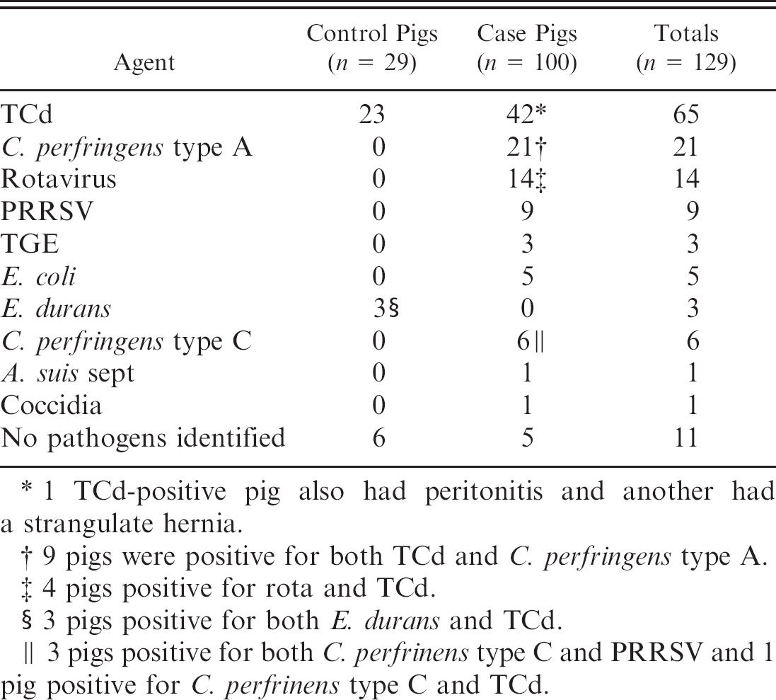
1 TCd-positive pig also had peritonitis and another had a strangulate hernia.
9 pigs were positive for both TCd and C. perfringens type A.
4 pigs positive for rota and TCd.
3 pigs positive for both E. durans and TCd.
3 pigs positive for both C. perfrinens type C and PRRSV and 1 pig positive for C. perfrinens type C and TCd.
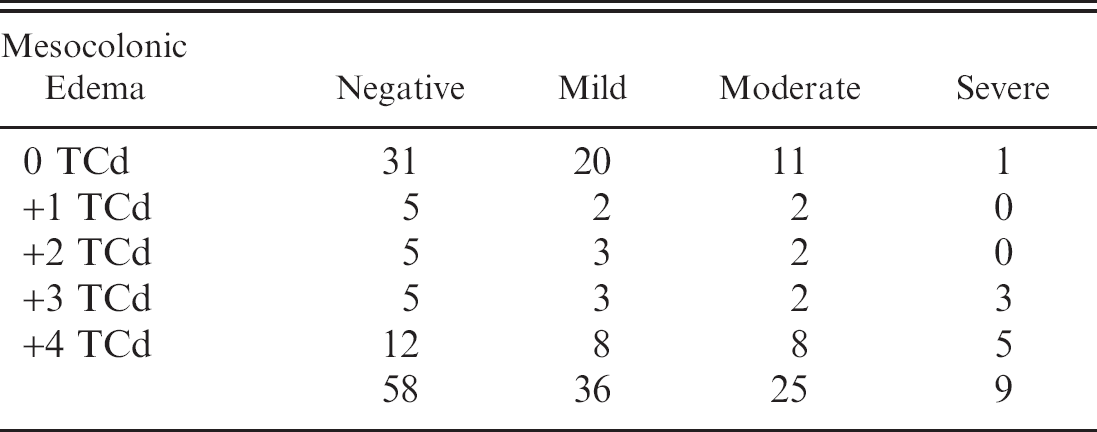
Edema of the mesocolon (Fig. 1) was observed in 70/129 (54%) of the piglets in this study, including 11/29 (38%) control piglets and 59/100 (59%) scouring piglets. Edema of the mesocolon was identified in 38/ 65 of TCd-positive piglets and 32/64 of TCd-negative pigs (Table 4). The positive predictive value of TCd as an indicator of mesocolonic edema was 54%. There was no statistically significant association between TCd and mesocolonic edema, even at high toxin levels (+3, +4). If piglets lack mesocolonic edema, the negative predictive value that they also lack toxin was calculated to be 53%.
The severity of mesocolonic edema was compared to TCd levels (Table 4). Eighty-nine% (8 of 9) of the piglets with severe mesocolonic edema had high (+3, +4) TCd. In piglets with severe mesocolonic edema, the lesion was significantly (P ≤ 0.001) associated with the presence of high levels of TCd.
Microscopic lesions identified in the cecum and colon of study piglets included loss of goblet cells with increased crypt mitotic activity, an infiltrate of neutrophils in the lamina propria, and multifocal erosions (Fig. 2). A piglet was considered to have colitis when any of these changes were observed, alone or in combination. Erosive lesions and infiltrates of neutrophils were always seen in conjunction with loss of goblet cells.
Typhlitis and colitis were each observed in 79/129 piglets, including 18/29 control pigs and 61/100 scouring piglets. Typhlitis was identified in 32/64 TCd-negative and 47/65 TCd-positive piglets. Colitis was observed in 30/64 TCd-negative and 49/65 TCd-positive piglets. There was a statistically significant association between both colitis (P ≤ 0.01) and typhlitis (P ≤ 0.025) and TCd. The PPV for colitis and typhlitis TCd as an indicator of TCd was 62% and 60% respectively.
Comparison of C. difficile toxin (TCd) levels and diarrhea scores.
Microscopic lesions in the colon were broken down into separate components, which included, loss of goblet cells, an infiltrate of neutrophils into the lamina propria, and multifocal erosive lesions. Each component was evaluated in regard to its association with TCd. Loss of goblet cells with increased crypt mitotic activity was identified in the colon of 79/129 (61%) of the piglets, including 49/65 (75%) TCd-positive piglets. An infiltrate of neutrophils was observed in 38/129 animals, of which 29/38 (76%) were TCd-positive. Multifocal erosive lesions were detected in 21 of the piglets, of which 16/21 (76%) were TCd-positive. Overall 29/65 (45%) TCd-positive piglets had neutrophils in the lamina propria of the colon and 16/65 (25%) TCd-positive piglets had erosive lesions. The positive predictive value of loss of goblet cells and TCd was 63%, neutrophils in the lamina propria and TCd was 76%, and erosions and TCd was 76%. TCd was significantly associated with each type of colonic lesion (P ≤ 0.001).
When other causes of severe colitis, such as Clostridium. perfringens type C and Actinobacillus suis, were excluded, 22/28 (79%) of the piglets with moderate to severe colitis were TCd-associated, 32/36 (89%) of the piglets with neutrophils in the lamina propria were TCd associated, and 16/17 (94%) of the piglets with erosions were TCd-associated.
Discussion
For the bulk of this discussion, data from the control group and case pigs will be combined. The rationale for combining this data is that if C. difficile toxins are the disease causing principal in CDAD, then there should be a correlation between the presence or absence of the toxins and clinical signs and lesions. Differences between control and study populations will be discussed.
This study supports previous work 35 demonstrating that C. difficile and its toxins are quite common in the colon of piglets less than a week of age. The organism was isolated from the large intestine of 51% (66/129) of study piglets and TCd were detected in the colon contents of 50% (65/129). Though C. difficile was isolated from a similar percentage of small intestines (47%), the detection of TCd in only 2% (3/129) of small intestine samples would indicate that conditions are generally only favorable for the production of detectable levels of toxin in the large intestine. Similar to the situation in man, culture alone may not be indicative of disease as the organism was isolated from 18% (23/129) of piglets in which toxin was not detected. 10
One of the primary purposes of this study was to critically evaluate gross and microscopic lesions to assess their value in determining whether C. difficile testing is warranted. Results indicate that the absence of gross evidence of diarrhea should not be used to exclude C. difficile testing. When animals with additional enteric pathogens were eliminated, TCd-positive piglets were significantly more likely to have normal to pelleted feces (59%) compared to the population of TCd-negative piglets (25%). This association was particularly apparent at high toxin levels (+4), where 72% (18/25) of +4 TCd-positive piglets had pelleted to normal feces.
Comparison of C. difficile toxin (TCd) levels and mesocolonic edema.
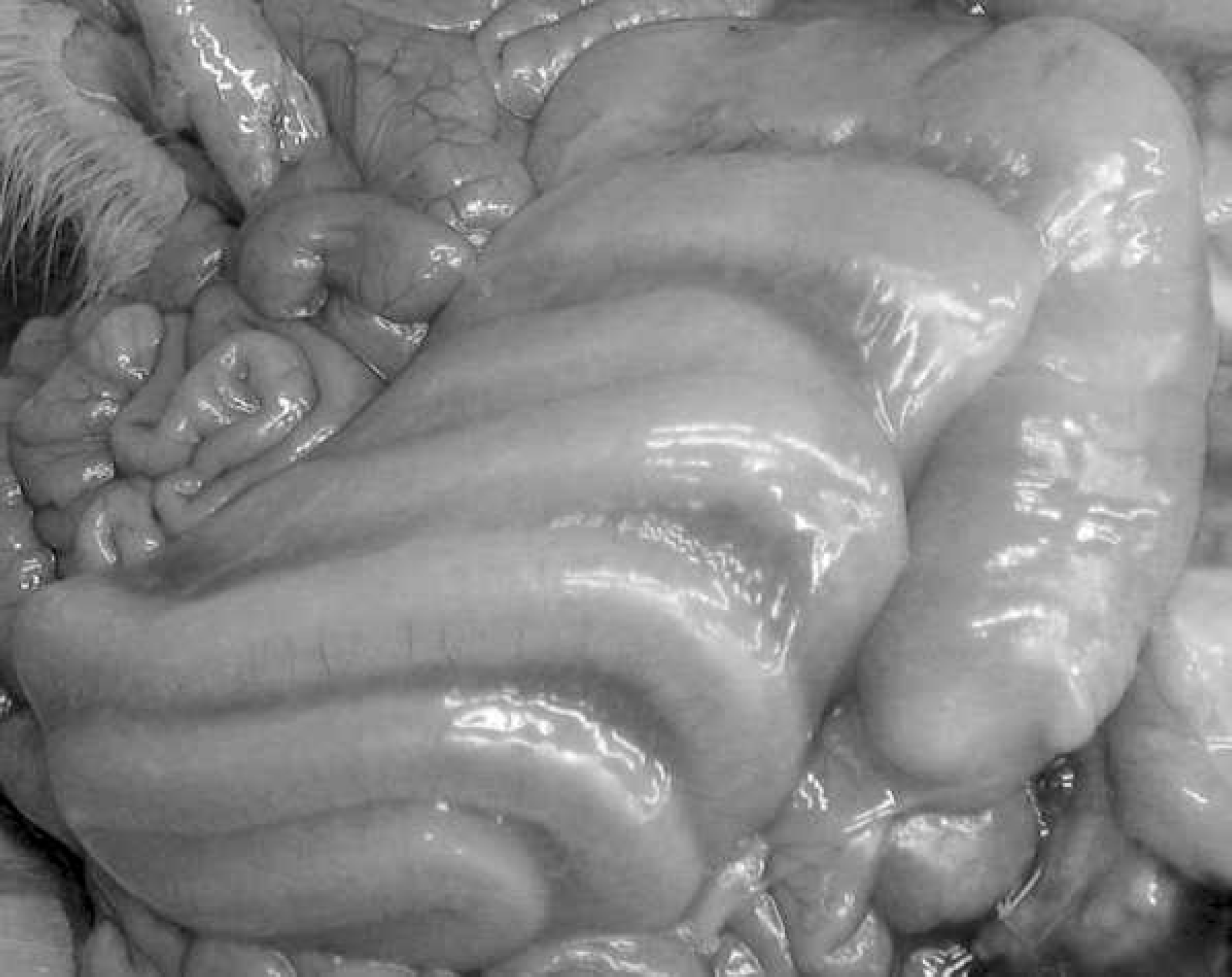
Moderate edema of the mesocolon.
This study reinforces the limited use of mesocolonic edema as a predictor of C. difficile toxins. Edema of the mesocolon was observed in 59% (38/65) of TCd-positive piglets and in half (32/64) of the toxin-negative pigs, yielding a positive predictive value for mesocolonic edema as a predictor of TCd of only 54%. A statistically significant association between mesocolonic edema and TCd was only identified when mesocolonic edema was severe, where 89% of piglets with severe edema of the mesocolon had high levels of TCd (+3, +4).
In the original study, the positive predictive value of colitis as an indicator of TCd was 84%. 35 A reasonable association between typhlocolitis and TCd was also identified in this study. The PPV of colitis or typhlitis as an indicator of TCd was 62% and 60% respectively. Microscopic lesions in the colon were broken down into several components to try and determine if specific features were of greater value in predicting the presence of TCd. Loss of goblet cells with increased crypt mitotic activity was the most common change observed in the colon (79/129) and had the lowest predictive value (63%) for the presence of TCd. An infiltrate of neutrophils in the lamina propria was less common (38/129), but had a higher predictive value for the presence of TCd (76%). Erosive/ulcerative colonic lesions were uncommon (21/129), and also had a positive predictive value for the presence of TCd of 76%.
Lesions are rarely specific for a single disease and their true value is to assist in establishing differential diagnoses. When other documented causes of severe colitis, such as Actinobacillus suis septicemia and C. perfringens type C were excluded, 79% of the colitis cases graded as moderate to severe were TCd-associated, 89% of the colitis cases with an infiltrate of neutrophils in the lamina propria were TCd-associated, and 94% of the cases with erosive lesions were TCd-associated. Particularly when other infectious causes of colitis have been excluded, the identification of a suppurative and erosive colitis in a neonatal piglet is strong endorsement for C. difficile testing,
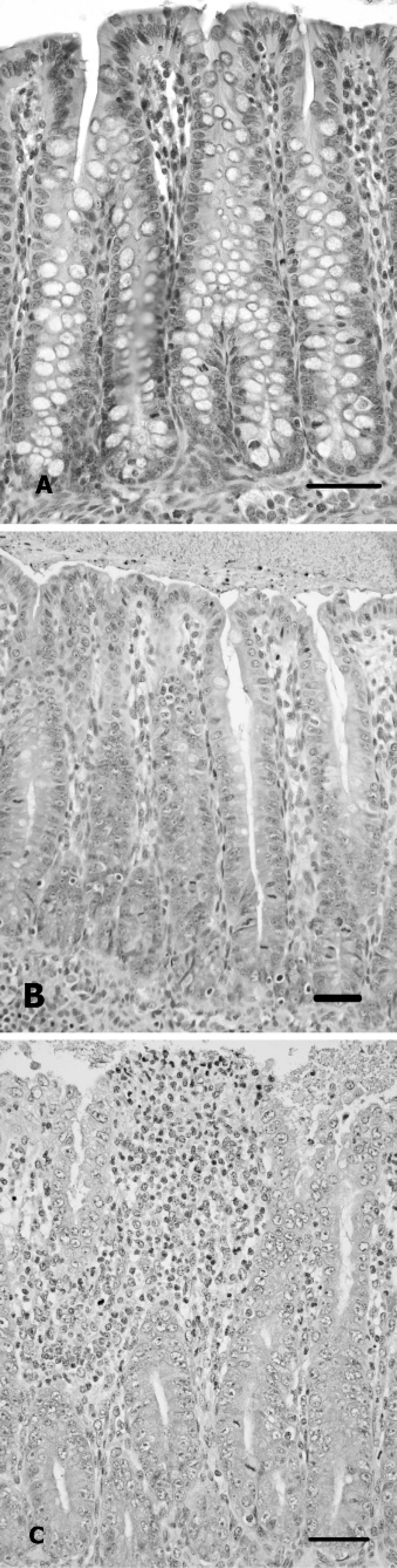
Microscopic lesions observed in the colon of C. difficile toxin (TCd) positive piglets;
One of the most startling findings of this study was the high number (79%) of apparently healthy control piglets positive for TCd. One possible explanation for this finding is that C. difficile in piglets may mimic the situation in human infants which appear to lack toxin receptors and do not develop disease despite the presence of the organism and high levels of toxin in their colon. 5,9,13 Information to date would argue against this theory. The disease has been experimentally reproduced in CDCD pigs (Songer, personal communication). Toxin A receptors have been identified in piglets, 17 and a significant association between TCd, severe mesosolonic edema and colitis/ typhlitis has been identified in this and previous studies. 28,35 Control piglets were not without lesions, as 70% of TCd-positive control animals had colitis, even though only 17% (5/29) had gross evidence of diarrhea. In general, control piglets tended to have lower levels of toxin than case piglets, and the lower average TCd levels may help to explain the lack of overt clinical disease in many of the control piglets. These results suggest that there is a need to explore the possibility that C. difficile infection may be an important subclinical disease in young swine by comparing the performance of TCd-positive and TCd-negative piglets.
There remain a number of troubling inconsistencies with regard to our understanding of the clinical signs and lesions associated with C. difficile infection in neonatal piglets. Particularly problematic are the piglets with +3or +4 TCd in colon contents that lack mesocolonic edema and typhlocolitis. This study did not provide insight into the cause for these inconsistencies. Possible explanations include the potential that factors produced by the organism, in addition to the toxin A and toxin B, may play a role in the pathogenesis of CDAD in swine. 1
Footnotes
a.
Premier Rotoclone, Meridian Diagnostics, Inc, Cincinnati, OH.
b.
C. difficile tox A/B II kit, TechLab, Blacksburg, VA.