
Abstract
Long non-coding RNAs are classified as a kind of RNA, which are longer than 200 nucleotides in length and cannot be translated into proteins. Multiple studies have demonstrated that long non-coding RNAs are involved in various cellular processes, including proliferation, differentiation, cell death, and metastasis. Among numerous long non-coding RNAs, we focus on Sprouty4-Intron 1 (SPRY4-IT1), a well-known long non-coding RNA that is overexpressed in various kinds of tumor tissues and cell lines. Accumulating evidences show that SPRY4-IT1 was dysregulated in various cancers, including melanoma, breast cancer, esophageal squamous cell carcinoma, non–small cell lung cancer, gastric cancer, colon cancer, and hepatocellular carcinoma, and amplification of SPRY4-IT1 was associated with different clinicopathological features of cancer patients. Importantly, SPRY4-IT1 exerts important roles in tumor progression and metastasis. However, detailed molecular mechanisms of SPRY4-IT1 in cancer progression and metastasis were poorly understood. In this review, we have focused on the characteristics of SPRY4-IT1 and illustrated the biological function and mechanism of SPRY4-IT1 in cancer development.
Keywords
Introduction
As we know, cancer has become a major public health problem all over the world. In the year 2015, there are approximately 1,658,370 new cancer cases and 589,430 cancer-related deaths in the United States. 1 Moreover, these numbers are growing constantly due to lack of advanced therapy for cancer. 2 Therefore, it is urgent to explore precise diagnostic biomarkers and effective therapeutic targets.
It was once thought that long non-coding RNAs (lncRNAs) were the transcriptional noise and do not work in the cellular life circles. 3 However, recent studies reported that lncRNAs can regulate the expression of downstream genes through transcriptional and posttranscriptional regulation, and dysregulation of lncRNAs contributes to cancer initiation, progression, and metastasis.3–5 Enforced expression of some cancer-promoting lncRNAs can enhance the proliferation and migration in cancer cells through regulating cell cycle protein and promoting epithelial–mesenchymal transition (EMT) process.6,7 But overexpression of some anti-cancer lncRNAs can inhibit the development of cancer, including proliferation inhibition, migration, suppression, and apoptosis induction.8,9
In many cancer-promoting lncRNAs, we chose lncRNA Sprouty4-Intron 1 (SPRY4-IT1) as the topic for discussion and summarize its oncogenic roles in the development of cancers. Moreover, the related clinicopathological characteristics and molecular functions of this lncRNA in cancers are presented in Tables 1 and 2.
Overexpression of SPRY4-IT1 is associated with clinicopathological features.

SPRY4-IT1: Sprouty4-Intron 1; TNM: tumor–node–metastasis; ER: estrogen receptor; AFP: alpha-fetoprotein; AST: aspartate aminotransferase; ALT: alanine aminotransferase.
The characteristics of SPRY4-IT1 in various cancers.

SPRY4-IT1: Sprouty4-Intron 1; CDK1: cyclin-dependent kinase 1; CDC20: cell-division cycle protein 20; DR5: death receptor 5; DPPIV: dipeptidyl peptidase-IV; XIAP: X-linked inhibitor of apoptosis protein; DGAT2: diacylglycerol O-acyltransferase 2; GPAT3: glycerol-3-phosphate acyltransferase 3; ZNF703: zinc finger protein 703; EMT: epithelial–mesenchymal transition; TGF-β: transforming growth factor beta; MMP: matrix metalloproteinase; EZH2: enhancer of zeste homolog 2; TNM: tumor–node–metastasis; ER: estrogen receptor; AFP: alpha-fetoprotein; AST: aspartate aminotransferase; ALT: alanine aminotransferase; MCM: minichromosome maintenance.
Identification of SPRY4-IT1
SPRY4 is a member of Sprouty (SPRY) family and regarded as a tumor suppressor in cancer development. 29 Overexpression of SPRY4 can inhibit cell growth, migration, and invasion in non–small cell lung cancer (NSCLC), breast cancer, and endometrial cancer cells.29–31 LncRNA SPRY4-IT1 (GenBank ID AK024556) was first identified in the melanoma cells, and it was derived from the second intron of SPRY4 gene. SPRY4-IT1 is 708 bp in length and located at the human chromosomes 5q31.3. Interestingly, SPRY4-IT1 has particular secondary structural features, which contains several long hairpins. 24 The secondary structure of SPRY4-IT1 was predicted by ViennaRNA Web services (Figure 1). Further experiments demonstrated that the expression level of SPRY4-IT1 is closely associated with SPRY4 expression in normal human tissues and patient samples. However, knockdown of SPRY4-IT1 cannot change the expression of SPRY4, and SPRY4 also cannot regulate SPRY4-IT1 expression.24,25 Two explanations are responsible for this phenomenon. First of all, SPRY4-IT1 is predominantly located in the cytoplasm, but SPRY4 is mainly located in the nucleus. Second, SPRY4-IT1 decays slower than SPRY4 RNA in both the nucleus and cytoplasm. Recently, it is reported that SPRY4-IT1 was augmented in melanoma, breast cancer, NSCLC, and so on. Moreover, suppression of SPRY4-IT1 may increase apoptosis rate through altering lipin 2 in lipid metabolism. 25 Enforced expression of SPRY4-IT1 can promote cell proliferation through upregulating the cell proliferation marker MKI67 and minichromosome maintenance genes [MCM2,MCM3,MCM4 and MCM5] (MCM2–5). Overexpression of SPRY4-IT1 can also upregulate the anti-apoptotic genes X-linked inhibitor of apoptosis protein (XIAP) and baculoviral IAP repeat–containing 7 (livin) and downregulate the tumor suppressor dipeptidyl peptidase-IV (DPPIV), thus resulting in apoptosis. 23 In addition, SPRY4-IT1 can increase the migration ability via facilitating EMT process. 12 In general, SPRY4-IT1 can promote the cancer progression through affecting the phenotypes of cancer cells and molecular pathways.

The locus of lncRNA SPRY4-IT1 in human chromosome and the structure of SPRY4-IT1. (a) LncRNA SPRY4-IT1 is located at the human chromosome 5q31.3. (b) SPRY4-IT1 is a transcript of SPRY4, which is derived from the second intron of SPRY4. SPRY4-001 and SPRY4-002 are the alternately spliced isoforms of SPRY4. (c) The website of ViennaRNA Web services was used to predict the secondary structure of SPRY4-IT1. The proportion of color represents the confidence of each base and shades of red represent strong confidence.
SPRY4-IT1 in various cancers
Melanoma
Melanoma is the most aggressive skin malignant tumor, resulting in about 9940 deaths in Americans. Moreover, the incidence of melanoma is rising rapidly, and about 73,870 new cases are diagnosed in the United States every year. Surgical operation is the best therapy for the initial melanoma, and there are good postoperative outcomes in these patients. 32 However, there is no effective treatment for the advanced melanoma due to rapid progression and distant metastasis, with approximately 16% 5-year survival rate.33,34 Therefore, a better understanding of molecular pathways in melanoma initiation and biomarkers can detect melanoma at an early stage. Identification of new molecular markers can provide potential melanoma therapies.
LncRNA SPRY4-IT1 was originally identified in the melanoma cells and tissues. It is derived from the second intron of SPRY4 gene, but it cannot affect SPRY4 expression. Decreased SPRY4-IT1 expression can inhibit melanoma cell proliferation, motility, and invasion and increase the rates of apoptotic cells. 24 Liu et al. 10 investigated the clinical significance of SPRT4-IT1 in melanoma patients and found that SPRY4-IT1 was upregulated in the blood of melanoma patients compared with that in healthy volunteers. High SPRY4-IT1 expression was positively correlated with tumor site and tumor–node–metastasis (TNM) stage and negatively associated with overall survival (OS) rates. To detect the potential mechanisms of SPRY4-IT1 in the progression of melanoma cells, Mazar et al. 25 used mass spectrometry to investigate the SPRY4-IT1-associated proteins. They found that lipin 2 was pulled down specifically after SPRY4-IT1expression was decreased. Lipin 2 is a kind of phosphatidic acid phosphatase (PAP), which is responsible for the triglyceride synthesis, and dephosphorylation of phosphatidic acid (PA) could produce diacylglycerol (DAG).35,36 Knockdown of lipin 2 led to loss of SPRY4-IT1, but downregulation of SPRY4-IT1 increased the levels of lipin 2 and PAP enzymatic activity. Moreover, knockdown of SPRY4-IT1 changes a series of lipid species, including reduced PA and phosphatidylcholine, as well as increased acyl carnitines and triacylglycerol (TAG). Interestingly, repression of SPRY4-IT1 increased the expression of glycerol-3-phosphate acyltransferase 3 (GPAT3) and diacylglycerol O-acyltransferase 2 (DGAT2) enzymes which play important roles in DAG and TAG synthesis (Figure 2). To examine how SPRY4-IT1 contributes to differentiation and migration of melanoma cells, Zhao et al. detected the molecular changes during gain or loss of function of SPRY4-IT1. They found that overexpression of SPRY4-IT1 in normal human melanocytes affects multi-nuclear and multi-dendrite cells, which is a sign of carcinogenesis. Gene ontology enrichment analysis revealed that SPRY4-IT1 could regulate chromatin and cell cycle pathway genes in normal human melanocytes. Moreover, SPRY4-IT1 can increase cell proliferation through increasing a series of proliferative pathway genes, including MCM2, MCM3, MCM4, MCM5, antigen-MKI67, cyclin-dependent kinase 1 (CDK1), and cell-division cycle protein 20 (CDC20). As expected, SPRY4-IT1 can reduce the apoptosis rate of melanoma cells, and a series of apoptosis-related protein were modulated, including downregulation of pro-apoptotic protein TRAIL-R2 (death receptor 5 (DR5)) and DPPIV and upregulation of livin and XIAP (Figure 3). 23

Knockdown of SPRY4-IT1 may induce apoptosis through lipin 2–mediated variation of a series of lipid species, including inhibited expression of phosphatidic acid and phosphatidylcholine and increased expression of acyl carnitines and fatty acylchains. Moreover, suppression of SPRY4-IT1 could increase the levels of DGAT2 and GPAT3 enzymes, which may block the conversion of DAG to TAG.

Overpression of SPRY4-IT1 in normal human melanocytes leads to multi-nuclear and multi-dendrite cells. Moreover, SPRY4-IT1 could increase cell proliferation through increasing a series of proliferative pathway genes, including MCM2, MCM3, MCM4, MCM5, antigen-MKI67, cyclin-dependent kinase 1 (CDK1), and cell-division cycle protein 20 (CDC20). In addition, SPRY4-IT1 could reduce the apoptosis rate of melanoma cells, and a group of apoptosis-related protein was modulated, including downrelgulation of pro-apoptotic protein DR5 and DPPIV and upregulation of livin and X-linked inhibitor of apoptosis protein (XIAP).
In conclusion, lncRNA SPRY4-IT1 can promote the melanocytic transformation through interfering with lipid metabolism and a group of molecular mechanisms. SPRY4-IT1 may serve as a cancer-promoting gene in the initiation and progression of melanoma.
Breast cancer
Breast cancer is the most common cancer in women, especially in developed country, and it is the first cause of death in women. 37 The 5-year survival of localized breast cancer is about 98% due to earlier diagnosis and treatment, but 20%–30% of breast cancer will progress to metastasis and remain incurable.38,39 Prevention, diagnosis, and treatment of breast cancer have become a major public health concern. Therefore, further understanding of molecular mechanisms governing progression and metastasis of breast cancer will explore effective prognostic biomarker and therapeutic target for breast cancer.
Shi et al. 11 first reported that lncRNA SPRY4-IT1 is overexpressed in breast cancer tissues and cell lines. In addition, they observed that overexpression of SPRY4-IT1 is significantly correlated with tumor grade and tumor size. Interestingly, the expression level of SPRY4-IT1 in estrogen receptor negative (ER−) breast cancer tissues is higher than that in ER positive (ER+) breast cancer tissues. To investigate the biological roles of SPRY4-IT1 in breast cancer, Shi et al. 11 downregulated SPRY4-IT1 in MDA-MB-231 and MDA-MB-435S cells and overexpressed SPRY4-IT1 in MCF-7 cells. They found that knockdown of SPRY4-IT1 decreased the cell colony formation ability, promoted G1 arrest, and caused apoptosis in breast cancer cells. Moreover, knockdown of SPRY4-IT1 disrupted the expression of cyclin D1 protein and increased the Bax protein and Bcl-2 protein levels (Figure 4). Further microarray analysis in SPRY4-IT1 knockdown cells exhibited a series of changed genes. In those changed genes, zinc finger protein 703 (ZNF703) displayed the most substantial change in response to SPRY4-IT1 knockdown. ZNF703 is a zinc finger transcription factor belongs to NET/NLZ family and contributes to the development and progression of various cancer, including NSCLC, breast cancer, gastric cancer (GC), and colorectal cancer (CRC).40–42 Moreover, ZNF703 is involved in the activation of the Akt/mammalian target of rapamycin (mTOR) signaling pathway in breast cancer cell lines. 43 Therefore, Shi et al. 11 detected the potential roles of ZNF703 in breast cancer cell lines and discovered that ZNF703 is augmented in breast cancer tissues and cell lines. Moreover, SPRY4-IT1 exerted its oncogenic effects, such as proliferation promotion and apoptosis inhibition, via upregulating ZNF703.

Knockdown of SPRY4-IT1 disrupted the expression of cyclin D1 protein and increased the Bax protein and Bcl-2 protein levels in breast cancer. Furthermore, microarray analysis in SPRY4-IT1-knockdown cells exhibited a series of changed genes. In those changed genes, ZNF703 displayed the most substantial change in response to SPRY4-IT1 knockdown.
Taken together, SPRY4-IT1 promoted proliferation and inhibited apoptosis in breast cancer cells thorough upregulating ZNF703. However, the molecular mechanisms and downstream targets associated with SPRY4-IT1 and ZNF703 are to be investigated in further studies.
Esophageal squamous cell carcinoma
Esophageal cancer is one of the most lethal malignancies with high mortality rate in the worldwide. 2 Esophageal squamous cell carcinoma (ESCC) and esophageal adenocarcinoma (EAC) are the major forms of esophageal cancer, which occurred in different human races. 44 ESCC is the leading subtype of esophageal cancer in Asia, while EAC is more predominant in the United States.2,45 Despite rapid development of systematic treatment, including surgical operation, radiotherapy, and chemotherapy, the prognosis of ESCC patients remains unsatisfactory, with a 5-year survival rate of about 25%–30%.46,47 Therefore, there is an urgent need to explore new molecular markers which may act as tumor markers or therapeutic targets.
SPRY4-IT1 is upregulated in ESCC tissues and cell lines. High expression of SPRY4-IT1 is closely associated with tumor grade, TNM stage, lymph node metastasis, and clinical stage, which lead to poor prognosis of ESCC patients. Moreover, suppression of SPRY4-IT1 expression could decrease the ability of proliferation, migration, and invasion in ESCC cells. Knockdown of SPRY4-IT1 also could increase the apoptosis rate of ESCC cells and inhibit ESCC cell growth in vivo. 13 Xue-Liang et al. 14 reported the above results and further found that silencing of SPRY4-IT1 could decrease ESCC cell proliferation via downregulating the expression of ZNF703. Meanwhile, downregulation of SPRY4-IT1 could obviously block the EMT process via increasing the level of E-cadherin and reducing the level of vimentin. 12 However, overexpression of SPRY4-IT1 can lead to opposite effects and promote the EMT process of ESCC cells. Upregulation of SPRY4-IT1 increased the expression of vimentin and fibronectin and decreased the expression of E-cadherin and ZO-1. 26 EMT is a developmental trans-differentiation process in which epithelial cells lose the cell–cell adhesion and apical–basal cell polarity and were changed into mesenchymal stem cells. 48 According to the biological processes, EMT is classified into three types: type-1, type-2, and type-3. Type-1 EMT is related with embryogenesis. Type-2 operates in the process of wound healing. Type-3 participates in the progression of cancer development and metastasis.49,50 Many evidences demonstrated that EMT contributes to cancer progression, metastasis, tumor microenvironment crosstalk, and drug resistance.51,52 EMT-induced cancer cells have been changed a lot at the molecular level and display increased expression of mesenchymal genes (fibronectin, N-cadherin, and vimentin) and decreased expression of epithelial genes (E-cadherin and ZO-1).52,53 In many cases, gain of N-cadherin and loss of E-cadherin are unique features of EMT. In order to investigate the role of transforming growth factor beta (TGF-β: in induced EMT in ESCC, Zhang et al. 26 detected the roles of SPRY4-IT1 in TGF-β-induced EMT of ESCC cells and found that SPRY4-IT1 exerted a positive role in TGF-β-induced EMT of ESCC cells. In addition, overexpression of SPRY4-IT1 could significantly improve the expression and nuclear translocation of the transcription factor snail in ESCC cells through direct interaction. Further experiments demonstrated that snail plays critical roles in SPRY4-IT1-induced EMT in ESCC cells (Figure 5).
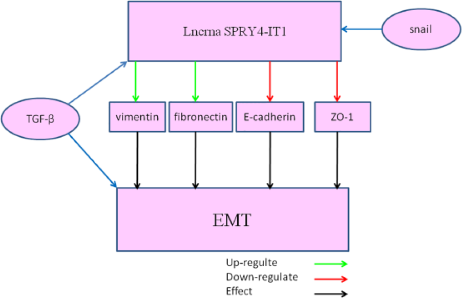
Enforced expression of SPRY4-IT1 can promote EMT process via increasing the expression of vimentin and fibronectin and decreasing the expression of E-cadherin and ZO-1. Moreover, the transcription factor TGF-β and snail can promote the EMT process through interacting with SPRY4-IT1.
These results suggest that the expression level of SPRY4-IT1 is closely associated with the prognosis of ESCC patients, and SPRY4-IT1 plays an important role in ESCC progression via promoting EMT process. Therefore, SPRY4-IT1 may act as a novel prognostic marker and potential therapeutic target for ESCC.
NSCLC
Lung cancer is the leading cause of cancer-related deaths in the world with less than 15% 5-year survival rate. 54 NSCLC is the main form of lung cancer, which is classified as squamous cell carcinoma, adenocarcinoma, and large cell carcinoma. 55 Despite recent advances in the examination and new therapies for lung cancer, the prognosis of NSCLC remains unsatisfied, and metastasis and recurrence are the main causes for poor prognosis. Hence, it is essential to gain insight into the mechanisms involved in lung carcinogenesis and metastasis.
Sun et al. 56 found that the expression of SPRY4-IT1 was lower in NSCLC tissues compared with adjacent histologically normal tissues, and downregulation of SPRY4-IT1 was significantly associated with tumor size, TNM stage, and lymph node metastasis. Moreover, they also discovered that downregulation of SPRY4-IT1 was an independent predictor of poor survival for NSCLC. Further functional analyses showed that amplification of SPRY4-IT1 can inhibit NSCLC cell proliferation, migration, and invasion. In addition, enforced expression of SPRY4-IT1 can promote NSCLC cell apoptosis in vitro and suppress cell metastasis in vivo. Interestingly, the expression of SPRY4-IT1 was negatively associated with enhancer of zeste homolog 2 (EZH2) expression in NSCLC tissues. Furthermore, chromatin immunoprecipitation assay revealed that EZH2 can repress SPRY4-IT1 expression via directly binding to the promoter region of SPRY4-IT1 and mediating H3K27me3 modification. Meanwhile, knockdown of EZH2 can lead to NSCLC cell growth arrest in vitro and in vivo and impair NSCLC cell migration and invasion. At last, they found that EZH2 promoted NSCLC cell proliferation, invasion, and EMT process via repressing the expression of SPRY4-IT1. However, Hu et al. 57 discovered that SPRY4-IT1 was overexpressed in the plasma of NSCLC patients compared with that of healthy volunteers. Amplification of SPRY4-IT1 in the plasma of NSCLC patients was closely correlated with the size of tumor. Receiver operating characteristic curve (ROX) analysis revealed that SPRY4-IT1 may act as a potential diagnostic biomarker for NSCLC. In short, SPRY4-IT1 may exert different roles in different stages of NSCLC. It means that SPRY41 may inhibit the progression of low stage of NSCLC and promote the progression of high stage of NSCLC.
GC
GC is the fourth most common malignancies, and its mortality ranked the second in all cancers. 58 In many GC patients, their diseases are diagnosed at an end stage and usually followed by lymphatic metastasis, distant metastasis, and complications. 59 Although great efforts have been made to improve the clinical outcomes of GC, advanced GC patients usually have a poor prognosis with a relatively low 5-year survival rate. 60 Thus, more sensitive GC-related biomarkers for diagnosis and prognosis evaluation are imperatively needed.
Xie et al. 61 found that the expression of SPRY4-IT1 was significantly downregulated in GC tissues and cell lines compared with normal control. They also found that downregulation of SPRY4-IT1 was significantly correlated with GC tumor size, advanced pathological stage, deeper depth of invasion, and lymphatic metastasis. In addition, GC patients with low SPRY4-IT1 expression had poorer disease-free survival and OS. Overexpression of SPRY4-IT1 could obviously inhibit GC cell proliferation, migration, and invasion. Moreover, upregulation of SPRY4-IT1 could inhibit GC cell tumorigenesis and metastasis in vivo. Similar with previous study, SPRY4-IT1 could influence the EMT process of GC cells. However, Peng et al. 15 reported that SPRY4-IT1 is upregulated in GC tissues and cell lines, and upregulation of SPRY4-IT1 was significantly correlated with tumor size, invasion depth, distant metastasis, and TNM stage. In addition, silencing of SPRY4-IT1 can suppress the proliferation, migration, and invasion of GC cells, and knockdown of SPRY4-IT1 could inhibit the expression levels of cyclin D1, matrix metalloproteinase 2 (MMP2), and MMP9. The above results from Xie and Peng are controversial, and how to explain these results is an important problem. We suspect that their GC cells are derived from different stages of GC tissues and the expression level of SPRY4-IT1 in those GC cells may be different. It means that the expression level of SPRY4-IT1 in SGC-7901 cells which are derived from T1 GC tissues is different to that in other stages of GC tissues.
CRC
CRC is one of most prevalent malignancies and the fourth leading cause of cancer death in the world.2,62 Cancer metastasis is the major reason for high mortality of CRC patients. 63 Although advanced treatments are prevalently used to improve the prognosis of CRC patients, cancer relapse and metastasis are still inevitable. 64 The lack of effective tumor biomarkers for tumor progression and metastasis is responsible for the failure of CRC therapy. Therefore, identifying novel diagnostic and prognostic biomarkers is an urgent task for the early detection and therapy of CRC.
The expression level of SPRY4-IT1 was significantly elevated in CRC tissues and cell lines compared with the corresponding normal tissues and cell lines.16,17 In addition, upregulation of SPRY4-IT1 was positively associated with TNM stage, depth of invasion, metastasis, and tumor size.16,17 Overexpression of SPRY4-IT1 can predict poor prognosis of CRC patients, and SPRY4-IT expression was an independent prognostic indicator for OS. 16 Further experiments demonstrated that silencing of SPRY4-IT1 can inhibit cell proliferation by altering cell cycle progression and promote cell migration by accelerating EMT process. Western blot assay showed that suppression of SPRY4-IT1 can obviously increase vimentin expression and inhibit E-cadherin expression. Besides, repression of SPRY4-IT1 induced apoptosis in CRC cells. 17 All these findings implied that SPRY4-IT1 may exert its important roles in CRC progression and metastasis. Therefore, it has the potential to become an effective diagnostic and prognostic marker for CRC.
Hepatocellular carcinoma
Hepatocellular carcinoma (HCC) is the fifth most commonly diagnosed malignant tumor, and it is the third leading cause of cancer-related deaths worldwide. 65 Aggressiveness and invasiveness are the main characteristics of HCC, resulting in delayed diagnosis and poor prognosis in HCC patients. 66 However, the current treatments cannot improve the outcome of advanced HCC notably, and the 5-year survival rate of HCC is only about 7%. 67 Alpha-fetoprotein (AFP) in serum is usually served as a diagnostic marker for HCC, but AFP may present negative result in 40% of early-stage and 15%–30% of advanced-stage HCC patients. 67 At present, the lack of understanding of HCC formation and progression prevents us to explore novel diagnostic biomarkers and effective treatments. Therefore, novel tumor biomarkers for HCC are urgently needed.
Jing et al. 18 performed the quantitative reverse transcription polymerase chain reaction (qRT-PCR) assay in 87 pairs of HCC tissues and found that SPRY4-IT1 was remarkably elevated in tumor tissues compared with non-tumor tissues. The high level of SPRY4-IT1 was positively associated with tumor differentiation, tumor size, and TNM stage. Furthermore, SPRY4-IT1 level in plasma of HCC patients was upregulated compared with healthy controls and associated with the levels of alanine aminotransferase (ALT) and aspartate aminotransferase (AST). Zhou et al. 27 found that SPRY4-IT1 was significantly increased in HCC cells, and overexpression of SPRY4-IT1 can promote cell proliferation and increase the cell invasion ability via promoting EMT progression. Moreover, overexpression of SPRY4-IT1 can suppress Twist1 and vimentin expression and increase E-cadherin expression. However, knockdown of SPRY4-IT1 could inhibit cell proliferation and invasion. They further demonstrated that SPRY4-IT1 can attract EZH2 and H3K27me3 to E-cadherin promoter and lead to gene suppression. Besides, silencing of SPRY4-IT1 can obviously inhibit tumor growth in vivo (Figure 6). In conclusion, SPRY4-IT1 may serve as a novel plasma marker for HCC.

Overexpression of SPRY4-IT1 can promote cell proliferation and strengthen the cell invasion ability via promoting EMT progression. Moreover, overexpression of SPRY4-IT1 can suppress Twist1 and vimentin expression and increase E-cadherin expression. SPRY4-IT1 can attract EZH2 and H3K27me3 to E-cadherin promoter and lead to gene suppression. Besides, silencing of SPRY4-IT1 can obviously inhibit tumor growth in vivo.
Other cancers
SPRY4-IT1 is also upregulated in glioma, gallbladder cancer (GBC), renal cancer, and bladder cancer.19–22,28 Further experiments have demonstrated that SPRY4-IT1 can promote cell proliferation, migration, and invasion and inhibit apoptosis.
Liu et al. 28 found that SPRY4-IT1 expression was higher in the glioma tissues and cell lines compared with normal brain tissues and normal human astrocytes. Suppression of SPRY4-IT1 expression can inhibit the proliferation and migration ability of glioma cells. They also found that knockdown of SPRY4-IT1 could block the EMT process, thus resulting in migration inhibition. Zhou et al. 19 investigated the clinical correlation of SPRY4-IT1 and found that high expression level of SPRY4-IT1 in glioma tissues was associated with World Health Organization (WHO) grade and tumor size. Further data analysis revealed that high SPRT4-IT1 expression level was an independent prognostic factor for OS, which could lead to poorer prognosis.
Yang et al. 20 showed that SPRY4-IT1 expression was increased in GBC tissues and cell lines. Moreover, gain or loss of function suggested that SPRY4-IT1 can promote GBC cell proliferation and accelerate the migration rate through affecting EMT process.
SPRY4-IT1 can also promote the progression of urologic neoplasm. SPRY4-IT1 was overexpressed in renal clear cell carcinoma tissues and cell lines. Moreover, increased SPRY4-IT1 expression was positively associated with histological grade, tumor stage, lymphatic metastasis, distant metastasis, and poorer prognosis. Similar to previous results, knockdown of SPRY4-IT1 can significantly inhibit cell proliferation and migration of renal cancer cells. 21 In consistent with the results in renal clear cell carcinoma, SPRY4-IT1 expression was increased in the bladder cancer tissues and cell lines. High expression level of SPRY4-IT1 was positively with tumor stage, histological grade, lymph node metastasis, and poorer prognosis. As expected, knockdown of SPRY4-IT1 decreased the proliferation and migration in bladder cancer cells. 22
Discussion
Cancer is a complex disease, which is accompanied with mutations in genes.68,69 LncRNAs can affect the expression of downstream genes through regulating the transcription of protein-coding gene promoter region and inducing chromatin remolding or/and histone modification.70,71 Moreover, some lncRNAs can modulate the activities of proteins via binding to its domain area. 72 Therefore, lncRNA is the important regulator in human cancer progression, and many lncRNAs are dysregulated in the development of cancer.73–75 LncRNA SPRY4-IT1, a well-known cancer-promoting lncRNA, contributing to the cancer development, and its characteristics were investigated. SPRY4-IT1 is derived from the second intron of SPRY4 gene and mainly located in the cytoplasm. High expression level of SPRY4-IT1 in tumor tissues is closely associated with clinical features, including tumor size, TNM stage, lymphatic metastasis, and distant metastasis. In addition, augmented expression of SPRY4-IT1 in plasma results in poor prognosis in cancer patients. But the exact concentration of SPRY4-IT1 in blood from healthy individuals is needed to be detected. Furthermore, the cancer patients involved in the SPRY4-IT1 study is not enough, which resulted in unconvincing results in clinical application. We also know that overexpression of SPRY4-IT1 can promote cancer cell proliferation, invasion, and migration and inhibit apoptosis, and knockdown of SPRY4-IT1 can generate the opposite effects. Inspiringly, a group of genes and pathways which are related with cell proliferation, migration, invasion, and apoptosis are discovered. However, detailed molecular mechanism of SPRY4-IT1 is still poorly understood. For instant, SPRY4-IT1-related microRNA and SPRY4-IT1-related lncRNA were reported, and the relation between SPRY4-IT1 and traditional oncogenes is needed to be examined in depth. Second, the roles of SPRY4-IT1 in the most common pathogenesis of cancer (DNA methylation, histone modification, and chromosome translocation) are unclear. Last but not least, SPRY4-IT1 seems to employ multiple mechanisms to exert its function. The mechanism by which SPRY4-IT1 controls those processes in different types of cancer cell is unclear.
In conclusion, the current studies suggest that lncRNA SPRY4-IT1 may act as a tumor biomarker and therapeutic target for cancer diagnosis and treatment. However, the research of SPRY4-IT1 is still in its infancy. Further study on the clinical application of SPR4-IT1 and the HOTAIR-related pathways is needed. More or less, combination use of SPR4-IT1 and conventional tumor markers may be a promising direction of clinical diagnosis.
Footnotes
Acknowledgements
The authors appreciated the donors, whose names were not included in the author list, but who participated in this program.
Declaration of conflicting interests
The author(s) declared no potential conflicts of interest with respect to the research, authorship, and/or publication of this article.
Funding
The author(s) disclosed receipt of the following financial support for the research, authorship, and/or publication of this article: This work was supported by the National Key Basic Research Program of China (973 Program) (2014CB745201).