
Abstract
The cell division cycle 20, a key component of spindle assembly checkpoint, is an essential activator of the anaphase-promoting complex. Aberrant expression of cell division cycle 20 has been detected in various human cancers. However, its clinical significance has never been deeply investigated in non-small-cell lung cancer. By analyzing The Cancer Genome Atlas database and using some certain online databases, we validated overexpression of cell division cycle 20 in both messenger RNA and protein levels, explored its clinical significance, and evaluated the prognostic role of cell division cycle 20 in non-small-cell lung cancer. Cell division cycle 20 expression was significantly correlated with sex (p = 0.003), histological classification (p < 0.0001), and tumor size (p = 0.0116) in non-small-cell lung cancer patients. In lung adenocarcinoma patients, overexpression of cell division cycle 20 was significantly associated with bigger primary tumor size (p = 0.0023), higher MKI67 level (r = 0.7618, p < 0.0001), higher DNA ploidy level (p < 0.0001), and poor prognosis (hazard ratio = 2.39, confidence interval: 1.87–3.05, p < 0.0001). However, in lung squamous cell carcinoma patients, no significant association of cell division cycle 20 expression was observed with any clinical parameter or prognosis. Overexpression of cell division cycle 20 is associated with poor prognosis in lung adenocarcinoma patients, and its overexpression can also be used to identify high-risk groups. In conclusion, cell division cycle 20 might serve as a potential biomarker for lung adenocarcinoma patients.
Keywords
Introduction
Lung cancer is currently both the most common cancer and the leading cause of cancer-related deaths worldwide. 1 Two main histological types are included: non-small-cell lung cancer (NSCLC) and small-cell lung cancer (SCLC). NSCLC constitutes about 80% of all lung cancers, and lung adenocarcinoma (LUAD) has been the most common subtype of NSCLC in recent years. Despite advances in lung cancer therapies, prognosis of NSCLC is still unfavorable, with an overall 5-year survival rate less than 15%.2–4 Therefore, further investigation on identification of prognostic markers and potential drug targets is eagerly needed to provide better prognosis and individualized treatment.
Cell division cycle 20 (CDC20), an important spindle assembly checkpoint (SAC) protein, activates anaphase-promoting complex (APC/C) to initiate anaphase. It consists of 499 amino acids with C-terminal WD40 domain for protein binding, serving as the substrate recognizing subunit of APC/C.5–7 In addition to regulating cell cycle, recent evidence has demonstrated that CDC20 also plays an important role in carcinogenesis and cancer progression and CDC20 might become a promising therapeutic target. 8 Abnormal expression of CDC20 may thus disorder APC/C activation and promote premature anaphase, which often results in mitotic abnormalities and aneuploidy in both daughter cells.9–11 Some microarray studies have already reported overexpression of CDC20 in various tumors such as tumors of the oral cavity,9,12 stomach, 13 pancreatic duct, 14 bladder, 15 and ovary. 16 Additionally, some specific genes including CDC20 were consistently correlated with total functional aneuploidy and were predictive of poor outcomes in several cancers by a computational method. 17 Although overexpression of CDC20 has been reported in human NSCLC, 18 to the best of our knowledge, there is no study using The Cancer Genome Atlas (TCGA) database to explore the clinical significance of CDC20 in NSCLC.
Our study validated the aberrant expression of CDC20 and explored its clinical and biological significance in NSCLC. Moreover, we concluded that CDC20 might play different clinical roles in different histological subtypes.
Methods
Expression evaluation and analysis
Three datasets named TCGA_LUNG_exp_HiSeqV2-2015-02-24, TCGA_LUAD_exp_HiSeqV2-2015-02-24, and TCGA_LUSC_exp_HiSeqV2-2015-02-24 were downloaded at the website of the UCSC Cancer Browser (https://genome-cancer.ucsc.edu/).19,20 These datasets contain a list of cancer-related characteristic information of 108 paired NSCLC tissue samples, 57 pairs of LUAD and 51 pairs of lung squamous cell carcinoma (LUSC) tissues (the tumor and paired normal tissue from a same patient), respectively. A list of 171 genes with highest expression correlation (Pearson’s r value > 0.7) with CDC20 was extracted from the TCGA LUNG dataset and then was submitted to DAVID Bioinformatics Resource 6.7 (https://david.ncifcrf.gov/tools.jsp)21,22 for REACTOME pathway enrichment analysis to assess the molecular role of CDC20 in NSCLC. All the messenger RNA (mRNA) expression values in TCGA LUNG database were normalized and the values of CDC20 expression of paired tissue samples were obtained from the file “genomicMatrix.” Then files named “clinical_data” in datasets were used to analyze the association between the CDC20 expression with some certain clinical characteristics. To check the protein expression of CDC20 in NSCLC and normal lung tissues, the Human Protein Atlas (HPA, http://www.proteinatlas.org/) was used following their guidelines.23–26
Prognosis analysis
An online database named Kaplan–Meier Plotter (http://kmplot.com) 27 was used to determine the relevance of CDC20 mRNA expression to overall survival (OS) in NSCLC. Currently, the website has breast cancer, 28 lung cancer, 27 ovarian cancer, 29 and gastric cancer databases. NSCLC patients in the database were identified from Cancer Biomedical Informatics Grid (caBIG; http://cabig.cancer.gov/; microarray samples are published in the caArray project), the Gene Expression Omnibus (GEO, https://www.ncbi.nlm.nih.gov/geo/), and TCGA (http://cancergenome.nih.gov/) lung cancer database. The database was established using gene expression data and survival information of 1926 NSCLC patients downloaded from GEO, European Genome-phenome Archive (EGA), and TCGA. Briefly, CDC20 was entered into Kaplan-Meier Plotter (http://kmplot.com) to obtain Kaplan–Meier survival plots. The number at risk is shown below the main plot. The hazard ratio (HR), 95% confidence interval (CI), and log-rank p-value were also displayed on the webpage.
Cell lines, cell culture, and small-interfering RNA transfection
H1299, H1975, and A549 cells were obtained from American Type Culture Collection (ATCC, Manassas, VA, USA), while human bronchial epithelial (HBE) cell and SPC-A-1 cells were gifted by Dr Zhibin Hu. All cells were grown in RPMI 1640 media (KeyGEN, Nanjing, China) supplemented with 10% fetal bovine serum (FBS) and penicillin/streptomycin and cultured at 37°C in a humidified incubator containing 5% CO2. Transfection was performed following the small-interfering RNA (siRNA) sequences’ transfection protocol for Lipofectamine RNAiMAX (Invitrogen, Carlsbad, CA, USA). Nonsense RNAi (nsRNA) was used as a negative control for CDC20 siRNA. Transfection efficiency was evaluated by quantitative real-time (qRT) polymerase chain reaction (PCR) and western blot. Two siRNAs were designed: the sequences were as follows: siRNA-1 for CDC20: 5′-GGAGCUCAUCUCAGGCCAUUU-3′ (sense), 5′-AUGGCCUGAGAUGAGCUCCUU-3′ (antisense); siRNA-2 for CDC20: 5′-ACCAACCCAUCACCTCAGUTT-3′ (sense), 5′-ACUGAGGTGAUGGGTTGGUTT-3′ (antisense). And the following nonsense siRNA was used as negative control: 5′-UUCUCCGAACGUGUCACGUTT-3′ (sense), 5′-ACGUGACACGUUCGGAGAATT-3′ (antisense).
RNA extraction and qRT-PCR
Total RNA was extracted from cultured cells using TRIzol reagent (Invitrogen) according to the manufacturer’s instruction. For RT-PCR, 1000 ng total RNA was reverse-transcribed to a final volume of 20 µL complementary DNA (cDNA) using a Reverse Transcription Kit (Takara, Dalian, China; cat. no. RR036A). qRT-PCR analysis was performed with SYBR Select Master Mix (Applied Biosystems, Foster City, CA, USA; cat. no. 4472908). The primers for CDC20 and β-actin are as follows: CDC20 forward, 5′-TCGCATCTGGAATGTGTGCT-3′ and reverse, 5′-CCCGGGATGTGTGACCTTTG-3′; β-actin forward, 5′-TGACGTGGACATCCGCAAAG-3 and reverse, 5′-CTGGAAGGTGGACAGCGAGG-3. The qRT-PCR data collection was performed using a QuantStudio™ 6 Flex Real-Time PCR System (Applied Biosystems, Foster City, CA, USA), and the qRT-PCR reaction included an initial denaturation step at 95°C for 10 min, followed by 40 cycles of 92°C for 15 s and 60°C for 1 min. Each sample was run in triplicate and the relative expression was calculated and normalized using the 2−ΔΔCt method.
Cell proliferation assays
The cell proliferation was monitored using a Cell Counting Kit-8 (CCK-8; KeyGEN). Cells were plated in 96-well plates at a density of 2000 cells in 100 µL per well, and the absorbance was measured at 450 nm with an ELx-800 Universal Microplate Reader (BioTek, Winooski, VT, USA). Each experiment was repeated in quadruplicate independently. For colony formation assay, a total of 100 transfected cells were placed in a fresh six-well plate and maintained in media containing 10% FBS, replacing medium every 3 or 4 days. After 2 weeks, cells were fixed with 4% paraformaldehyde and stained with 0.1% crystal violet. Visible colonies were then counted. For each treatment group, wells were assessed in triplicate.
Statistical analysis
Three TCGA datasets and three online databases as previously described were used to extract data, analyze correlation, and evaluate prognosis. Fisher’s F-test, Student’s t-test, χ2 test, and Pearson’s correlation test were performed using SPSS software version 20.0(SPSS, Inc., Chicago, IL, USA). p <0.05 was considered statistically significant. The data graphs were made by GraphPad Prism 6.0 software (GraphPad Software, Inc., La Jolla, CA, USA).
Results
CDC20 plays an important role in cell cycle in NSCLC
A REACTOME pathway enrichment analysis of the resulting gene signature was performed. Most of the genes co-expressed with CDC20 were enriched in mitosis and checkpoints in cell cycle pathway ( Figure 1(a)). And the other terms were DNA replication, DNA repair, and APC/C-CDC20-mediated degradation of cyclin A and Nek 2A. The result strongly indicated that CDC20 plays an important molecular role in cell cycle especially in mitosis and checkpoints in NSCLC.

CDC20 plays an important role in cell cycle and is highly expressed in NSCLC tissues. (a) REACTOME pathway enrichment analysis indicates that CDC20 plays an important molecular role in mitosis and checkpoints in cell cycle in NSCLC. (b)–(d) CDC20 is overexpressed in 99.1% (107 of 108) of the lung cancer tissues with an average overexpression 1.82-fold change (p < 0.0001) as compared to paired normal tissues (1.76-fold change in LUAD and 1.88-fold change in LUSC tissues) in TCGA LUNG database. (e) CDC20 IHC staining is not detected in normal lung tissues but is positive in tumor tissues.
Overexpression of CDC20 was observed in NSCLC tissues
When we focused on the paired NSCLC tissues from the three TCGA datasets, we found that CDC20 expression was 1.82-fold overexpressed in lung cancer tissues (1.76-fold change in LUAD and 1.88-fold change in LUSC, all the p-value <0.0001; Figure 1(b)– (d)). As shown in Figure 1(e), CDC20 protein immunohistochemistry (IHC) staining was not detected in normal lung tissues in the HPA, but was expressed in all the 11 NSCLC tumor tissues (high staining: 2, medium: 4, low: 5; antibody product code: HPA045842).
CDC20 overexpression was significantly correlated with more aggressive clinical characteristics in NSCLC
After further analyzing the file “clinical_data” in TCGA_LUNG_exp_HiSeqV2-2015-02-24 dataset, 104 patients with full-scale clinical data were extracted from the 108 patients as previously mentioned. Among the 104 patients, the median age at the time of diagnosis was 67 years (range: 42–86 years), 56.7% of the patients were men, 57 cases were LUAD, and 47 cases were LUSC. We chose the median expression value of 9.71 as the cut-off value and then the 104 patients were divided into two groups: high-level CDC20 expression and low-level CDC20 expression. Then we explored the relationship between the CDC20 expression and clinicopathological features. We concluded that CDC20 expression was significantly correlated with tumor size (p = 0.0116), sex (p = 0.0030), and histological classification (p < 0.0001). However, CDC20 expression had no significant association with age (p = 0.6506), lymph node status (p = 0.6224), or tumor stage (p = 0.4173) in NSCLC (Table 1).
Correlations between CDC20 expression and clinical characteristics in NSCLC patients.

CDC20: cell division cycle 20; LUAD: lung adenocarcinoma; LUSC: lung squamous cell carcinoma.
Significant correlation.
Different roles of CDC20 were observed in LUAD and LUSC patients
In consideration of the conclusion that CDC20 expression was significantly correlated with tumor size and histological classification, we next explored the relationship between CDC20 expression and clinical characteristics in LUAD and LUSC, respectively. The results demonstrated that overexpression of CDC20 significantly correlated with bigger primary tumor size in LUAD (p = 0.0023) but showed no significant association in LUSC (p = 0.5912; Figure 2(a)). However, there was no significant association between CDC20 expression and lymph node status both in LUAD patients (p = 0.8645) and LUSC patients (p = 0.3561; Figure 2(b)).
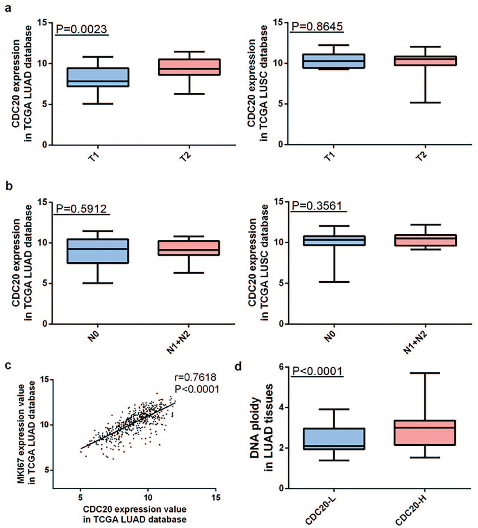
CDC20 shows significant association with T stage in LUAD but not in LUSC. (a) Overexpression of CDC20 significantly correlates with bigger primary tumor size in LUAD (p = 0.0023) but no significant association was observed in LUSC (p = 0.5912). (b) No significant association was observed between CDC20 expression and lymph node status in both LUAD patients (p = 0.8645) and LUSC patients (p = 0.3561). (c) CDC20 expression is significantly correlated with MKI67 expression in LUAD tissues (r = 0.7618, p < 0.0001). (d) Overexpression of CDC20 in LUAD is significantly associated with higher DNA ploidy level (DNA ploidy in CDC20-L: 2.389 ± 0.0712; DNA ploidy in CDC20-H: 2.943 ± 0.0831, p < 0.0001).
CDC20 expression was significantly correlated with MKI67 expression and tumor DNA ploidy level in LUAD tissues
Considering that high expression of CDC20 was observed in LUAD tissues and its overexpression was significantly associated with bigger tumor size only in LUAD, then we used the dataset TCGA_LUAD_exp_HiSeqV2-2015-02-24 to further analyze the correlation between CDC20 and MKI67 in LUAD dataset to assess whether CDC20 is a promising proliferation marker for LUAD patients. CDC20 and MKI67 expression profiles of 510 LUAD patients were extracted and then Pearson’s correlation test was used. As shown in Figure 2(c), CDC20 expression was significantly correlated with MKI67 expression in LUAD tissues (r = 0.7618, p < 0.0001).
Since CDC20 acts as an important protein in SAC regulating mitosis 10 and aneuploidy is a hallmark of carcinogenesis, 30 we then explored the relationship between CDC20 expression and tumor DNA ploidy by further exploring “clinical_data” in TCGA_LUAD_exp_HiSeqV2-2015-02-24 dataset. Totally, 170 LUAD patients’ DNA ploidy levels were recorded in the dataset. We chose the median CDC20 expression value of 8.9435 as the cut-off value and then the patients were divided into two groups: high CDC20 expression group (CDC20-H, n = 85) and low CDC20 expression group (CDC20-L, n = 85). The F-test and t-test results noted that CDC20 overexpression in LUAD was significantly associated with higher DNA ploidy level (DNA ploidy in CDC20-L: 2.389 ± 0.0712; DNA ploidy in CDC20-H: 2.943 ± 0.0831, p < 0.0001; Figure 2(d)).
Overexpression of CDC20 indicates poor prognosis in LUAD patients
We next evaluated the prognostic value of CDC20 in Kaplan-Meier Plotter as mentioned in section “Methods.” The desired Affymetrix ID is valid: 202870_s_at (CDC20). Survival curves are plotted for all NSCLC patients (n = 1928), for LUAD patients (n = 866), and for LUSC patient (n = 675), respectively. High expression of CDC20 was found to be correlated with poor OS in all NSCLC patients followed for 20 years (HR = 1.82, CI: 1.6–2.07, p < 1e−16; Figure 3(a)). Furthermore, high expression of CDC20 was found to be correlated with poor OS in LUAD patients (HR = 2.39, CI: 1.87–3.05, p = 8.6e−13) but not in LUSC patients (HR = 1.12, CI: 0.89–1.42, p = 0.34; Figure 3(b) and (c)).

CDC20 shows different prognostic value in different histological types. (a) Hyper-expression of CDC20 is positively correlated with poor overall survival (OS) in NSCLC patients followed for 20 years (HR = 1.82, CI: 1.6–2.07, p < 1e–16). (b) and (c) Hyper-expression of CDC20 is correlated with poor OS in LUAD patients (HR = 2.39, CI: 1.87–3.05, p = 8.6e−13) but not in LUSC patients (HR = 1.12, CI: 0.89–1.42, p = 0.34). (d)–(f) Hyper-expression of CDC20 is significantly associated with poor OS in LUAD patients who underwent successful surgery (HR = 4.32, CI: 1.76–10.57) and smoked (HR = 4.26, CI: 2.44–7.43) but does not reach a significant level in LUAD patients who never smoked (HR = 1.31, CI: 0.58–2.96).
To evaluate the prognostic value of CDC20 in other subgroups, we examined the OS in both LUAD and LUSC patients in three independent groups: only surgical margins negative, smoked, and never smoked. High expression of CDC20 was significantly associated with poor OS in LUAD patients who underwent successful surgery (HR = 4.32, CI: 1.76–10.57; Figure 3(d)) and smoked (HR = 4.26, CI: 2.44–7.43; Figure 3(e)) but did not reach a significant level in LUAD patients who never smoked (HR = 1.31, CI: 0.58–2.96; Figure 3(f), Table 2). However, there was no significant association between CDC20 expression and prognosis in LUSC patients who were divided into any subgroup as mentioned above (Table 2).
Prognostic value of CDC20 in NSCLC patients.

HR: hazard ratio; CDC20: cell division cycle 20; CI: confidence interval; LUAD: lung adenocarcinoma; LUSC: lung squamous cell carcinoma.
Significant correlation.
Knockdown of CDC20 inhibits LUAD cells’ proliferation in vitro
To choose appropriate cellular models for further investigation, we compared the expression level of CDC20 in different LUAD cell lines. CDC20 was upregulated in LUAD cell lines when compared with normal HBE cells using qRT-PCR (Figure 4(a)). To investigate the biological function of CDC20 in vitro, two different effective siRNAs were used to knockdown CDC20 (Figure 4(b)). As shown in Figure 4(c), CCK-8 assay revealed that knockdown of CDC20 inhibited proliferation of both A549 and H1299 cells. Moreover, si-CDC20 transfected group had significantly fewer colonies than si-NC group (Figure 4(d)).

Knockdown of CDC20 inhibited LUAD cells’ proliferation and colony formation in vitro. (a) Expression level of CDC20 in HBE and LUAD cell lines. (b) A549 and H1299 cell lines were chosen as appropriate cellular models for further investigation, and two different effective siRNAs were used to knockdown CDC20. (c) Knockdown of CDC20 inhibited both A549 and H1299 cells’ proliferation. (d) Colony numbers of A549 and H1299 cells transfected with si-CDC20 were less than those transfected with si-NC.
Discussion
Human CDC20, a homolog of CDC20 protein, is generally considered as one of the major regulators in mitosis. It is responsible for activation of the APC/C complex, an E3 ubiquitin ligase, which targets mitotic proteins such as securin and cyclin B for degradation by the 26S proteasome to allow cells to exit mitosis. 31 CDC20 overexpression has been reported in many cancers in recent years.16,17,32 And such overexpression was associated with failure in SAC activity and consequent mitotic abnormalities and aneuploidy which may promote carcinogenesis.9,10 Although the role of CDC20 in tumorigenesis and prognosis in several cancers has been deeply investigated,12,14,15,33–35 the method of analyzing TCGA database to explore the clinical significance of CDC20 has never been reported in NSCLC.
In recent years, bioinformatics analysis has become a novel method to predict and explore potential oncogenes.36–38 In our study, three TCGA datasets and three online databases were used to explore its biological and clinical role in NSCLC. REACTOME pathway enrichment analysis confirmed that CDC20 is an important molecule in mitosis and cell cycle checkpoint in NSCLC. TCGA datasets revealed hyper-expression of CDC20 in mRNA level and IHC staining in HPA validated CDC20 overexpression in protein level in NSCLC. After exploring the TCGA lung dataset, we concluded that CDC20 expression was significantly associated with tumor size, sex, and histological classification in NSCLC patients, but no significant association was observed with age, lymph node status, or tumor stage.
After further analysis, we found that overexpression of CDC20 was significantly correlated with higher MKI67 expression and higher DNA ploidy level in LUAD patients. In addition, CDC20 overexpression was significantly correlated with bigger primary tumor size in LUAD, but not in LUSC. Then, the prognostic value of CDC20 was explored in an online database providing prognosis information. In subsequent subgroup analysis, CDC20 also showed significant prognostic value in LUAD, but not in LUSC patients.
Our results indicated that CDC20 may play an important role in the malignancy of NSCLC especially LUAD. In addition, knockdown of CDC20 inhibited LUAD cells’ proliferation in vitro. Overexpression of CDC20 might be a useful marker for poor prognosis in LUAD patients. On the other hand, high CDC20 expression could also serve as a biomarker to identify high-risk subgroups in LUAD patients. However, in LUSC patients, no clinical significance was observed at least in mRNA level.
Footnotes
Acknowledgements
R.S., Q.S., and J.S. contributed equally to this work.
Declaration of conflicting interests
The author(s) declared no potential conflicts of interest with respect to the research, authorship, and/or publication of this article.
Funding
This work was supported by the Natural Science Foundation of Jiangsu Province (no. BK2012482), National Natural Science Foundation of China (no. 81472702), and Jiangsu Provincial Special Program of Medical Science (no. BL2012030).