
Abstract
Background
Doxorubicin (DOX), an anthracycline-based chemotherapeutic agent, has been widely used as a first-line treatment for several cancers, including hematological and solid tumors, but its clinical utility is limited by severe cardiotoxicity and nephrotoxicity.
Purpose
This study aims to evaluate the protective effects of pumpkin oil (PO), olive oil (OO), and their combinations against DOX-induced nephrotoxicity in rats.
Materials and Methods
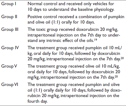
A total of six groups of rats were established for this experiment. The first group of rats served as a normal control and received only the vehicle for 10 days. The second group served as a positive control and received only the same pumpkin-to-OO ratio (1:1 v/v) orally daily for 10 days. The third group was a toxic group and received DOX 20 mg/kg intraperitoneally only on the fourth day. Groups four, five, and six were treated orally with pumpkin, olive, or a combination of both oils (10 mL/kg) daily for 10 days, followed by a single intraperitoneal dose of 20 mg/kg DOX on the 7th day.
Results
The toxic group exhibited a significant increase in oxidative stress thiobarbituric acid reactive substances (TBARS), pro-inflammatory cytokines (interleukin-1β (IL-1β) and tumor necrosis factor-α (TNFα)), apoptotic markers (caspase-3 and caspase-9), renal markers (triglyceride (TG), blood urea nitrogen (BUN), uric acid (UA), and creatinine (Cr)), and deoxyribonucleic acid (DNA) fragmentation while antioxidant (glutathione (GSH)) and antioxidant enzymes (glutathione peroxidase (GPx), glutathione reductase (GR), catalase (CAT), and superoxide dismutase (SOD)) were significantly reduced in comparison to the normal control. These alterations were notably reversed upon treatment with pumpkin and OO alone, as well as a combination, in comparison to the toxic group. These changes were also reflected in the histopathology of the kidney. No significant difference was observed between the positive and normal controls.
Conclusion
Thus, we can conclude that the potential synergistic effects of pumpkin and OO in mitigating oxidative stress and inflammatory cytokines, which may play a significant role as a native medicine in the future, with limitations.
Introduction
Doxorubicin (DOX), a powerful anthracycline antibiotic, is frequently used as an anti-neoplastic medication to treat a variety of cancers. However, there are specific side effects associated with DOX use, among which renal toxicity is a significant concern. This toxicity is considered related to the ability of the drug to initiate processes that may stimulate cellular damage, causing oxidative stress and inflammation that can finally lead to organ failure. 1 Since a detoxification system becomes essential for protecting the body against toxic compounds, any aberration of the function may lead to devastating consequences for overall health. 2 Understanding the mechanisms leading to DOX-induced renal toxicity is a priority.
DOX causes direct renal cell damage, especially in tubular epithelial cells, as it enters cells via active transport mechanisms, leading to a buildup within renal tissues. DOX, when metabolized, generates reactive oxygen species (ROS), and an excess of ROS disrupts intracellular antioxidant defense systems, leading to the propagation of oxidative stress. 3 Cellular damage and dysfunction are the results of this oxidative stress. Further, DOX causes the infiltration of inflammatory cells within the tissues of the kidneys, and chemokines and cytokines with inflammatory effects are released along with the process, leading to the attraction of the immune cells and the aggravation of tissue damage. DOX induces apoptosis in renal cells mainly through mitochondrial dysfunction and deoxyribonucleic acid (DNA) damage. The suppression of apoptotic pathways leads to cell death and contributes to the loss of functional kidney tissue. DOX-induced nephrotoxicity usually displays as acute renal failure, which is an accelerated decline in renal function. 4 Identifying possible therapeutic interventions, including those involving dietary supplementation such as pumpkin and olive oil (OO), might ease some of the adverse effects of this life-saving medication. 5 For a very long time, olive and pumpkin oils (POs) have been known due to their great potential for promoting better health. Nutrients believed to confer myriad health benefits, from anti-inflammatory effects to the development of antioxidant properties, are plentiful in them. 6
Pumpkin (Cucurbita maxima) is a unique plant, and its flesh and seeds have been used as food and folk medicine in several regions. Carotenoids, which are found in pumpkins, have been demonstrated to have antioxidant effects.7–9 The bioactive compounds of pumpkin, like phenolics and flavonoids, as well as tannins and antioxidant scavengers, are recognized for their prospective health benefits.10, 11 The research by Batool et al. found that pumpkins are rich in various bioactive compounds linked to anti-hypertensive, anti-bacterial, anti-diabetic, anti-carcinogenic, and anti-inflammatory properties. 12
These properties suggest that pumpkin may suppress the oxidative and inflammatory pathways, possibly helping to mitigate the adverse effects of the chemotherapy drug DOX. OO contains vitamin E, polyphenolic chemicals, and monounsaturated fats, which enhance its anti-inflammatory and antioxidant properties. These features suggest that it may reduce renal toxicity in male rats by improving some of DOX’s side effects. 13 Polyphenols display anti-inflammatory properties. 14 Additionally, OOs antioxidant prevents free radical generation and balances antioxidants. Utari et al. observed that extra virgin OO reduced cardiac biomarker upregulation in DOX-treated rats. If mixed with virgin coconut oil, it produces synergistic protective effects. 15 Moreover, a few previous studies have also shown that pumpkin and OO have potential therapeutic activities for renal toxicity.16, 17
The synergistic effects of pumpkin and OO in ameliorating kidney damage induced by DOX toxicity have not been demonstrated yet. Therefore, this study was designed to examine the therapeutic effects of the pumpkin and OO, individually and in combination, to mitigate oxidative stress and the inflammatory cytokine profile in the kidneys of rats.
Materials and Methods
Biochemical Kits and Drugs
Enzyme-linked immunosorbent assay (ELISA) assay kits for caspase-3 (ab39401), caspase-9 (ab65608), interleukin-1β (IL-1β) (ab100767), and tumor necrosis factor-α (TNFα) (ab100784) were obtained from Abcam, UK. Photometric kits for triglycerides (TGs), uric acid (UA), blood urea nitrogen (BUN), and creatinine (Cr) were obtained from Randox, Ardmore, 55 Diamond Road, Crumlin, Co. Antrim, BT29 4QY, UK. DOX and other chemical reagents were obtained from Sigma–Aldrich, 3050 Spruce Street, St. Louis, MO 63103, USA. Pumpkin seed oil and OO (Al Jouf brand; botanical source: Al Jouf region, Saudi Arabia; cold-pressed; glass vial; stored at room temperature) were purchased locally. DNA extraction kit and other molecular dyes and buffers were purchased from Beijing Solarbio Science and Technology Co., Ltd., Building 85, Liandong U Valley, Majuqiao, Tongzhou District, Beijing 101102, China.
Gas Chromatography–Mass Spectrometry (GC–MS) Analysis
100 µL of each oil sample was mixed with 900 µL methanol (MS grade) and vortexed for 1 min, and filtered through a 0.45 µm syringe filter directly into a 2 mL GC–MS vial for analysis. 2 µL of each sample was analyzed by GC–MS (QP2010 Ultra, Shimadzu, Japan). The capillary column (Trace™ TR-5MS, 30 m length, 0.25 µL film thickness, 0.25 mm internal diameter, Thermo Scientific, USA) was used for chromatographic separation of components. The injection port temperature was 270°C, and the carrier gas flow rate was 1.2 mL/min. The initial oven temperature was 70°C, followed by a 15°C ramping rate to 290°C, and held for 20 min. The total runtime is 34 min. The ion source and interface temperatures were 230°C and 300°C, respectively. The mass analyzer was running in scan mode for the m/z 40–500 mass range. LabSolutions GC–MS (Shimadzu, Japan) was used for data processing and analysis. 18
Animals
Wistar albino male rats weighing 150–180 g were used and housed in sterile enclosures at 22°C ± 2°C under a 12-h daylight/dark cycle. Furthermore, the animals were supplied with uniform food and water throughout the experiment. The study followed NCBE and ARRIVE guidelines and was approved by the Standing Committee for Scientific Research, University of Jazan (REC-45/05/845).
Study Design
Sample Preparation (Kidney Tissue)
At the end of the experiment, rats were euthanized using ketamine (50 mg/kg)/xylazine (5 mg/kg) via intraperitoneal injection. Blood was collected by cardiac puncture, and the kidneys were excised for biochemical and histopathological analysis. 21 Kidney tissues were homogenized to produce a 10% homogenate in phosphate-buffered (0.1 M) solution and pH (7.4). The homogenate was centrifuged for 10 min at 4°C at 800 rpm to eliminate cell debris, and the first supernatant was collected. The lipid peroxidation assay was conducted in the first supernatant. After that, the second supernatant was produced by centrifuging the homogenate for 15 min at 4°C at 10,500 rpm. In the second supernatant, all antioxidant enzyme assays were carried out. 22
Serum Test
The kidney function markers were carried out in blood serum. Whole blood was collected without an anti-coagulant, allowed to clot at room temperature, and centrifuged at 1,000–2,000 × g for 10–15 min to obtain serum, which was stored at 2°C–8°C until analysis. This serum was used for kidney function markers such as UA, TGs, BUN, and Cr using standard protocols of diagnostic testing kits.
Antioxidant Test
The method of glutathione (GSH) estimation, as Jollow et al. analyzed, 23 is simple and sensitive for the measurement of antioxidant power in biological sample analyses. Such a method will help determine the levels of oxidative stress that are occurring in any cell or tissue where GSH helps in scavenging various ROS and, therefore, protects the cell from being damaged by oxidative species. 0.5 mL of supernatant 2 and 0.5 mL of sulfosalicylic acid (4%) are used in the process. The samples were incubated for 1 h at 4°C, and then they were centrifuged for 15 min at 4°C at 3,000 rpm. Take 0.5 mL of the supernatant and add 2 mL of phosphate buffer (0.1 M, pH 7.4) and 0.5 mL of 5,5′-dithiobis (2-nitrobenzoic acid) (DTNB) to produce a total volume of 3.0 mL. In the spectrophotometer, the yellow color was created at 412 nm.
The lipid peroxidation assay is a method that determines the effect of antioxidants on the formation of lipid hydroperoxides, a principal cause of cellular damage. 24 To conduct this assay, a few specific reagents and chemicals are to be used with a detailed procedure by Utley et al. 25 A metabolic shaker is used in the process. For 1 h, 0.5 mL of homogenate was kept at 37°C, and then at 0°C for 1 h. After the incubation period, 0.5 mL of trichloroacetic acid (TCA) and TBA were added to the mixture, and it was spun for 10 min at 4,000 × g. Following that, test tubes containing the supernatant were heated for 10 min. The developed pink color was tested at 535 nm using a spectrophotometer. TBARS was measured using a value of 1.56 × 10⁵ M−1 cm−1 as the molar extinction coefficient. The result was represented as nanomoles of TBARS formed per gram of protein per hour.
Glutathione peroxidase (GPx) was tested using Mohandas et al. 26 GPx activity was determined using the molar extinction coefficient of 6.22 × 10³ M−1 cm−1, and it was represented as nmol nicotinamide adenine dinucleotide phosphate (NADPH) oxidized/min/mg protein. Glutathione reductase (GR) was estimated by Carlberg and Mannervik. 27 GR was measured using NADPH disappearance, and the molar extinction coefficient of 6.22 × 10³ M−1 cm−1 was used to compute nmol of NADPH oxidized/min/mg protein.
The Habig et al. method was used to estimate glutathione S-transferase (GST). 28 Phosphate buffer, reduced GSH, 1-chloro-2,4-dinitrobenzene (CDNB), and post-mitochondrial supernatant (PMS) were included in each sample’s reaction mixture. Enzymatic activity was measured by measuring absorbance at 340 nm every minute for 3 min. The molar extinction coefficient of 9.6 × 10³ M−1 cm−1 was used to compute the number of CDNB conjugates formed/min/mg protein.
Catalase (CAT) plays a part in defining the radical scavenging activity. 29 Therefore, the procedure of this assay basically consists of several steps. First, reconstitute the CAT sample at the required concentration and then dilute it with the mixing buffer. After that, 2 mL of phosphate buffer (0.1 M, pH 7.4), 0.95 mL of hydrogen peroxide (H2O2) (20 mM), and 0.05 mL of PMS (10%) were mixed and incubated at room temperature. By using a spectrophotometer, the reaction mixture was measured at 520 nm. Calculate the reduction of the H2O2 concentration in terms of the CAT activity observed after the incubation period. A molar extinction value of 43.6 × 10³ M−1 cm−1 was used to measure the CAT activity in nanomoles of H2O2 consumed/min/mg protein.
The ability to dismutase superoxide anions, which are typically one of the many ROS, is associated with the antioxidant assay of superoxide dismutase (SOD). 30 The assay that follows can be crucial in revealing SOD’s antioxidant properties. SOD activity was assessed using the Stevens et al. protocol 31 by keeping an eye on the maintenance of (–)-epinephrine autooxidation at pH 10.4 for 3 min at 480 nm. 1 mM (–)-epinephrine, 0.2 mL of 10% PMS, and 50 mM glycine buffer (pH 10.4) were present in the reaction mixture. By adding (–)-epinephrine, the reaction was started. At a molar extinction value of 4.02 × 10³ M−1 cm−1, the enzyme activity was given in terms of nmol (–)-epinephrine kept from oxidation per minute per mg of protein.
Cytokines Assay
To analyze TNFα, IL-1β, and caspase-3 and caspase-9 in biological materials, a standard protocol of Abcam was followed using an ELISA reader (Elx800™ Biotech, USA). The horseradish peroxidase (HRP) colorimetric detection system at 405 nm and caspase-antigen contacts (immunoassay) were used to determine the targets of the ELISA analytical biochemical approach.32, 33
DNA Extraction (Solarbio Kits)
Genomic DNA was extracted from kidney tissue using a Solarbio tissue DNA extraction kit following the manufacturer’s protocol.34 Briefly, 25 mg of kidney tissue was homogenized in phosphate buffer (0.1 M, pH 7.4) and centrifuged to collect the cell pellet. The pellet was lysed using solution A with RNase A and Proteinase K, followed by incubation to ensure complete digestion. After adding solution B and ethanol, the lysate was loaded onto an adsorption column, washed twice, and centrifuged to remove impurities. DNA was finally eluted using preheated elution buffer, incubated briefly, and collected by centrifugation for further analysis.
Quantification of DNA by the NanoDrop Technique
After DNA extraction by the Solarbio kit for tissue samples, we used the NanoDrop ND-2000 spectrophotometer to quantify DNA. 35 A widely used technique for estimating nucleic acid concentration is to measure the absorbance of the sample at 260 nm.
Gel Electrophoresis (Agarose Gel)
Agarose gel electrophoresis was performed to analyze DNA fragmentation. Agarose (1 g) was dissolved in 50 mL tris-acetate-EDTA (TAE) buffer, heated, cooled, and mixed with ethidium bromide before being poured into a casting tray with combs and allowed to solidify. The gel was placed in an electrophoresis chamber containing TAE buffer, and DNA samples (16 µL) mixed with loading dye (2 µL) were loaded into the wells. Electrophoresis was carried out at 120 V for 5 min, followed by 95 V for 25 min. DNA bands were visualized under ultraviolet (UV) light based on fragment size migration toward the positive electrode. 36
Renal Histology
Each group’s kidney tissue sample was stabilized in 10% formalin and thereafter subjected to dehydration in gradient alcohol and xylene solutions. Blocks were then created when the tissue had been embedded in paraffin. Using a Leica microtome, more tissue was sectioned at a thickness of 5 µm. Hematoxylin (H) and eosin (E) staining were then applied for pathological analysis using a light microscope with a 40× magnification. 22
Statistical Analysis
Data were evaluated using GraphPad Prism 9 software, utilizing one-way analysis of variance (ANOVA) and post hoc Tukey’s test (mean ± standard deviation).
Results
GC–MS Analysis
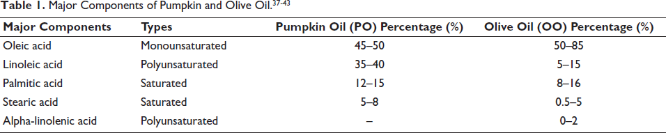
The chemical constituents of pumpkin and OO were determined using the built-in software tools with a set value of 900 in GC–MS. All the discovered components found in PO are listed in the chromatogram (Figure 1S and Table 1S, available online as supplementary material). OO chromatogram and components are listed in Figure 2S and Table 2S (available online as supplementary material), along with their percentage relative content, retention duration, and molecular formula. There are many components that were found, and some of the major components of pumpkin and OO are given in Table 1.
Effects of Pumpkin and OO on Serum Markers
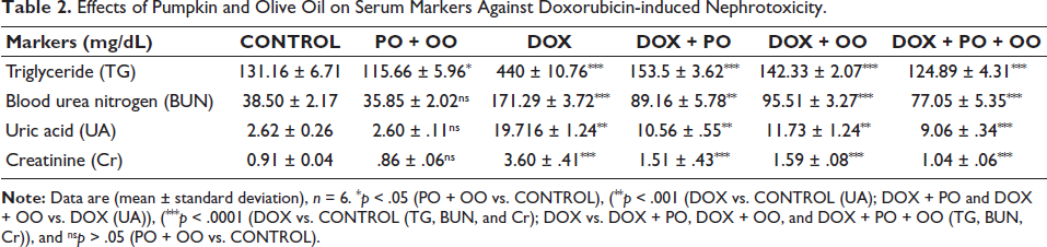
Table 2 shows how serum markers significantly increased (p < .001) for UA and (p < .0001) for TG, BUN, and Cr in Group III (DOX) compared to the control. They significantly decreased (p < .001) for BUN and UA and (p < .0001) for TG and Cr in Group IV (DOX + PO) compared to Group III (DOX). Group V (DOX + OO) significantly decreased (p < .001) for UA and (p < .0001) for TG, BUN, and Cr compared to Group III (DOX). Group VI (DOX + PO + OO) significantly decreased serum markers (p < .0001) compared to Group III (DOX). No changes were noticed in Group II (p > .05), except for a significant decrease in TG (p < .001).
Effects of Pumpkin and Olive Oil on Serum Markers Against Doxorubicin-induced Nephrotoxicity.
Effects of Pumpkin and OO on Antioxidant Enzyme
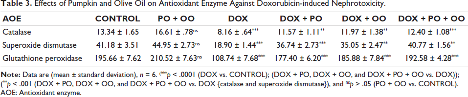
In the DOX treatment group, the levels of antioxidant enzymes (CAT, SOD, GPx, and GR) were significantly lower (p < .0001) than in the control (Table 3). The treatment of PO in group (DOX + PO) increased (p < .001) significantly for CAT and (p < .0001) for other antioxidants. Similarly, the treatment with the OO group (DOX + OO) significantly increases (p < .001) all antioxidant enzymes (p < .0001) compared to the DOX group. Treatment with both pumpkin and OO in group (DOX + PO + OO) significantly enhanced (p < .001) for SOD and (p < .0001) for CAT, GPx, and GR compared to the DOX group. The positive control group (PO + OO) did not show any significant differences (p > .05) from control.
Effects of Pumpkin and Olive Oil on Antioxidant Enzyme Against Doxorubicin-induced Nephrotoxicity.
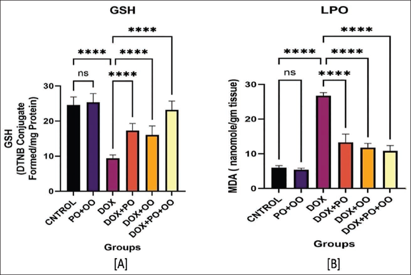
Effect of Pumpkin and OO on Oxidative Stress
In Figure 1A, the DOX group showed a significant increase in lipid peroxidation levels than control (p < .0001). Treatment with PO (DOX + PO), OO (DOX + OO), and their combination (DOX + PO + OO) significantly decreased lipid peroxidation levels compared to the DOX group (p < .0001). While the combination of both oils did not show any noticeable (p > .05) changes compared to the control.
In Figure 1B, the reduced GSH level was significantly lower in the DOX group compared to the normal control (p < .0001). Treatment with PO (DOX + PO), OO (DOX + OO), and a combination of both oils (DOX + PO + OO) showed a significant increase in GSH levels compared to the DOX (p < .0001). While the combination of both oils did not show any noticeable (p > .05) changes compared to control.
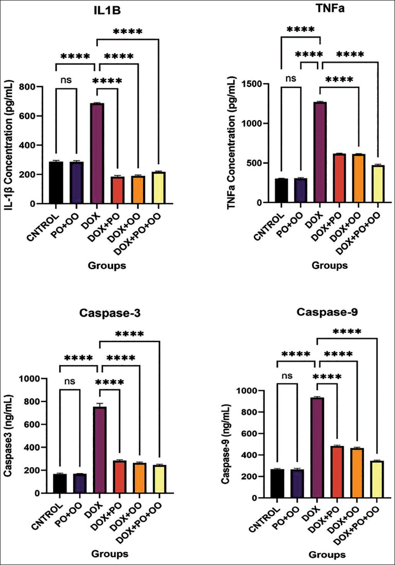
Effects of Pumpkin and OO on Cytokines and Apoptotic Markers
The effects of DOX on TNF-α, IL-1β, caspase-3, and caspase-9 are shown in Figure 2. DOX administration in the DOX group significantly (p < .0001) increased cytokine levels (TNF-α and IL-1β) compared with the CONTROL group. Similarly, apoptotic markers (caspase-9 and caspase-3) also increased significantly in the DOX administration group. The treatment with PO in group DOX + PO, OO in group DOX + OO, and combined oil in group DOX + PO + OO significantly reduced these cytokines (p < .0001) and apoptotic markers. There were no statistically significant (p > .05) differences observed between the positive control group PO + OO and CONTROL.
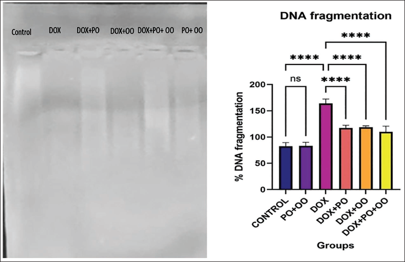
Effects of DNA Fragmentation Produced by DOX
DOX treatment caused marked DNA damage, as evidenced by a significantly higher percentage of DNA fragmentation in the DOX group compared to the control group, as observed by gel electrophoresis and confirmed by NanoDrop analysis (Figure 3). Treatment with PO, OO, and their combination markedly reduced DNA fragmentation and smearing, indicating a protective effect against DNA damage (p < .0001 vs. DOX). No DNA fragmentation was observed in the normal control or positive control (PO + OO) group.
Effects of Pumpkin and Olive Oil on Gel Electrophoresis Deoxyribonucleic Acid (DNA) Fragmentation, and the Bar Graph Represents the % DNA Fragmentation Quantification by the NanoDrop ND-2000 Spectrophotometer. % DNA Fragmentation was High in the Doxorubicin (DOX) Group Versus the CONTROL Group (****p < .0001). The Treatment with Oil Represented a Significant Improvement (****p < .0001) Versus DOX. No Significant (ⁿˢp > .05) Changes were Observed Between the Normal Control and the Positive Control (PO + OO).
Histopathological Examination
Renal histopathology examined under a microscope revealed that the normal control group (CONTROL) had normal renal tubule shapes and intact glomeruli (Figure 4A). However, it was found that giving DOX to the DOX group disrupted the renal architecture and caused kidney dysfunction. DOX caused significant degenerative alterations in the glomerular basement membrane and lesions in the renal tubules. These lesions were seen as edema, coagulative necrosis, vacuolation, congestion, and hemorrhage (Figure 4C).
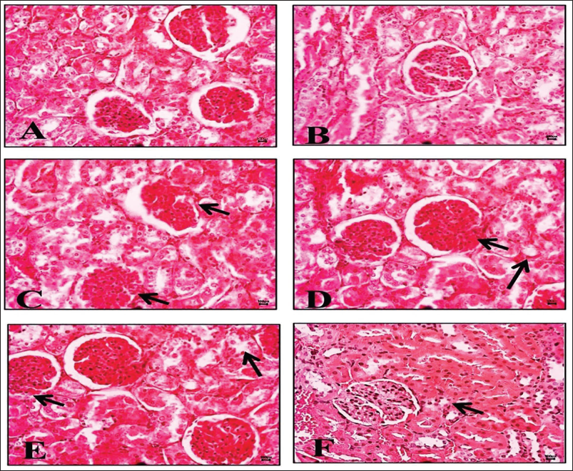
However, after treatment with PO, OO, and a combination of both oils (PO + OO), the severities were minimized, and the glomeruli, vacuolization, and morphology of the proximal and distal tubules were improved compared to the DOX group (Figure 4D–4F). The positive control group (PO + OO) did not exhibit significant lesions, and its histology remained unchanged and similar to that of the normal control (Figure 4B).
Discussion
DOX is a widely used chemotherapeutic agent for the treatment of several cancers, including lymphoma, breast cancer, and leukemia. Its anti-cancer efficacy results from DNA intercalation and inhibition of topoisomerase enzymes, leading to cytotoxicity in rapidly dividing cells. Despite its effectiveness, DOX use is limited by serious adverse effects, particularly nephrotoxicity, which may worsen with cumulative dosing. DOX-induced renal injury is largely attributed to oxidative stress, inflammation, and structural damage to glomeruli and renal tubules, ultimately impairing kidney function. 37
DOX-induced renal injury through various interconnected mechanisms. Following cellular uptake creates the drug accumulates in renal tissues, disrupting normal cellular metabolism. A major contribution to this toxicity is the excessive production of ROS, which overwhelms endogenous antioxidant defenses and induces oxidative stress. 37 DOX simultaneously triggers inflammatory responses, leading to the secretion of cytokines and chemokines that worsen tissue injury. This leads to the attraction of immune cells and the aggravation of tissue damage. 38
PO is the most profound source of phytochemicals that account for carotenoids, tocopherols, and polyphenols obtained from pumpkin nuts. 39 Strong antioxidants found in carotenoids, particularly beta-carotene, possess strong antioxidant activity and effectively scavenge free radicals, thereby reducing oxidative damage. In addition, polyphenols exert anti-inflammatory effects by modulating inflammatory signaling pathways and suppressing pro-inflammatory cytokine production. These properties suggest that PO may serve as a protective agent against oxidative stress and inflammation-mediated renal injury.
The cornerstone of the Mediterranean diet is OO, which has a wealth of monounsaturated fats, polyphenols, and tocopherols. Compounds like oleocanthal exhibit anti-inflammatory effects similar to non-steroidal anti-inflammatory drugs (NSAIDs). 40 In addition, OO also brings a rich source of polyphenols, for example, hydroxytyrosol, which are found to have highly effective antioxidant action and protect against cellular damage caused by oxidative stress. 41 The study looks for improved antioxidant and inflammatory properties in the combination of pumpkin and OO. The interaction of phytochemicals from both oils produces a synergistic effect that makes them more effective in suppressing inflammation and oxidative stress.
In this study, DOX-treated rats showed significantly elevated plasma Cr, BUN, and UA levels compared with controls, confirming renal dysfunction. Treatment with pumpkin and OO individually significantly improved these renal biomarkers, while combined oil treatment produced a more pronounced protective effect, suggesting superior renoprotection.
GSH is a key intracellular antioxidant involved in ROS detoxification. Depletion of GSH increases susceptibility to lipid peroxidation and membrane damage. 42 ROS-induced cellular damage mediates lipid peroxidation and is significantly influenced by GSH depletion. 33 Our study showed that administering DOX significantly reduced GSH and increased malondialdehyde (MDA), indicating enhanced oxidative stress. In contrast, treatment with pumpkin and OO significantly restored GSH levels and reduced lipid peroxidation, demonstrating strong antioxidant activity and protection against free radical-mediated renal damage.
DOX also significantly reduced antioxidant enzymes (GPx, GR, S-transferase, CAT, and SOD). This reduction is likely due to DOX’s renal tubular damage. GPx converts H2O2 into water using GSH, while GR regenerates reduced GSH from its oxidized form, maintaining redox homeostasis. 43 Consistent with the previous reports, DOX significantly restored antioxidant enzyme activities, indicating recovery of the renal antioxidant defense systems.
Oxidative stress is closely associated with inflammatory signaling. Tumor necrosis factor (TNF-α) is a central regulator of inflammatory cytokine production and plays a key role in inflammatory disorders. 44 TNF-α stimulates the release of other cytokines, such as IL-1β and IL-6, 45 which contribute to renal inflammation and tissue injury. 46 DOX also activates the caspase-dependent apoptotic pathway, leading to programmed cell death. 47 In this study, DOX significantly increased TNF-α, IL-1β, caspase-3, and caspase-9 levels, while treatment with pumpkin and OO markedly reduced these inflammatory and apoptotic markers, reflecting their anti-inflammatory and anti-apoptotic effects.
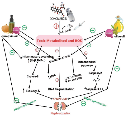
Caspase-9 and caspase-3 are key mediators of mitochondrial apoptosis. DOX-induced ROS damages mitochondria, resulting in cytochrome c release and apoptosome formation, which activates caspase-9 and subsequently caspase-3. Activated caspase-3 leads to DNA fragmentation and renal cell death. In this study, oil treatment significantly attenuated caspase activation, indicating suppression of apoptosis. The proposed mechanism is illustrated in Figure 5.
Mechanistic Approach of Doxorubicin-mediated Renal Injury and Protective Effects of Pumpkin and Olive Oils.
DNA fragmentation patterns are an accurate metric for assessing apoptosis. 48 Endonucleases and proteolytic enzymes break down DNA and chromatin into a smear-like pattern following cell death. 49 Treatment with pumpkin and OO significantly reduced DNA fragmentation as confirmed by gel electrophoresis and NanoDrop analysis. Histopathological examination further supported these findings, showing that combined oil treatment improved renal architecture, reduced tubular degeneration, and prompted tissue regeneration.
Overall, this study demonstrates that pumpkin and OO, when administered individually and more effectively in combination, provide significant protection against DOX-induced nephrotoxicity through antioxidant, anti-inflammatory, and anti-apoptotic mechanisms in rats. However, this study has limitations and was conducted in an animal model; therefore, further clinical investigations are necessary to confirm its relevance to human therapy.
Conclusion
The current study concludes that a combined administration of pumpkin and OO effectively attenuates DOX-induced nephrotoxicity in rats by mitigating oxidative stress, suppressing inflammatory cytokines, inhibiting the apoptotic pathway, and reducing DNA fragmentation. These protective effects were reflected in improved renal biomarkers and histopathology. The combined treatment showed superior efficacy due to synergistic antioxidant and anti-inflammatory actions of its bioactive components, with no observed adverse effects, supporting its safety against DOX-induced renal injury.
Footnotes
Abbreviations
BUN
Authors Contribution
MFA: Conceptualization and project administration, writing—original draft preparation, funding acquisition, reviewing editing; AMAZ, MAAK: Writing—original draft preparation; SM, AH, SA, AAA, MEO, SI, AAD, SM, YM: Reviewing editing; MEO, WM, SM, AMAH, SA, AMJ, WM, SI, AAA; MAQ, YM, AAD: Methodology, software, formal analysis, data curation.
Data Availability Statement
Data related to this research have been provided in the manuscript.
Declaration of Conflicting Interests
The authors declared no potential conflicts of interest with respect to the research, authorship, and/or publication of this article.
Ethical Approval
This project was approved by the Standing Committee for Scientific Research, Universityof Jazan (REC-45/05/845).
Funding
The authors disclosed receipt of the following financial support for the research, authorship, and/or publication of this article: This project was funded by the Deanship of Graduate Studies and Scientific Research at Jazan University, Saudi Arabia (Project Number: RG24-M022).
Informed Consent
The participant has provided informed consent for the submission of the article to the journal.
Supplementary Material
References
Supplementary Material
Please find the following supplemental material available below.
For Open Access articles published under a Creative Commons License, all supplemental material carries the same license as the article it is associated with.
For non-Open Access articles published, all supplemental material carries a non-exclusive license, and permission requests for re-use of supplemental material or any part of supplemental material shall be sent directly to the copyright owner as specified in the copyright notice associated with the article.