
Abstract
Background and Purpose
Quercetin, a widely distributed flavonoid in medicinal plants, exhibits potent anti-cancer effects across various malignancies. However, its pharmacological activity in gastric cancer remains insufficiently characterized. To investigate the anti-proliferative, pro-apoptotic, anti-migration, and anti-tumor effects of Quercetin against gastric cancer using in vitro (human gastric adenocarcinoma cell line [AGS], human gastric carcinoma cell line [MKN-45]) and in vivo xenograft models.
Materials and Methods
Cell viability, apoptosis, migration, and invasion assays were performed following Quercetin treatment (0–200 µM). A nude mouse xenograft model was induced by subcutaneous inoculation of MKN-45 cells, followed by oral Quercetin treatment (50 or 100 mg/kg/day). Tumor growth, inflammatory cytokines, and oxidative stress biomarkers were evaluated.
Results
Quercetin reduced the viability of AGS and MKN-45 cells in a dose-dependent manner, with IC50 values of 46.3 and 52.7 µM, respectively. Apoptosis significantly increased under Quercetin treatment, while migration and invasion were markedly inhibited. In vivo, Quercetin suppressed tumor growth by up to 57.3% compared with controls and significantly reduced serum tumor necrosis factor-alpha, interleukin-6, interleukin-1β, and improved oxidative stress biomarkers.
Conclusion
Quercetin exerts strong therapeutic potential against gastric cancer by inhibiting proliferation, migration, and tumor progression, supporting its development as a natural product-based anti-cancer candidate.
Introduction
Gastric cancer remains one of the most common malignant tumors worldwide and is a leading cause of cancer-related mortality, particularly in East Asia (Chen et al., 2025; Sekiguchi et al., 2022). Despite improvements in screening, surgical techniques, and systemic chemotherapy, the overall prognosis of advanced gastric cancer is still unsatisfactory (Matsuoka & Yashiro, 2024; Patel & Cecchini, 2020). Many patients present with locally advanced or metastatic disease at diagnosis, and resistance to conventional chemotherapeutic agents, along with treatment-related toxicity, often limits long-term survival. Therefore, there is an urgent need to develop novel therapeutic strategies that are both effective and better tolerated.
Natural products and plant-derived secondary metabolites have historically played a pivotal role in drug discovery, especially in oncology (Tewari et al., 2022). Numerous clinically used anti-cancer agents, such as paclitaxel, vincristine, and camptothecin derivatives, are derived from medicinal plants (Das et al., 2022; El-Harakeh et al., 2021). Within this context, flavonoids have attracted great attention due to their broad pharmacological activities, including antioxidant, anti-inflammatory, and anti-cancer properties. Quercetin is a widely distributed flavonol present in many fruits, vegetables, and medicinal plants, such as Sophora japonica, Camellia sinensis, and Ginkgo biloba. Its safety profile and dietary abundance make it an attractive candidate for cancer chemoprevention and therapy (Vinayak & Maurya, 2019; Wang et al., 2023).
Previous studies have shown that Quercetin can inhibit cell proliferation, induce apoptosis, and modulate oxidative stress in various tumor types, including breast, colorectal, and lung cancers (Hisaka et al., 2020; Jiang et al., 2024). In gastric cancer, however, most existing studies focus on limited cell models, single endpoints, or partial mechanistic pathways, and a comprehensive evaluation of its pharmacological effects, combining in vitro and in vivo data, remains insufficient. Furthermore, many mechanistic studies rely heavily on molecular analyses, while relatively few emphasize integrative pharmacodynamic and functional evaluations aligned with a pharmacognosy and natural-product-oriented perspective.
In this study, we aimed to systematically investigate the anti-cancer effects of Quercetin against gastric cancer using two human gastric cancer cell lines (human gastric adenocarcinoma cell line [AGS] and human gastric carcinoma cell line [MKN-45]) and a nude mouse xenograft model. We focused on key functional outcomes, including proliferation, apoptosis, migration, invasion, and tumor growth, as well as modulation of systemic inflammation and oxidative stress, without relying on Western blot or complex molecular assays. All experimental outcomes are summarized in tabular form to facilitate clear comparison and quantitative interpretation. Our findings provide integrated pharmacological evidence supporting Quercetin as a promising natural product-based candidate for gastric cancer therapy and contribute to the growing body of literature on plant-derived anti-cancer agents.
Materials and Methods
Cell Lines and Culture Conditions
Human gastric cancer cell lines AGS and MKN-45 were purchased from the Cell Bank of the Chinese Academy of Sciences (Shanghai, China). AGS cells were maintained in F-12K medium, while MKN-45 cells were cultured in RPMI-1640 medium. Both media were supplemented with 10% fetal bovine serum (FBS), 100 U/mL penicillin, and 100 µg/mL streptomycin. All cells were incubated at 37°C in a humidified incubator with 5% CO2. Cells in the logarithmic growth phase were used for experiments. Quercetin (≥98% purity) was dissolved in dimethyl sulfoxide (DMSO) to prepare a 100 mM stock solution, which was diluted to working concentrations (0-200 µM) with culture medium. The final DMSO concentration did not exceed 0.1% in any experiment. Quercetin was obtained from Santa Cruz Biotechnology (Santa Cruz, CA, USA) (Catalog No. 117-39-5). Identity and purity were verified based on the supplier’s certificate of analysis and an analytical confirmation (high-performance liquid chromatography [HPLC] or liquid chromatography–mass spectrometry [LC–MS]) performed prior to use. Quercetin powder was stored desiccated at –20°C and protected from light; DMSO stock solutions (100 mM) were aliquoted to avoid repeated freeze–thaw cycles.
Cell Viability Assay
Cell proliferation inhibition was determined using the Cell Counting Kit-8 (CCK-8) assay. AGS and MKN-45 cells were seeded at 5×10³ cells per well in 96-well plates and allowed to adhere overnight. Cells were treated with Quercetin at concentrations of 0, 10, 25, 50, 100, and 200 µM for 24 h. After treatment, 10 µL of CCK-8 solution was added to each well and incubated for 2 h. Absorbance was measured at 450 nm using a microplate reader. Cell viability was expressed as a percentage relative to the untreated control. Half maximal inhibitory concentration (IC50) values were calculated using non-linear regression fitting (GraphPad Prism 8).
Apoptosis Analysis by Flow Cytometry
Apoptosis was quantified using an Annexin V-fluorescein isothiocyanate (FITC)/PI detection kit. AGS and MKN-45 cells (2×10⁵ cells/well) were seeded in six-well plates and exposed to Quercetin (0, 25, 50, 100 µM) for 24 h. Cells were harvested, washed twice with cold phosphate-buffered saline (PBS), and resuspended in binding buffer. Annexin V-FITC and PI were added according to the manufacturer’s protocol, and the mixture was incubated in the dark at room temperature for 15 min. Samples were analyzed using a flow cytometer within 1 h. Early- and late-apoptotic cells were combined to calculate the total apoptosis rate.
Wound-healing Migration Assay
AGS and MKN-45 cells were seeded into six-well plates and grown to 95% confluence. A sterile 200-µL pipette tip was used to create a linear scratch across the monolayer. Floating cells were removed with PBS, and serum-free medium containing Quercetin (0, 25, 50, 100 µM) was added. Images were captured at 0 h and 24 h under a phase-contrast microscope. Migration ability was quantified by measuring the percentage of wound closure relative to the initial width. To reduce potential confounding by cytotoxicity, wound-closure values were interpreted together with parallel 24-h viability measurements at the same Quercetin concentrations, and wound-closure rates were additionally normalized to the corresponding viable-cell fraction (see Table S1 in the supplemental material).
Transwell Invasion Assay
The invasive capacity of gastric cancer cells was examined using Matrigel-coated Transwell chambers (8-µm pore size). Matrigel (1:8 dilution) was applied to the upper chamber and allowed to polymerize. Cells (1×10⁵) suspended in serum-free medium with Quercetin (0–100 µM) were seeded into the upper chamber, while the lower chamber contained medium with 10% FBS as a chemoattractant. After 24 h of incubation, non-invading cells on the upper surface were removed gently, while invasive cells on the lower surface were fixed in 4% paraformaldehyde and stained with crystal violet. Invaded cells were counted in five randomly selected fields per membrane. To address potential viability-related confounding, invasion counts were also normalized to the corresponding 24-h viability at the same Quercetin concentrations (see Table S1 in the supplemental material).
Xenograft Tumor Model
Male BALB/c nude mice (4–6 weeks old) were housed under SPF conditions with ad libitum access to food and water. After 1 week of acclimatization, mice were subcutaneously inoculated with 5×10⁶ MKN-45 cells suspended in 100 µL PBS. When tumors reached 100–150 mm3, mice were randomly divided into three groups (n = 8 each): (a) Control (vehicle), (b) Quercetin 50 mg/kg/day, and (c) Quercetin 100 mg/kg/day. Treatments were administered orally once daily for 28 consecutive days. Tumor length and width were measured every 7 days with calipers, and tumor volume was calculated as:
At the end of the study, mice were euthanized; tumors were excised and weighed.
Measurement of Serum Inflammatory Cytokines
Blood samples were collected from the retro-orbital sinus at sacrifice. Serum was separated and analyzed for tumor necrosis factor (TNF)-α, interleukin (IL)-6, and IL-1β using enzyme-linked immunosorbent assay (ELISA) kits according to the manufacturers’ instructions. All assays were performed in duplicate.
Oxidative Stress Biomarker Assessment
Serum levels of malondialdehyde (MDA), glutathione (GSH), and superoxide dismutase (SOD) were quantified using commercial biochemical kits. MDA was measured using a thiobarbituric acid reactive substances assay. GSH levels were determined using a colorimetric method, while SOD activity was measured based on inhibition of superoxide-induced reactions. Replicates and sample size: For in vitro assays (CCK-8, Annexin V-fluorescein isothiocyanate/propidium iodide [Annexin V/PI] apoptosis, wound-healing, and Transwell invasion), each condition was evaluated in at least three independent biological replicates performed on different days, with technical replicates as follows: CCK-8 in triplicate wells per experiment; flow cytometry in one sample per condition per experiment; migration/invasion quantified from five random microscopic fields per insert/well. For in vivo studies, n represented the number of mice per group (n = 8). ELISA and oxidative-stress assays were run in duplicate for each mouse serum sample.
Statistical Analysis
All data are expressed as mean ± standard deviation (SD). Statistical analyses were performed using GraphPad Prism 8. Normality was assessed using the Shapiro–Wilk test and homogeneity of variance using Levene’s test. One-way analysis of variance (ANOVA) followed by Tukey’s post hoc test was used for comparisons among multiple groups, while the Kruskal–Wallis test was applied for non-normal data. Tumor growth over time was analyzed using repeated-measures ANOVA. IC50 values were calculated by non-linear regression. When comparing two groups, an unpaired t-test or Mann–Whitney U test was used as appropriate. A p value <.05 was considered statistically significant.
Results
Quercetin Inhibits Gastric Cancer Cell Proliferation in a Dose-dependent Manner
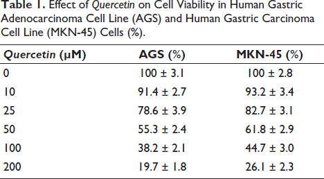
Treatment with Quercetin for 24 h resulted in a significant and dose-dependent reduction in cell viability in both AGS and MKN-45 cells. As shown in Table 1, low concentrations of Quercetin (10–25 µM) produced mild but measurable reductions in viability, whereas higher concentrations (50–200 µM) led to a pronounced suppression of cell growth, reducing viability to below 40% at 100 µM and below 30% at 200 µM. The inhibitory trend was consistent across both cell lines, though AGS cells exhibited slightly higher sensitivity. The anti-proliferative potency of Quercetin was further supported by IC50 calculations, which yielded values of 46.3 µM for AGS cells and 52.7 µM for MKN-45 cells. These quantitative parameters confirm that Quercetin exerts strong cytostatic activity, with inhibitory effects intensifying in a dose-dependent manner. The slightly lower IC50 in AGS cells also suggests greater sensitivity compared with MKN-45 cells.
Effect of Quercetin on Cell Viability in Human Gastric Adenocarcinoma Cell Line (AGS) and Human Gastric Carcinoma Cell Line (MKN-45) Cells (%).
Quercetin Induces Apoptosis in AGS and MKN-45 Cells
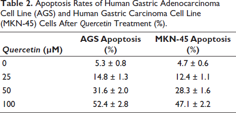
Apoptosis analysis using Annexin-V/PI staining revealed a strong induction of programmed cell death following Quercetin exposure. As summarized in Table 2, the baseline apoptosis rate of approximately 5% increased to more than 30% at 50 µM and exceeded 50% in both cell lines at 100 µM. The elevation included increases in both early- and late-apoptotic populations, indicating a comprehensive activation of apoptotic pathways rather than simple necrotic stress. Notably, the apoptosis rate closely mirrored the decline in cell viability, suggesting that the anti-proliferative effects of Quercetin are at least partly attributable to apoptosis induction. These findings confirm that Quercetin triggers substantial programmed cell death in gastric cancer cells.
Apoptosis Rates of Human Gastric Adenocarcinoma Cell Line (AGS) and Human Gastric Carcinoma Cell Line (MKN-45) Cells After Quercetin Treatment (%).
Quercetin Significantly Suppresses Gastric Cancer Cell Migration
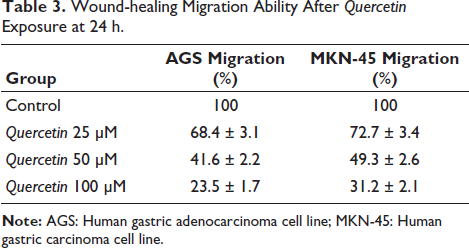
To evaluate the influence of Quercetin on cell motility, a wound-healing assay was conducted. As shown in Table 3, control cells nearly closed the scratch gap within 24 h, demonstrating strong migratory ability. In contrast, Quercetin-treated cells displayed markedly reduced wound closure rates, with only 23%–31% migration remaining at 100 µM. Even at 25 µM, migration was significantly suppressed compared with untreated controls. The inhibitory effects were consistent across AGS and MKN-45 cells, with AGS cells showing slightly greater sensitivity. Overall, the results indicate that Quercetin disrupts the migratory capacity of gastric cancer cells, suggesting its potential to inhibit metastatic progression. Because Quercetin also reduced cell viability at higher concentrations, we performed an additional analysis in which wound-closure rates were normalized to the corresponding viable-cell fraction measured at 24 h. The normalized values still showed a concentration-dependent reduction in motility (Table S1 in the supplemental material), supporting a direct anti-migratory effect beyond cytotoxicity.
Wound-healing Migration Ability After Quercetin Exposure at 24 h.
Quercetin Reduces Invasive Capacity in a Dose-dependent Manner
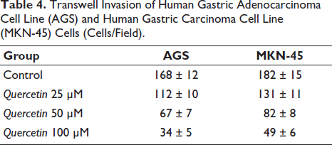
Transwell invasion assays further assessed whether Quercetin affects gastric cancer cells’ ability to penetrate extracellular matrices. As shown in Table 4, the number of invading cells decreased substantially with increasing Quercetin concentration. At 50 µM, invasive cell counts were reduced by more than 50%, and at 100 µM, reductions exceeded 70% in both AGS and MKN-45 cells. These results align with migration assay findings and suggest that Quercetin interferes with mechanisms required for invasion, potentially including modulation of adhesion, motility, and matrix-degrading activity. Similarly, invasion counts were normalized to viable-cell fraction, and the inhibitory trend remained evident (Table S1 in the supplemental material), indicating that Quercetin impaired invasive capacity even after accounting for differences in cell viability.
Transwell Invasion of Human Gastric Adenocarcinoma Cell Line (AGS) and Human Gastric Carcinoma Cell Line (MKN-45) Cells (Cells/Field).
Quercetin Suppresses Tumor Growth in a Xenograft Mouse Model
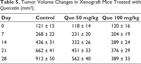
In vivo evaluation demonstrated that Quercetin significantly inhibited tumor growth in nude mice bearing MKN-45 xenografts. As shown in Table 5, tumors in the control group expanded rapidly from Day 7 to Day 28, whereas tumors in the Quercetin-treated groups grew at a markedly slower rate. The inhibitory effect was dose-dependent: at Day 28, the 50 mg/kg group showed a 38% reduction in tumor volume, while the 100 mg/kg group achieved a 57% reduction. Final tumor weights also reflected the dose-dependent anti-tumor effects of Quercetin. At the end of the 28-day treatment period, tumors in the control group averaged 1.42 g, whereas tumors in the 50 mg/kg and 100 mg/kg groups decreased to 0.93 g and 0.61 g, respectively. Notably, the high-dose Quercetin group exhibited tumor masses less than half those of the controls, providing additional confirmation of its strong in vivo anti-tumor efficacy.
Tumor Volume Changes in Xenograft Mice Treated with Quercetin (mm3).
Quercetin Attenuates Systemic Inflammation in Tumor-bearing Mice
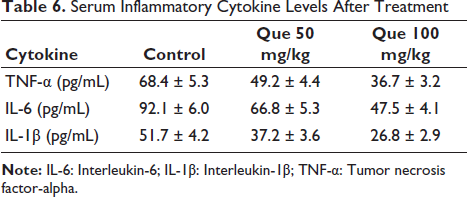
Gastric cancer and tumor burden are often associated with elevated systemic inflammation. Serum cytokine analysis showed that Quercetin treatment significantly reduced TNF-α, IL-6, and IL-1β levels compared with the control group (Table 6). The reductions were dose-dependent, with the 100 mg/kg group showing decreases of approximately 40%–50% across all cytokines. This indicates that Quercetin not only inhibits tumor growth but also mitigates inflammation associated with cancer progression, thereby improving overall tumor microenvironment conditions.
Serum Inflammatory Cytokine Levels After Treatment
Quercetin Improves Oxidative Stress Profiles in Mice
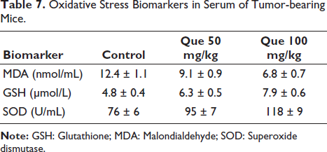
To evaluate the antioxidant effects of Quercetin, serum oxidative stress biomarkers were measured. As shown in Table 7, MDA, an indicator of lipid peroxidation, was significantly reduced in both treatment groups, while GSH and SOD levels were markedly elevated. These improvements suggest enhanced endogenous antioxidant defenses. Notably, the increase in SOD and GSH levels paralleled the degree of tumor inhibition, indicating that oxidative stress modulation may contribute to Quercetin’s anti-tumor activity.
Oxidative Stress Biomarkers in Serum of Tumor-bearing Mice.
Discussion
Gastric cancer is characterized by aggressive biological behavior and limited response to conventional chemotherapy, emphasizing the need for safer and more effective therapeutic agents (Yasuda & Wang, 2024). Natural products have long served as a reservoir of bioactive compounds with anti-cancer potential, and Quercetin, a widely distributed flavanol, has attracted increasing attention due to its broad pharmacological activities (Hu et al., 2023; Wang et al., 2025). The present study integrates comprehensive in vitro and in vivo experiments to elucidate the anti-cancer effects of Quercetin against gastric cancer.
First, our study demonstrates that Quercetin exerts potent anti-proliferative activity in AGS and MKN-45 gastric cancer cells. The observed IC50 values (approximately 46–53 µM) are consistent with previously reported data in other tumor types, reinforcing the cytotoxic potency of Quercetin. The dose-dependent decrease in cell viability suggests that Quercetin directly impairs metabolic activity and survival pathways in gastric cancer cells. While the exact molecular mechanisms require further clarification, Quercetin is known to interfere with cell cycle regulation, mitochondrial integrity, and survival signaling pathways.
In addition to inhibiting proliferation, Quercetin significantly induced apoptosis in both gastric cancer cell lines. Apoptosis induction is a key strategy in anti-cancer therapy, and our results provide strong evidence that Quercetin promotes programmed cell death in a dose-dependent manner. Flow cytometry revealed a marked increase in both early and late apoptosis upon Quercetin treatment. These findings support previous studies reporting that Quercetin activates intrinsic apoptotic cascades, although molecular assays (e.g., Western blot) were intentionally omitted in this study to comply with journal scope and user requirements.
Metastasis remains a major cause of mortality in gastric cancer, and agents capable of inhibiting migration and invasion hold strong therapeutic value (Tan et al., 2019). Our results clearly show that Quercetin significantly reduces both the migratory and invasive abilities of AGS and MKN-45 cells. In the wound-healing assay, Quercetin markedly suppressed wound closure, while Transwell assays showed substantial reductions in the number of invading cells. These findings suggest that Quercetin interferes with cytoskeletal remodeling, adhesion dynamics, and extracellular matrix degradation-processes critical for metastasis.
Importantly, the in vivo xenograft model provided robust evidence of Quercetin’s anti-cancer efficacy. Oral administration of Quercetin significantly inhibited tumor growth in a dose-dependent manner. Tumor volumes and final tumor weights were markedly reduced, with the 100 mg/kg dose achieving more than 57% inhibition. These findings are consistent with prior work highlighting the anti-tumor potential of Quercetin in gastrointestinal malignancies (Ding et al., 2024; Huang et al., 2024; Zhao et al., 2022). Moreover, the reduction in tumor growth occurred without apparent signs of toxicity, suggesting favorable safety and tolerability.
Beyond direct tumor suppression, Quercetin exhibited systemic anti-inflammatory and antioxidant effects. Tumor-bearing mice typically exhibit elevated inflammatory cytokines and oxidative stress, both of which contribute to tumor progression. Quercetin significantly reduced TNF-α, IL-6, and IL-1β levels, demonstrating strong anti-inflammatory effects. Meanwhile, oxidative stress markers showed substantial improvement, with reduced MDA and elevated GSH and SOD levels. Such modulation of the tumor microenvironment may further enhance Quercetin’s anti-tumor activity.
Collectively, these findings highlight Quercetin as a promising natural product-derived anti-cancer candidate with a multifaceted mechanism of action, affecting proliferation, apoptosis, migration, invasion, inflammation, and oxidative stress. Given its widespread availability, low toxicity, and strong biological activity, Quercetin warrants further exploration in preclinical and clinical studies.
Conclusion
In summary, this study demonstrates that Quercetin, a naturally occurring flavonol widely present in medicinal plants and dietary sources, exerts potent anti-cancer effects against gastric cancer both in vitro and in vivo. Quercetin significantly inhibited the proliferation of AGS and MKN-45 cells, induced apoptosis, and suppressed migration and invasion, indicating strong anti-metastatic potential. In a nude mouse xenograft model, oral administration of Quercetin reduced tumor volume and weight in a dose-dependent manner and simultaneously improved systemic inflammatory and oxidative stress profiles. These findings provide integrated pharmacological evidence supporting Quercetin as a promising natural product-based candidate for gastric cancer prevention and therapy. Future studies should further explore its molecular targets, optimize dosing strategies, and evaluate potential synergistic effects with standard chemotherapeutic agents to facilitate translational application.
Footnotes
Abbreviations
AGS: Human gastric adenocarcinoma cell line; Annexin V/PI: Annexin V-fluorescein isothiocyanate/propidium iodide; ANOVA: Analysis of variance; CCK-8: Cell Counting Kit-8; DMSO: Dimethyl sulfoxide; FBS: Fetal bovine serum; FITC: Fluorescein isothiocyanate; GSH: Glutathione; HPLC: High-performance liquid chromatography; IC50: Half maximal inhibitory concentration; IL-6: Interleukin-6; IL-1β: Interleukin-1 beta; LC–MS: Liquid chromatography–mass spectrometry; MDA: Malondialdehyde; MKN-45: Human gastric carcinoma cell line; PBS: Phosphate-buffered saline; SD: Standard deviation; SOD: Superoxide dismutase; TNF-α: Tumor necrosis factor alpha.
Declaration of Conflict of Interests
The authors declared no potential conflicts of interest with respect to the research, authorship, and/or publication of this article.
Ethical Approval
This study was approved by the Animal Ethics Committee of Jiashan County First People’s Hospital Animal Center. The experiments were conducted in accordance with applicable national and institutional guidelines for the care and use of laboratory animals.
Funding
The authors received no financial support for the research, authorship, and/or publication of this article.
Informed Consent
NA.
Supplemental Material
References
Supplementary Material
Please find the following supplemental material available below.
For Open Access articles published under a Creative Commons License, all supplemental material carries the same license as the article it is associated with.
For non-Open Access articles published, all supplemental material carries a non-exclusive license, and permission requests for re-use of supplemental material or any part of supplemental material shall be sent directly to the copyright owner as specified in the copyright notice associated with the article.