
Abstract
Gastric cancer is one of the major cancers threatening people’s lives worldwide. Recent studies showed that Gypsophila oldhamiana gypsogenin (GOG) exhibits inhibition effects and cytotoxic activities against different cell lines. The aim of this study was to explore the inhibitory effect and dose response of GOG on gastric cancer cell line NCI-N87 and to provide the theoretical basis for clinical anti-tumor therapy. The experiments showed that GOG could inhibit the proliferation and promote the apoptosis of human gastric cancer cell line NCI-N87. GOG could dose dependently reduce the expression of vascular endothelial growth factor (VEGF) and matrix metalloprotein (MMP)-9 proteins, while increase the expression of caspase-3 and Bax proteins. Compared with model group, tumor volume (TV), relative tumor volume (RTV), and relative tumor increment rate (T/C) in the mid-dose and high-dose GOG groups were significantly reduced, and the inhibition rate (IR) in the two groups was significantly increased. The results indicated that the anti-tumor effect of GOG on gastric cancer cells may be related with the downregulation of caspase-3 and Bax and the upregulation of MMP-9 and VEGF.
Introduction
Gastric cancer is one of the major cancers threatening people’s lives worldwide, and its morbidity and mortality, respectively, rank the fourth and the third of all the cancer around the world. 1 Etiologies leading to gastric cancer are various, including Helicobacter pylori infection and the interaction of bacteria, host, and environment.2,3 The pathogenesis of gastric cancer mainly concentrated in the abnormal expression of key protein, which is caused by proto oncogene activation and anti-oncogene inactivation. 3 Since gastric cancer is systemic with high recurrence rate and unapparent clinical manifestations, patients are often diagnosed only when the cancer is at an advanced stage; anti-cancer drugs are still considered as a standard treatment. 4 However, most of the current anti-cancer drugs have obvious side effects with a poor perceived prognosis. 5 So, looking for a new anti-tumor drug with high efficiency and low toxicity is a big difficulty in the field of drug development and treatment. Natural extracts or products have attracted attention and become the focus of anti-cancer drug research because over two-thirds of novel anti-cancer drugs discovered in recent decades are of natural origin. 6
Gypsophila oldhamiana is a herb belonging to carnation plant classification, which is widely distributed in the north of China. 7 In traditional medicine, its roots are usually used as a folk medicine for the treatment of fever, consumptive disease, and infantile malnutrition. 8 Gypsophila oldhamiana gypsogenin (GOG) is the natural triterpenoid saponins component extracted from the root of G. oldhamiana. Recent studies showed that GOG exhibits inhibition effects and cytotoxic activities against different cell lines. Hong Bai et al. 9 isolated a new saponin from the roots of G. oldhamiana and found it to be highly active against three different cell lines: human colon cancer (HT29), human gastric cancer (SGC7901), and human hepatoma cells (PLC/PRF/5). Likewise, another research also proved that the triterpenoid saponin of G. oldhamiana inhibited tumor cell viability in vitro, delayed tumor growth in anterior flank and induced cell apoptosis.
However, research about the inhibition of GOG on gastric cancer cell line NCI-N87 has not been reported till now. Therefore, in this study, we investigated the inhibitory effect of GOG on gastric cancer cell line NCI-N87 by cell experiments and nude mice transplanted tumor model to find the possible mechanism of GOG inhibition.
Materials and methods
Materials
The NCI-N87 human gastric cancer cell line was purchased from Shanghai cell bank of Chinese Academy of Sciences (Shanghai, China) and cultured in RPMI 1640 (Carlsbad, California, USA) supplemented with 10% heat-inactivated fetal bovine serum (FBS), 100 μg/mL streptomycin, and 100 U/mL penicillin. The cells were maintained in a humidified atmosphere of 95% air/5% CO2 at 37°C. GOG (high-performance liquid chromatography (HPLC) ≥ 98%) was kindly provided by Shanghai Ruiqi Biotechnology, Co. Ltd (Shanghai, China). The antibodies against vascular endothelial growth factor (VEGF), matrix metalloprotein (MMP)-9 caspase-3, and Bax were purchased from Cell signaling Technology Co. Ltd (Danvers, MA, USA), and goat anti-rabbit horseradish peroxidase–conjugated secondary antibodies were from Wuhan Boster Biological Engineering Co. Ltd (Wuhan, China).
Female mice were purchased from Jinan Pengyue Experimental Animal Breeding Co., Ltd. and animal production license number was SCXK (Lu) 2014-0007, and it was raised in a specific pathogen-free (SPF) environment. They were exposed to a 12-h light/dark cycle at 24 ± 2°C and relative humidity 40%–70% and allowed free access to water and diet. The animal experiments were approved by the animal experimentation ethics committee of the Hospital and performed within the guidelines for the Care and Use of Laboratory Animals.
Cell groups and proliferation
The cells were divided into five groups: untreated group (control), a low-dose GOG (L-GOG) treatment group (5 μmol/L), a medium-dose GOG (M-GOG) treatment group (15 μmol/L), a high-dose GOG (H-GOG) treatment group (25 μmol/L), and cisplatin group (25 μmol/L). After treatment with different doses of GOG and cisplatin (25 μmol/L) for 24 and 48 h, respectively, 10 μL of Cell Counting Kit-8 (CCK-8) solution was added, and cells were incubated at 37°C for 4 h. The culture medium in the control group was without GOG. The optical density (OD) value of each hole was measured at 450 nm by the microplate reader (Molecular Devices, Sunnyvale, CA, USA), and then, the cell proliferation level was calculated according to the OD value.
Cell apoptosis
The cells were seeded in the six-well plate at a density of 1 × 10⁵ per well and then treated with the above-mentioned treatment. The final concentrations of GOG were 5, 15, and 25 μmol/L. Cell apoptosis was monitored by flow cytometry (CyAn ADP9; Beckman Coulter, Fullerton, CA, USA) according to the Annexin V-FITC/PI Apoptosis Detection Kit (BD Biosciences, San Diego, CA, USA).
Establishment of a tumor-bearing nude mice model
Under sterile conditions, NCI-N87 cells in the logarithmic growth phase were selected and adjusted as a cell suspension with a concentration of 1 × 10⁷ mL; 0.2 mL cell suspension was inoculated subcutaneously into the right flank of nude mice, and the growth state of subcutaneous tumor was observed continuously and regularly. The tumor diameter reached 4 mm and considered as successful of tumor-bearing nude mice models.
Animal groups
A total number of 30 female BALB/c nude mice aged 4–6 weeks and weighting 16 ± 1 g were used in the experiment. After the subcutaneous tumor model was established, the inoculated mice were randomly divided into five independent groups (n = 6): a control model group (Model), a cisplatin treatment group (Cisplatin), an L-GOG group, an M-GOG group, and an H-GOG group. Three GOG groups were given intraperitoneal injections with 8, 12, and 16 μmol/kg GOG, respectively, which was dissolved with saline. While the Model group was intraperitoneally injected with the same volume of saline, and the cisplatin group was intraperitoneally injected with 84 μmol/kg cisplatin. All treatments were continued for 15 days with GOG once a day or were injected with cisplatin every 3 days. Then, the mental state, activity, diet, and tumor growth of the nude mice were observed every day.
Body weight change of the nude mice and calculation of tumor inhibition rate
Mice were weighed at the first and the last days of intraperitoneal administration, respectively. And the long diameter and short diameter of the tumor were measured every 3 days. On the day after the final administration, mice were anesthetized with 0.75% pentobarbital sodium intraperitoneally, and then, mice were sacrificed by cervical dislocation to excise the tumor tissue and record the tumor weight. Finally, the tumor growth state and anti-tumor effect were evaluated by correlation index, such as tumor volume (TV), relative tumor volume (RTV), relative tumor increment rate (T/C), and the inhibition rate (IR) were calculated using the following formula: TV = ½ Length×Width2; RTV = final TV/initial TV; T/C (%) = RTV in treatment group/RTV in the control group × 100%; and IR%= (average tumor weight (in the control group − in experimental group))/average tumor weight in control group × 100%.
Histopathologic examination
After weighting and measuring size, a fraction of tumor tissues of each group was fixed in 4% paraformaldehyde for 24 h and processed routinely by embedding in paraffin. Then, the paraffin-fixed tissue specimens were stained with hematoxylin and eosin (HE staining) and mounted on glass slides. A light microscopy was used to observe the pathologic changes of tumor tissue.
Western blot analysis
The total protein of cells or the tumor tissues of each group was extracted by radioimmunoprecipitation assay (RIPA) cell lysis buffer containing proteinase inhibitor cocktail. Bicinchoninic acid assay was used to determine the protein concentrations. Proteins were resolved by sodium dodecyl sulfate–poly acrylamide gel electrophoresis (SDS-PAGE) and then transferred onto polyvinylidene fluoride membranes. Membranes were sealed with 5% skimmed milk powder and incubated with primary antibodies against VEGF, MMP-9, caspase-3, and Bax (dilution, 1:1000) overnight at 4ºC. Subsequently, the membranes were incubated with goat anti-rabbit horseradish peroxidase–conjugated secondary antibodies (dilution, 1:3000) for 1 h at 37ºC; the electrochemiluminescence (ECL) reagent (Bio-Rad, USA) was used after washing the membrane. Image J 1.46 analysis software was used to analyze the western blot strip.
Statistical analysis
Data statistical analysis was performed by SPSS 19.0, and all data were expressed as mean ± SD. Differences between groups were analyzed by one-way analyses of variance (ANOVAs) and Dunnett’s test was used for multiple comparisons. P < 0.05 was considered to indicate statistical difference.
Results
Effects of GOG on the cell proliferation
The effects of different doses of GOG on cells proliferation were shown in Figure 1. Compared with the control group, the cell proliferation of the cells in L-GOG group increased after treatment with 24 h, while the cell proliferation decreased after 48 h. The difference was not statistically significant (P > 0.05). This indicates that the effect of low doses of GOS on cells is limited. With the increase in GOG dose, the cell proliferation decreased gradually, which was significantly different from the control group (P < 0.05). H-GOG treatment group showed the lowest cell proliferation, but it was still higher than that of cisplatin group.

Effects of GOG on the cell proliferation, and GOG inhibited the NCI-N87 cell proliferation (*P < 0.05 and **P < 0.01 vs control group).
Effects of GOG on the cell apoptosis
The effects of different doses of GOG on apoptosis were shown in Figure 2. The cell apoptosis rate increased with the increase in the dose of GOG. The M-GOG and H-GOG treatment groups were significantly higher than the control group (P < 0.01). The apoptosis rate of H-GOG treatment group was lower than that of cisplatin group. The cell apoptosis rate of the 48 h treated group was increased compared with 24 h.

Effects of GOG on the cell apoptosis, and GOG promoted the NCI-N87 cell proliferation (*P < 0.05 and **P < 0.01 vs control group).
Effects of GOG on the protein expression of the cell
Cell proliferation and apoptosis results showed that cell culture was effective for 48 h, so the expression of protein was detected at 48 h. As shown in Figure 3, the expression of MMP-9 and VEGF in each group was lower than that in control group (P < 0.05, P < 0.01). The expression of caspase-3 and Bax in each group was significantly higher than that in control group (P < 0.05, P < 0.01).

Effects of GOG on the expression of the NCI-N87 cell protein at 48 h. GOG reduced the expression of MMP-9 and VEGF proteins and increased the expression of caspase-3 and Bax proteins: (a) western blot for caspase-3, MMP-9, Bax, and VEGF in each group and (b) densitometry analysis of caspase-3, MMP-9, Bax, and VEGF expression based on western blot data. Data are expressed as mean ± SD (*P < 0.05 and **P < 0.01 versus control group).
Effects of GOG on the body weight of tumor-burdened nude mice
The change in body weights of the tumor-burdened nude mice in different groups was shown in Table 1. The average body weights of the tumor-burdened nude mice of the five groups were similar, and the mice do not require significantly different treatment (P > 0.05). Meanwhile, animals’ diet, activity, and mental state were normal. After 15 days of treatment, there were also no significant differences in the body weights of all the five groups (P > 0.05). When subtracting the tumor weight, we found that body weights of GOG-treated groups were still in close proximity to that of the control model group (P > 0.05). Compared with the model group, the body weight of the cisplatin group was slightly reduced, but there was no statistical significance (P > 0.05).
Effect of GOG on body weight of nude mice from each group.

GOG: Gypsophila oldhamiana gypsogenin; L-GOG: low-dose GOG treatment group; M-GOG: medium-dose GOG treatment group; H-GOG: high-dose GOG treatment group.
Effects of GOG on the growth of gastric cancer xenografts
The change in TV, RTV, T/C (%), and IR (%) was calculated to reveal GOG’s effects on the growth of gastric cancer xenografts. As demonstrated in Figure 4(a), after 15 days of treatment, the TV of the M-GOG treatment group, H-GOG treatment group, and cisplatin group was all significantly smaller than that of the model group (P < 0.05, P < 0.01, and P < 0.001, respectively). However, there was no significant difference between the L-GOG and model groups (P > 0.05). The similar situation also happened in the case of RTV and T/C (%) of gastric cancer xenografts (Figure 4(b) and (c)). After 15 days of administration, tumor tissue was excised and weighted to calculate the IR (%). The results showed that (Figure 4(d)) compared with the model group, the tumor mass of the M-GOG, H-GOG, and cisplatin groups was all significantly decreased (P < 0.05, P < 0.01, and P < 0.001, respectively). The IR (%) of the above three groups was 10%, 22%, and 62%, markedly higher than the model group (P < 0.05, P < 0.01, and P < 0.001, respectively).
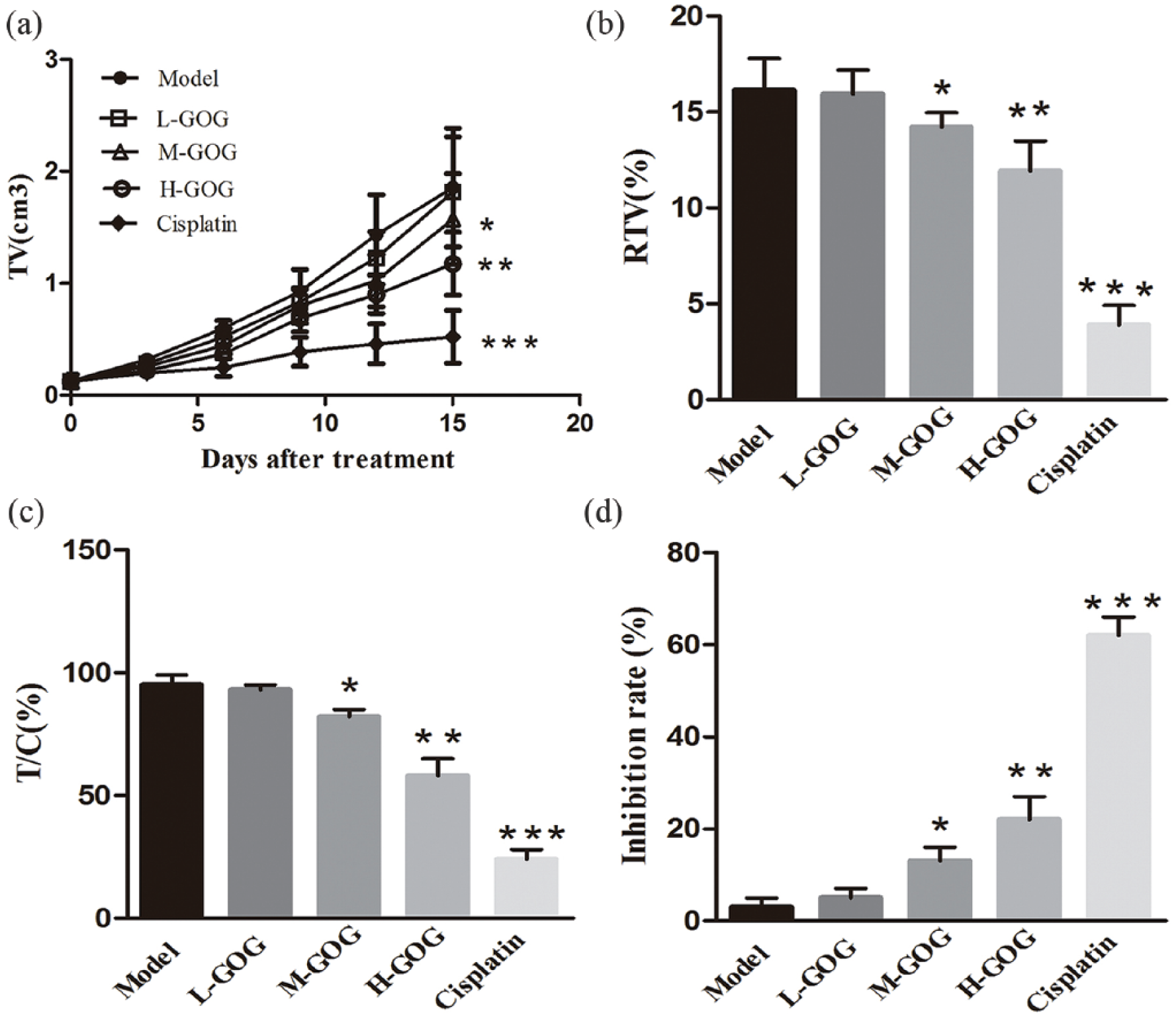
GOG suppressed the growth of gastric cancer xenografts in vivo. (a) Tumor growth curves for each group. (b) Relative tumor volume for each group at the end of the study. (c) Relative tumor growth rate for each group at the end of the study. (d) Inhibition rate for each group at the end of the study. Data are shown as mean ± SD (n = 6; *P < 0.05, **P < 0.01, and ***P < 0.001 vs model group).
Effects of GOG on the morphology of gastric cancer xenografts
The change in morphology of gastric cancer xenografts in different groups is shown in Figure 5. Histopathological examination showed that pathologic mitosis, necrosis, and neat arrangement were visible in the tumor cells of the control model group, showing irregular shape, large nucleoli, and dense cytoplasm (Figure 5(a)). And tumor cells in the L-GOG group showed morphology similar to those of the model group (Figure 5(b)). However, compared with the above two groups, tumor cells in the M-GOG, H-GOG, and cisplatin groups presented varying degrees of pathologic improvement. In fact, some of the symptoms were observed in cancer cells in these three groups, such as scattered arrangement, disappearing cellular structure, nuclear condensation, fragmentation, and apoptosis (Figure 5(c)–(e)).

Effect of GOG on histological changes of gastric cancer xenografts in nude mice: (a) model group, (b) low-dose GOG-treated group, (c) mid-dose GOG-treated group, (d) high-dose GOG-treated group, and (e) cisplatin-positive control group (original magnification, 400×).
Effects of GOG on the expression of gastric cancer xenograft protein
To explore GOG’s molecular mechanism of inhibiting gastric cancer proliferation, western blot was used to detect protein expression of angiogenesis central regulator VEGF, metastasis-related protein MMP-9, apoptosis key molecular caspase-3, and Bax. As shown in Figure 6, compared with the model group, the expression of MMP-9 and VEGF proteins in the M-GOG, H-GOG, and cisplatin groups was significantly decreased (P < 0.01, P < 0.001, and P < 0.001, respectively), while the level of caspase-3 and Bax proteins in the M-GOG, H-GOG and cisplatin groups was dose dependently increased (P < 0.05, P < 0.01, and P < 0.001, respectively).

Effect of GOG on the expression of caspase-3, MMP-9, Bax, and VEGF proteins in mice. GOG reduced the expression of MMP-9 and VEGF proteins and increased the expression of caspase-3 and Bax proteins. (a) Western blot for caspase-3, MMP-9, Bax, and VEGF in xenografts from each group. (b) Densitometry analysis of caspase-3, MMP-9, Bax, and VEGF expression based on western blot data. Data are expressed as mean ± SD (*P < 0.05, **P < 0.01, and ***P < 0.001 vs model group).
Discussion
Gastric cancer is one of the most common malignant tumors characterized as having high morbidity, high mortality, and high recurrence rate. 10 Many chemotherapeutic agents, such as cisplatin, 5-FU, paclitaxel, and doxorubicin, have been used for clinical treatment of patients for years. However, the overall outcome remains poor due to drug resistance, radiotherapy, or side effects. 11 In recent years, natural products are becoming the potential clinical drugs for cancer prevention and treatment, because of the outstanding effects of rising immunity and low toxicity on cancer patients.12,13 Triterpenoid saponins are one kind of natural medicines with cellular toxicity and chemical prevention activity, which have been reported to have significant anti-tumor and apoptosis promotion activities.14–16 GOG is the component of triterpenoid saponins extracted from G. oldhamiana. This study investigated the inhibitory effect of GOG on gastric cancer by using the nude mice model bearing cell line.
NCI-N87 cell line has high HER2 amplification and expresses high levels of HER2 protein and has shown efficacy in a cell line model that is driven by an amplified oncogene.17,18 The results of the study revealed that GOG could obviously reduce the proliferation of NCI-N87 cells in a dose-dependent manner. However, the anti-tumor effects of GOG were significantly weaker than those of cisplatin. This result was a little different from the viewpoint of Hong Bai, 9 who observed that cisplatin exhibited more active cytotoxicities against cancer cell lines than the GOG, but the differences were not significant. This could be due to GOG’s variant efficacy on different cell lines. Meanwhile, our results also found that GOG had no obvious weight loss effect on the tumor-burdened nude mouse, which meant that the cellular toxicity and side effects of GOG were low. Zhang et al. 19 studied the triterpenoid saponin-rich G. oldhamiana root extraction and found that it inhibits the proliferation and induces apoptosis of SMMC-7721 cells, but its cytotoxic effects on normal human hepatic L02 cells were much lower.
The abnormal proliferation and metastasis of tumor cells determine occurrence and development of tumor. 20 Angiogenesis is a key limiting step in tumor growth and metastasis. There are a lot of cytokines involved in the process of regulating angiogenesis, among which VEGF is one of the most important regulators. 21 VEGF and its receptor can strongly induce endothelial cells mitosis, increase the vascular permeability, promote proteinosis in the mesenchyme, and ultimately lead to the formation of new blood vessels, which can provide oxygen and nutrients for tumor growth. Excessive expression of VEGF is related to the development and prognosis of a variety of tumors including colon cancer, breast cancer, prostate cancer, and melanoma. And a growing number of studies have confirmed that blocking the VEGF signaling can inhibit tumor angiogenesis to achieve the purpose of inhibiting tumor growth.22,23 Therefore, VEGF, as the central regulator of angiogenesis, is becoming a kind of important targeting therapy of anti-angiogenesis. 24 MMPs are a group of zinc endopeptidase, which can degrade the extracellular matrix (ECM) and play a key role in the tumor invasion and transfer. MMP-9 can degrade type IV collagenase of ECM, 25 make tumor cells infiltrating and directly enter the lymphatic vessels, and eventually involve in tumor invasion and metastasis. 26 Recent studies showed that high expression of MMP-9 was found in gastric cancer, and its expression was positively related to the occurrence, invasion, lymph node metastasis, staging, and 5-year survival rate of tumor.27,28
Not only the abnormal proliferation but also the apoptosis ability of tumor cells plays important roles in the tumorigenesis and tumor progression. Caspase-3 may serve as one of the central segments of the apoptotic cascade and is considered to be the final effect protein of apoptosis. 29 In the process of cell apoptosis, starting signal of multi-factors can activate caspase-3, which will further cleavage different substrates, resulting in amplification of the cleaved protein cascade and ultimately leading to irreversible cells apoptosis. Hence, the activation degree of caspase-3 is often used as a reliable indicator of apoptosis judgment. 30 The expression and regulation of Bcl-2 family are also the key factors directly affecting the apoptosis process. It is divided into two major categories of apoptosis inhibitory protein and pro-apoptotic protein, among which Bcl-2 and Bax are the principal members, respectively. Bcl-2 was confirmed that its increased expression would cause tumor evasion and life extension.31–33 As the pro-apoptotic protein, on one hand, the high level of Bax forms heterodimer with Bcl-2 to block the apoptosis inhibition; on the other hand, it forms homodimer to promote apoptosis.
In this study, histopathological examination showed that GOG promoted the tumor tissue necrosis and cell apoptosis. Western blot testing in cell and xenografts tissue demonstrated that GOG could significantly reduce the expression of VEGF and MMP-9 proteins in a dose-dependent manner, while the level of caspase-3 and Bax proteins was dose dependently increased. From the results, we can see that GOG may inhibit the proliferation of gastric cancer by blocking angiogenesis and metastasis as well as inducing apoptosis.
In summary, in vitro experiments showed that GOG could inhibit cell proliferation and promote the mechanism of apoptosis, decrease the expression of VEGF and MMP-9 proteins, and increase the expression of caspase-3 and Bax proteins. In vivo experiments showed that GOG of middle and high dose could effectively inhibit the proliferation of gastric cancer by regulating the angiogenesis, metastasis, and apoptosis-related proteins. However, the anti-tumor effect of GOG was not as strong as the cisplatin. We expect that combination therapy with GOG and cisplatin may obviously reduce the side effects under the precondition of not decreasing the tumor inhibition potency. This research revealed that GOG could serve as an anti-cancer new drug of high efficiency and low toxicity.
Footnotes
Acknowledgements
Y. L. and X. L. contributed equally to this work.
Declaration of conflicting interests
The author(s) declared no potential conflicts of interest with respect to the research, authorship, and/or publication of this article.
Funding
The author(s) received no financial support for the research, authorship, and/or publication of this article.