
Abstract
Background
Rosa odorata Sweet var. gigantean (Coll. et Hemsl.) Rehd. et Wils (FOE), a sort of ethnodrug from the Yi nationality called “GU-GONG-GUO,” the root of which has been shown to have the effect of relieving diarrhea with astringents. Whether fruit extract (FOE) has therapeutic properties for related intestinal diseases is unclear.
Objectives
The study dedicates to expounding the results and mechanisms of FOE for treating ulcerative colitis (UC).
Materials and Methods
we used dextran sulfate sodium to induce UC in vivo and lipopolysaccharide to stimulate macrophage in vitro for the purpose of exploring and determining the effectiveness and potential mechanism of action of FOE.
Results
The weight loss ratio and Disease activity index score of FOE administration groups were obviously lower and the colon tissue morphology was significantly relieved. The levels of various proinflammatory cytokines in colon tissue were evaluated to be decreased. Meanwhile, the FOE administration group also alters oxidative stress factor levels. The levels of Nrf2 and HO-1 protein were once distinctly up-regulated, whereas the levels of NF-κB p65, p-IKK α/β, and Keap1 were dose-independently and prominently suppressed by FOE administration. In vitro, FOE significantly reduced the secretion of proinflammatory cytokines and inhibited the oxidant stress injury in macrophage cells induced by macrophage. The relative expressions of NF-κB p65, p-IKK α/β, and Keap1 proteins in FOE groups were memorably down-regulated, while the Nrf2 and HO-1 levels presented as a contrary tendency.
Conclusion
These results indicated that FOE has shown potential therapeutic efficacy on UC and might be considered an effective anti-inflammatory agent from natural sources.
Keywords
Introduction
Ulcerative colitis (UC) is a persistent non-specific inflammatory ailment of the digestive tract that influences the rectum and colon. It is a complex multifactorial and multivariable disease that is prone to cancer and Crohn’s disease (CD) and Inflammatory bowel disease (IBD) (Kobayashi et al., 2020). UC has a high incidence in European and American countries. Statistics indicated that the morbidity in the USA is as high as 2/1000, while that in Asian countries is relatively low. However, in recent years, the reported cases are increasing year by year and the recurrence rate is 72%, as well as the infected population, tends to be younger (Du & Ha, 2020). UC is easily transformed into colon cancer after repeated attacks and prolonged treatment. It has been identified by WHO as one of the most difficult chronic diseases to treat. The clinical manifestations of UC are mainly abdominal pain, diarrhea, tenesmus, and defecate bloody purulent stool (Zhang et al., 2021). Most scholars believe that the development of UC is caused by the interaction of a variety of factors, involving genetics, environment, immunity, spirit, and so on (Forman & Zhang, 2021; Guo et al., 2020). For the moment, research has shown that UC is closely related to the imbalance of oxidative stress response. Oxidative stress will overexpress oxygen free radicals, causing damage to fat, protein, and DNA, aggravating or inducing UC (Değer et al., 2021; Finamore et al., 2020). Meanwhile, research has proven that the expression of NF-κB protein in sufferers with UC is notably increased (Glassner et al., 2020; Liu et al., 2022a; Piotrowska et al., 2021).
Existing drugs for the remedy of UC mainly include immunosuppressants, biological agents, glucocorticoids, and amino acid salicylic acids. Nevertheless, these drugs can only control the inflammatory response and rapid relief of symptoms instead of achieving radical healing (Decara et al., 2020; Lu & Zhao, 2020; Tang et al., 2021). UC is a chronic recurrent disease, these drugs are easy to cause recurrence after discontinuation. Long-term use may cause adverse reactions such as nausea, vomiting, hyperthyroidism, Cushing’s syndrome, and so on. In severe cases, drug desensitization and intolerance may occur, and patients are also prone to cancer. Severe cases may appear as drug desensitization and intolerance (Duan et al., 2021; Yang et al., 2019). Accordingly, at present, a large number of scholars turn their research focus to traditional Chinese medicine and natural plant medicine, with a view to looking for a drug for UC with positive efficacy and slight side effect.
Rosa odorata Sweet var. gigantean (Coll. et Hemsl.) Rehd. et Wils (FOE), a kind of ethnodrug from the Yi nationality known as “GU-GONG-GUO.” At present, research on “GU-GONG-GUO” at domestic and overseas is basically the center of attention on the root. Whereas systematic researches on the fruit of “GU-GONG-GUO” are very few (Ma et al., 2010). FOE mainly contains triterpenoids and phenolic acids, which are also the main chemical constituents of the root. The pharmacological effects have been reported mainly in anti-inflammatory, antibacterial, antioxidant, anti-tumor, anti-diarrhea, and anti-virus (Li et al., 2020; Shi et al., 2017). Besides, The root has been established favorable impact on the hindrance of UC in mice via inhibiting infection and oxidative stress (Liu et al., 2022a,b). Thus the motive of the find out is to preliminarily discover the mechanism of FOE legislation on Nrf2 and NF-κB signaling pathway, and to lay a foundation for future research on targets that can simultaneously act on Nrf2 and NF-κB, regulate oxidative stress response and inflammatory response and thus has a therapeutic effect on UC.
Materials and Methods
Plant Materials and Extract Preparation
“GU-GONG-GUO” was identified as large-flowered Scabious fruit, originating from Qujing City, Yunnan Province. The fruit samples have been processed by using immersing one hundred g of chopped fruits in 600 mL of 60% ethanol answer for 2 h and extracted twice at reflux for 2 h every time, and the received options had been blended evenly. The extract was once centered to gain 23.7 g of powder extract. Based on the previous work of our research group, a total of 26 pearks were extracted by UPLC, and 21 components were identified in FOE extract based on UPLC-QTOF-MS data, UV-vis spectral data, retention time, and standard control. The 21 components were mainly flavonoids, including proanthocyanidin B3, proanthocyanidin B2, Taxifolin-3-O-glucoside, Apigenin-4′-O-glucoside,7-Methoxy-apigenin-4′-O-glucuronic acid, 2″-O-p-Coumaroylvitexin, and other flavonoids, as well as phenylalanine and its isomers (Liu et al., 2022a, b).
Animals
Forty-two SPF males were bought from SPF Biotechnology Co. Ltd. (Certificate No. SCXK 2011-0004). The experimental process was in accordance with the relevant guidelines of animal ethics.
DSS-induced Experimental Colitis
According to the random variety desk method, 42 mice have been divided into 6 groups, 7 mice in every crew: normal group (0.9% sodium chloride solution without colitis), model group, FOE high-dose (500 mg/kg−1), medium-dose (250 mg/kg−1) and low-dose (125 mg/kg−1) groups, sulfasalazine group (500 mg/kg−1), and the intragastric volume of mice was 10 mL/kg−1, for 7 consecutive days. After adaptive feeding for 1 week, the mice in the mannequin crew have been given 3% DSS answer as a substitute for consuming water for 7 consecutive days, which used to be modified as soon as each and every different day.
Disease Activity Index
The modifications of the physique weight of mice have been recorded each and every day, and the modifications of fecal qualities have been found and recorded. The fecal occult blood was once detected in accordance with the guidelines of the kit, and DAI was scored by reference (Del Sordo et al., 2022). The DAI rating of every crew used to be calculated in accordance with the components.
Macroscopic Assessment
After a week of treatment, mice were killed on the eighth day to take out blood, colonic contents, spleen, and colon tissues. The blood samples have been centrifuged to accumulate serum and saved at −80℃. We measured the length of the colons. Cutting off enough colon tissues, we got tissue homogenate for comparing the number of protein concentrations by BCA protein assay kit (PC0020, Solarbio).
Histology Method and Immunohistochemical Method Analysis
The paraffin blocks had been reduced constantly at 4 µm and positioned in an oven at 60℃ for 1 h. The experimental process was performed according to the kit instructions, including dewaxing, dehydrating, inactivating, anti-primary repair, sealing, adding primary antibody by drop, incubating secondary antibody, and DAB coloration development, counterstaining, dehydrating, transparentizing, and sealing. Five non-overlapping visible fields had been randomly chosen underneath a 200× mild microscope, and the common imperative absorbance AA of the goal protein was once decided by using the ImageJ software program.
Cell Culture
Normal mouse RAW246.7 cells had been cultured in DMEM medium and positioned in an incubator with 5% CO2 at 37℃. According to the increase of cells, the answer used to be modified as soon as 12−24 h. After the cells grew to 70%−80%, the cells have been digested with 0.25% trypsin-EDTA and separated by means of blowing and centrifugation for subculture. RAW264.7 cells have been subcultured as soon as about 4−6 days and then frozen in liquid nitrogen for standby application.
Cell Viability Assay
RAW264.7 cells had been cultured with distinctive FOE and encouraged with LPS (10 µg L−1) for the indicated time. The reserve solution of FOE was prepared in DMSO. In the experiment, FOE was used as the medium to dilute to the specified concentration, and the final concentration of DMSO was less than 0.5%. RAW264.7 cells (2.0 × 10⁹ cells L−1) had been handled with distinct concentrations of FOE.20 µL MTT (5 g L−1) was once brought to every well, and the supernatant was once discarded after addition incubation for 4 h. A 150 µL DMSO was once added, and the crystals have been utterly dissolved by way of shaking for 10 min. The absorbance used to be examined at 492 nm in a microplate reader.
Detection of the Levels of NO, SOD, and MDA
In strict accordance with the package instructions, the double antibody sandwich ELISA approach used to be used to detect. The kit, serum samples, and colon tissues to be tested were balanced to room temperature, the reference substance and washing solution were diluted, and the sample was added, incubated, washed, colored, and terminated as required. The OD fee of every nice was once measured with the aid of a microplate reader, and the Curve Expert 1.4 curve software program was once used for analysis. The attention of NO was once calculated from the well-known curve in accordance with the OD value of samples. The activity of SOD and the content of MDA were measured by chemical colorimetry according to the instructions.
Detection of the Levels of Related Inflammatory Factors
The serum of mice and tissue homogenate organized on ice had been gathered and the TNF-α, IL-6, and IL-1β degrees had been detected through the application package.
Western Blotting Analysis
The whole protein in colon tissue was once extracted, and the awareness of protein used to be quantified by way of the BCA method. After SDS-PAGE electrophoresis and electrical conversion, the essential antibodies (1:1000) had been introduced and then incubated at 4°C for a single day. The corresponding secondary antibody (1:10000) used to be introduced and incubated at room temperature for 2 h. GE gel imaging machine was once used for publicity and development. The grey fee of the goal band was once analyzed through the ImageJ Image evaluation system.
Statistical Analysis
Data were analyzed with GraphPad Prism statistical software, and the detection data were expressed as means ± SD. A value of p < 0.05 was considered statistically significant.
Results
Effect of FOE on Mouse Morphology and Organ Tissue
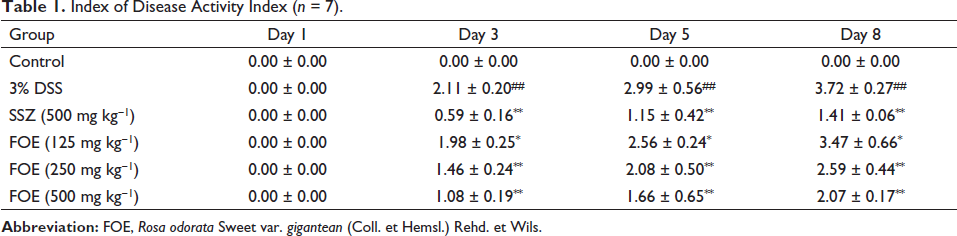
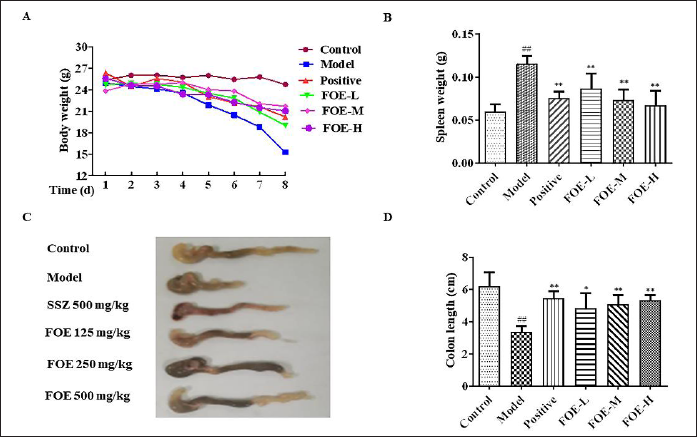
As proven in Table 1 and Figure 1, in contrast with the regular group, the physique weight of the mannequin team was once notably lowered (p < 0.01), and the DAI rating used to be notably elevated (p < 0.01). In the mannequin group, the physique weight loss charge and DAI rating of mice in the high, medium, and low dose FOE organizations and SSZ crew had been considerably diminished (p < 0.01), and the excessive dose FOE team had the high-quality effect.
Index of Disease Activity Index (n = 7).
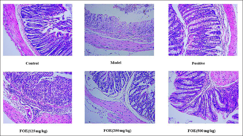
FOE Diminished Colonic Histopathology Method Changes
In the normal group, the colonic mucosal epithelium was intact, the submucosal blood vessels were clear, there was no congestion and edema, the glands and goblet cells were arranged neatly, and there was no ulcer formation. On the contrary, the intestinal mucosal epithelium was damaged, glands were disordered, some of them were necrotic or disappeared, crypts and goblet cells had been considerably reduced, a giant range of inflammatory cells have been infiltrated in the mucosa and submucosa, and ulcers were formed. The morphology of intestinal tissue of mice in different dose groups of FOE was gradually improved, the mucosal injury was alleviated, inflammatory cell infiltration was gradually reduced, and the morphology of goblet cells and crypts was relatively intact. Epithelial tissue cells were rarely shed in the mucosa of the SSZ group. There were abundant intestinal glands in the lamina propria and lymphocytes infiltrated topically. The degree of colonic pathological injury was improved in mice with UC treated with FOE, as presented in Figure 2.
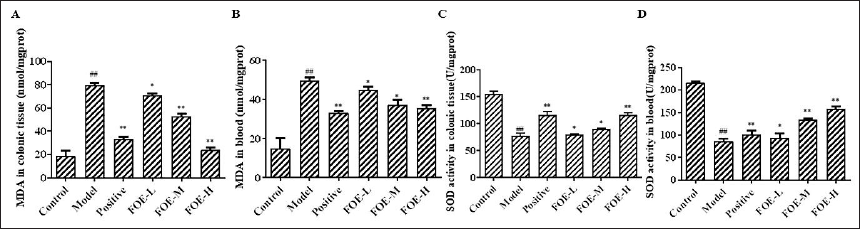
FOE Has an Anti-Oxidative Stress Effect
As proven in Figure 3, in contrast with the manipulated group, the MDA concentrations used to be increased, and the SOD exercise was once reduced. In the mannequin group, the MDA concentrations and FOE companies were once lowered, and the SOD endeavor was once extended (p < 0.05, p < 0.01). MDA and SOD have an important function in oxidative stress and are important markers of an oxidative stress response. Therefore, it can be concluded that FOE can regulate the oxidative stress response and have a therapeutic effect through antioxidant effect.
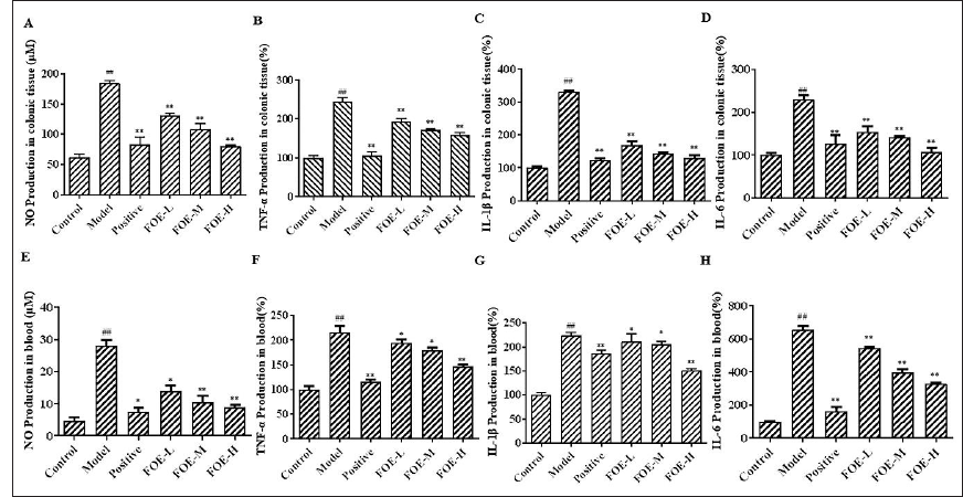
FOE Lowered the Manufacturing of Inflammatory Cytokines
Compared with the manipulated group, the expression of NO, TNF-α, IL-6, and IL-1β in the colon tissue and serum of mice in the mannequin crew used to be increased. Compared with the mannequin crew, the increase of inflammatory cytokines mentioned above was inhibited in the sulfasalazine group and FOE dose groups, which showed that FOE can play a therapeutic effect by reducing and regulating the infiltration of pro-inflammatory factors (Figure 4).
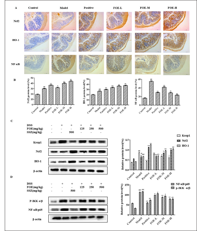
FOE with Respect to NF-κB and Nrf2 Signaling Pathway
Figure 5 shows that the fantastic expression of NF-κB p65 was once a good deal greater in the colon tissues of the mannequin team in contrast with that of the management group. However, FOE dramatically decreased the variety of nuclear NF-κB p65 advantageous cells. moreover, FOE elevated the number of Nrf2 in contrast with the mannequin group. Compared with the mannequin group, the protein expression ranges of Nrf2 and HO-1 in every crew have been enhanced, and the protein expression degrees of Nrf2 and HO-1 in the SSZ team and FOE high-dose team had been statistically significant. Moreover, the Nrf2 and HO-1 proteins used to be obviously rose, and the Keap1, p-IKK α/β, and p-NF-κB p65 were once reduced with the aid of FOE therapy.
FOE Attenuates Inflammatory and Oxidative Stress Factor Responses in RAW264.7 Cells
In comparison with the manipulated group, the degree of IL-6, TNF-α, and IL-1β in the supernatant of the LPS crew used to be substantially increased. After including 25, 50, and 100 µg mL−1 FOE, the degrees of pro-inflammatory cytokines have been lowered. Compared with the LPS group, 100 µg mL−1 FOE decreased the most significantly. The results suggested that LPS could induce inflammation in RAW264.7 macrophages, and FOE ought to appreciably inhibit the secretion of pro-inflammatory cytokines through LPS (Figure 6).
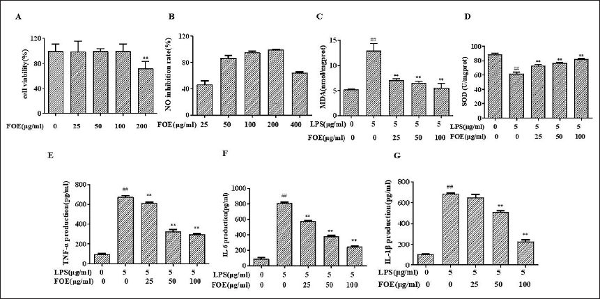
Figure 6 additionally indicated that the contents of MDA in the mannequin crew had been notably accelerated in contrast with the management group, whilst the pastime of SOD was once notably decreased; After FOE administration, the expressions of MDA decreased, whilst the exercise of SOD extended. In contrast with the LPS group, 100 µg mL−1 FOE had the most obvious effect. These effects advised that LPS can promote the oxidative stress response of RAW264.7 macrophages, and FOE can significantly inhibit the oxidative stress induced by LPS.
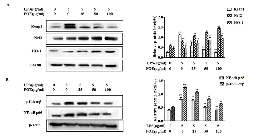
FOE Exerted Anti-Inflammatory Consequences by Using Nrf2 and NF-κB Pathway in vitro
The role of Western blotting confirmed that contrasted with the manipulate group, the relative expressions of Keap1, p-NF-κB, and p-IKKα/β in the mannequin crew had been substantially increased. After being dealt with FOE for 24 h, the relative expressions of p-IKK α/β, p-NF-κB, and Keap1 in 100 and 50 µg mL−1 companies had been substantially diminished in contrast with these in the mannequin crew, while the change of Nrf2 and HO-1 presented as a contrary tendency (Figure 7).
Discussion
In current years, applicable research has proved that immune elements are the essential purpose of the incidence and improvement of UC. The pathogenesis of UC involves the whole process of immune response (Wan et al., 2022; Wang et al., 2019a). In this study, we show that FOE has a therapeutic effect on DSS-induced UC, and the relationship between the changes of IKK/IκB/NF-κB and Nrf2/HO-1 proteins was explored, laying the experimental foundation to clarifying the biological mechanism of FOE in curing UC (Liu et al., 2022a, b). p50/p65 heterodimer is the most common form of NF-κB activation. Under physiological conditions, NF-κB binds to its inhibitor IκB.
If cells are affected by external stimuli, the IκB kinase complex (IKKs) is activated to phosphorylation and ubiquitination of IκB, leading to the separation of NF-κB and IκB into the nucleus and specific binding of corresponding target genes, inducing the expression of related genes (Wei et al., 2021). Promotes the release of large amounts of inflammatory factors. In addition, cytokines such as TNF-α can also be activated by phosphorylation of IKK by NF-κB-induced kinase, which in turn phosphorylates IκB, thereby activating NF-κB and leading to inflammatory signal amplification. The results have proven that NF-κB is exceptionally expressed in UC and induces massive expressions of pro-inflammatory factors, which play a vital position in UC (Bing et al., 2017; Chen et al., 2020; Wang et al., 2019). The IKK/IκB/NF-κB proteins are intently associated with the improvement and development of UC, and might also be a doable goal for the remedy of UC.
Recent research has located that oxidative stress overactivation is intently associated with the pathogenesis of UC. Under the regular physiological state, oxidation and anti-oxidation of the physique are in a relative stability country (Chen et al., 2021). Under the action of external baleful stimulating factors, the excessive activation of reactive oxygen free radicals in the body leads to the disorder of cell structure and function, Organism tissue damage, and ultimately induces and aggravates UC (Arab et al., 2021). In current years, Oxidative stress over activation in intestinal mucosa can injure the intestinal mucosal barrier. The mechanical barrier shaped by using the shut connection between epithelial cells is of utmost significance (Liu et al., 2021). Nrf2 is a central regulator of keeping intracellular redox homeostasis and one of the key cytokines concerned with oxidative stress response, which can reply to exogenous harm and pathogen molecules (Arab et al., 2021; Zhang et al., 2021; Zhu et al., 2021). Keap1 is a negative regulator of Nrf2. In the physiological state, Nrf2 mostly exists in the cytoplasm and binds with Keap1, which is inhibited. When stimulated by oxidative stress signals in the body, it is rapidly dissociated from Keap1 and transferred into the nucleus in a steady-state form, starting antioxidant responsive element (ARE) and combining with it to initiate transcription and translation of downstream multiple genes involving CAT, SOD, GSH, and HO-1. SOD activity and MDA level are important indexes reflecting the severity of oxidative damage (Huang et al., 2020; Wang et al., 2019a, b). In this way, the expression of antioxidants can be up-regulated to reduce the oxidative stress response caused by various injury factors. Nrf2/HO-1 pathway is one of the primary antioxidant stress response pathways in the body and is involved in many aspects of the tumor, apoptosis, inflammatory response, and mucosal repair.
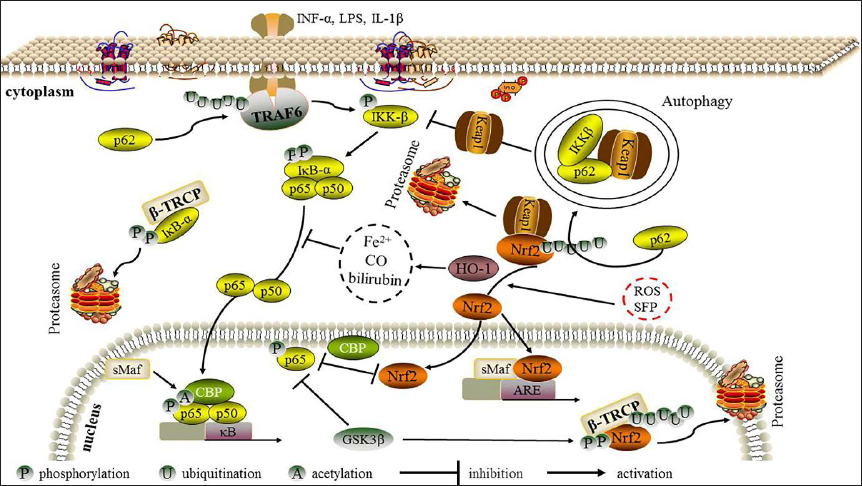
In recent years, both positive and negative regulation of NF-κB and Nrf2 pathways have been demonstrated many times, mainly involving two pathways. On the one hand, Nrf2 down-regulates pro-inflammatory factors such as IL-6 and TNF-α, reduces upstream activator of NF-κB and down-regulates NF-κB expression by transcription of downstream target genes of antioxidants (Fusco et al., 2021; Park et al., 2021; Van et al., 2020). The molecular mechanism of NF-κB and Nrf2 signaling interaction in UC is shown in Figure 8. This finds out about suggests that the essential objectives FOE acts on UC primarily based on NF-κB and Nrf2/HO-1 signaling crosstalk can be similarly explored in the future. Combined with the molecular mechanism of regulating the occurrence, it will in addition enlarge the medical utility of TCM in the prevention and cure of UC and contribute to the research and development of UC prevention and treatment drugs. This paper elucidated the therapeutic effect and mechanism of extract from FOE in the treatment of UC. Further studies are needed to check out the particular fantastic chemical compounds in the extract and determine the specific anti-inflammatory properties derived from the extracts of FOE.
Conclusion
The outcomes of this finding show that FOE plays a defensive position in DSS-induced colitis and LPS-stimulated RAW264.7 cells by way of inhibiting infection and oxidative stress. Additionally, Nrf2 used to be imperative in the development of colitis ‘to’ The Nrf2 signaling pathway is activated at the same time when colitis occurs. The current learn affords new reference natural medicine into the curative approach of colitis, and FOE would possibly be an achievable drug for the cure of colitis.
Footnotes
Summary
FOE can play a protective role against DSS-induced colitis and LPS-stimulated RAW264.7 cells by way of inhibiting infection and oxidative stress.
FOE exerts its anti-inflammatory effects through the NF-κB/Nrf2 signaling pathways.
FOE would possibly be an achievable drug for the remedy of colitis.
Abbreviations
FOE: The extract from fruits of Rosa odorata Sweet var. gigantea (Coll. et Hemsl.) Rehd. et Wils; UC: Ulcerative colitis; DSS: Dextran sulfate sodium; CD: Crohns’ disease; IBD, Inflammatory bowel disease; ARE: antioxidant responsive element.
Acknowledgment
National Natural Science Foundation of China and the National Key R&D Program of China for financial support.
Declaration of Conflicting Interests
The authors declared no potential conflicts of interest with respect to the research, authorship, and/or publication of this article.
Ethical Approval
The animal use protocol has been reviewed and approved by the Animal Ethical and Welfare Committee (AEWC). Approval No. IRM-DWLL-2020150
Informed Consent
Not applicable.
Funding
The work was supported by the National Natural Science Foundation of China (81673693) and the National Key R&D Program of China (2017YFD0201402) for financial support.