
Abstract
Determination of age of an individual for medicolegal purpose in civil and criminal cases mostly depends upon radiological data of appearance and fusion of ossifications centers. The X-ray of wrist is important in this regard to determine age in early adolescent age group. This study has been aimed to determine age of appearance and fusion of ossification centers around wrist joint for known aged person among Bengali population and compare them with standard age. Radiological films of wrist joint of 483 individuals (373 male and 110 females) have been studied in retrospective way. Subjects came for age estimation between the period 1 January 2017 to 31 December 2019. Data analyzed in Microsoft excel 10 and presented in the form of tables. The study revealed age of appearance and fusion of Pisiform, lower end of Radius, and lower end of Ulna in Bengali population which corroborate with other studies in some cases and do not match with some studies. This study will help to determine age of early adolescent individuals in Bengali population which vary from existing standard data to some extent due to various factors like nutritional status, environmental condition, food habits and socioeconomic status.
Introduction
Identification of human being is one of the most important aspects of criminal investigation. Estimation of age is one parameter of identification process and is also essential in various civil cases when actual age of a person is challenged because of insufficient or forged documents. Age can be estimated from skeletal remains of dead induvial and from radiological and dental data of both living and dead. Mostly, in children and adolescents, skeletal age and chronological age tally with each other. 1 In this part of the country, age estimation is often termed as “Ossification Test” by judiciary system as age of appearance and fusion of ossification centers of long bones around major joints of human body is considered to be reliable though it varies with different nutritional, developmental, and endocrinal factors which affect skeletal growth. 2
Notable works on the determination of age from such appearance and fusion of epiphyses in the long bones around different joints of the body namely elbow, wrist, pelvis, knee, shoulder, ankle, and others have been done in different parts of India and abroad. It is evident that there is significant difference in the age of epiphyseal union in different population even within a country. 3 Ossification centers around wrist joint are helpful mostly in determination of age among children and adolescents. 4 In this study, we have examined age of appearance and fusion of ossification centers around wrist joint like lower end of Radius and lower end of Ulna and also age of appearance of Pisiform bone. Three-hundred seventy three male and 110 female subjects have been examined between the age group of 7 and 21 years. Our objectives are to observe status of appearance and fusion of ossification centers around wrist for a known chronological age, to determine range of their appearance and fusion in males and females and to compare them with known standards specifically those mentioned in Galstaun chart. 5
Materials and Methods
It was a cross-sectional study done on 483 individuals (373 males and 110 females) who came for radiological age estimation in the Dept. of Forensic Medicine & Toxicology of Medical College Kolkata between 1 January 2017 and 31 December 2019. All cases within the mentioned time period were examined in a census sampling except those who fell under exclusion criteria, that is, those who did not belong to Bengali population, whose stated ages were not known and who had fracture or pathology of concerned bones. Lower end of Radius and Ulna were examined in respect to their status of appearance and fusion. Presence or absence of Pisiform bone was examined. Stated age was confirmed from age proof documents. Ossification centers of concerned bony structures were examined to observe Whether appeared Whether fusion not started Whether fusion started and in process Whether fused
The epiphyseal scar at the junction of epiphysis and metaphysis has been considered as indicator of complete fusion. The skiagrams examination and evaluation were performed by two observers blindly and independently without knowing about age or sex. The data were put in an excel sheet and the parameters were examined statistically in relation to their stated age and age of their appearance and fusion were determined.
Result
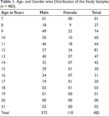
Age- and Gender-wise Distribution of the Study Samples (n = 483).
Appearance of Pisiform
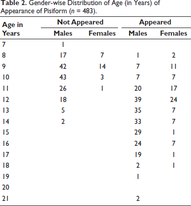
In females, Pisiform appeared in 77.27% of total population. Pisiform appeared in all subjects between 12 and 18 years of age, whereas 59.68% of female population showed appearance of Pisiform between 8 and 11 years age group. In males, 62.94% subjects showed appearance of Pisiform and all subjects between 15 and 21 years age group had the Pisiform bone on X-ray. Appearance started from eight years age. Between 8 years and 14 years of age, only 48.13% male population showed appearance of Pisiform (Table 2).
Gender-wise Distribution of Age (in Years) of Appearance of Pisiform (n = 483).
Appearance of Lower End of Radius
Lower end of Radius appeared in all 373 male subjects and 110 female subjects.
Fusion of Lower End of Radius
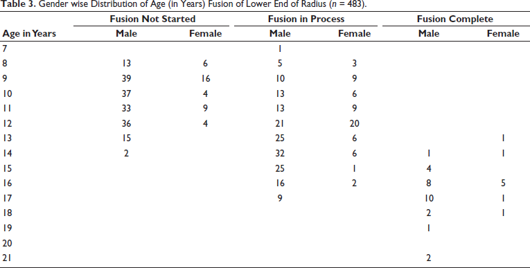
Among females, only nine (8.18%) aged between 13 and 18 years showed complete fusion of lower end of Radius. Lower end of Radius was in the process of fusion in 62 (56.36%) females aged between 8 and 16 years. Fusion did not start in 39 (35.45%) females aged between 8 and 12 years. One-hundred percent of female population had complete fusion above the age of 17 years and 71.43% females had complete fusion above the age of 16 years. Among males, only 37 (9.92%) subjects aged between 14 and 21 years showed complete fusion of lower end of Radius. Lower end of Radius was in the process of fusion in 170 (45.58%) males aged between 7 and 17 years. Fusion did not start in 175 (46.92%) males aged between 8 and 14 years. One-hundred percent of male population aged 18 or above had complete fusion, 52.63% in 17 years of age onward, 33.33% in 16 years of age onward, 13.79% in 15 years of age onward, and 2.86% in 14 years of age onward (Table 3).
Gender wise Distribution of Age (in Years) Fusion of Lower End of Radius (n = 483).
Appearance of Lower End of Ulna
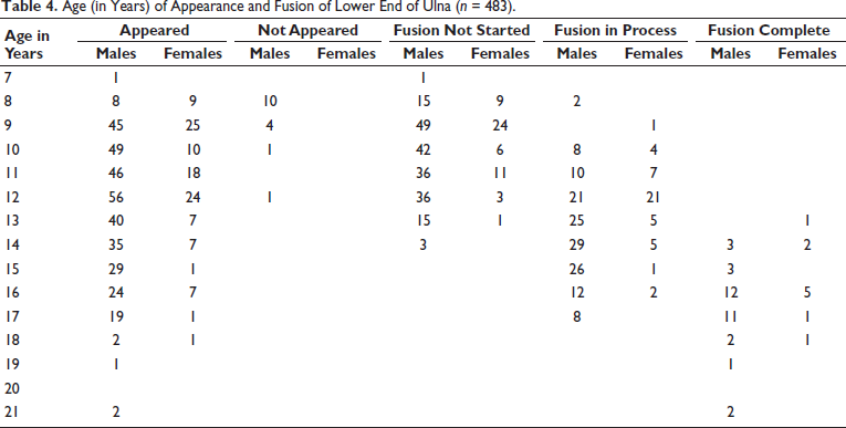
It appeared in all 110 female subjects, whereas 93.21% male population showed appearance of lower end of Ulna. One-hundred percent of male population had lower end of Ulna appeared above 13 years of age (Table 4).
Age (in Years) of Appearance and Fusion of Lower End of Ulna (n = 483).
Fusion of Lower End of Ulna
Among females, only 10 (9.09%) aged between 13 and 18 years showed complete fusion of lower end of Ulna. Lower end of Ulna was in the process of fusion in 46 (41.82%) females aged between 9 and 16 years. Fusion did not start in 54 (49.09%) females aged between 8 and 13 years. One-hundred percent of female population had complete fusion above 17 years of age and 71.43% females had complete fusion above 16 years of age, 28.57% females above 14 years of age and 14.28% females above 13 years of age. Among males, only 35 (9.38%) aged between 14 and 21 years of age showed complete fusion of lower end of Ulna. Lower end of Ulna was in the process of fusion in 141 (37.80%) males between 8 and 17 years of age group. Fusion did not start in 197 (52.82%) males between 7 and 14 years of age group. One-hundred percent of male population had complete fusion above 18 years of age, 57.89% males had complete fusion above 17 years of age, 50% males above 16 years of age, 10.34% males above 15 years of age, and 8.57% males above 14 years of age (Table 3).
Discussion
The study was aimed to guide medical professionals and law keeping authority in relation to estimation of skeletal age among Bengali population. The study was done to determine age of appearance and fusion of various ossification centers around wrist joint and to corroborate them with chronological age. Similar study has been attempted in Northern part (Punjab, Uttar Pradesh), Western Part (Rajasthan, Maharashtra), Southern part (Karnataka, Tamil Nādu), and eastern part (Manipur, West Bengal). In various foreign countries like USA, England, Australia, and Egypt, similar type of study has been carried out. In West Bengal, four studies have been conducted on this background—two in 1937, one in 1995, and one in 2013. In this study, we will compare our result among other similar studies across the country and also those took place in countries outside India. The results of different such studies are summarized in Tables 5 and 6.
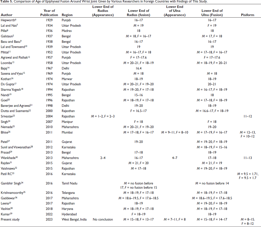
Comparison of Age of Epiphyseal Fusion Around Wrist Joint Given by Various Researchers in Foreign Countries with Findings of This Study.
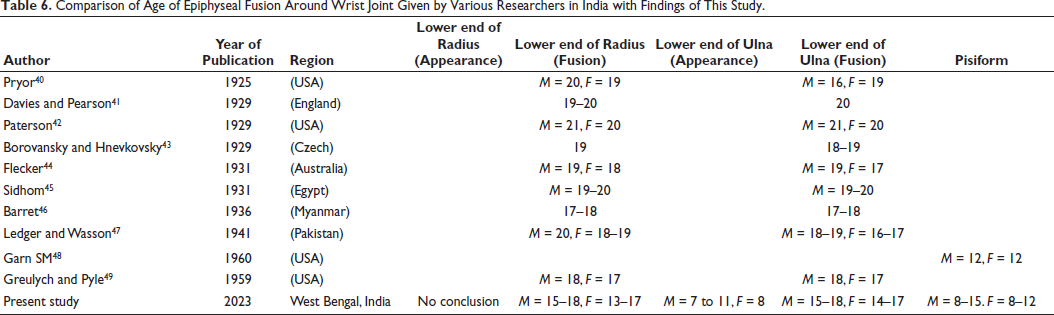
Comparison of Age of Epiphyseal Fusion Around Wrist Joint Given by Various Researchers in India with Findings of This Study.
We have studied radiological appearance and fusion of the ossification centers of bones around wrist joint including lower end of Radius, lower end of Ulna, and Pisiform. This combined and comprehensive approach have been seen in only few national and international studies as most of the studies have not included Pisiform bone. Moreover, all the earlier researchers specifically in West Bengal and other eastern parts of India have not included both male and female population while observing status of fusion of ossification centers around wrist joint together. Usually, appearance and fusion of these bony landmarks occur earlier in females as compared to males. Similar finding was observed in our study considering no female subject of seven years age was present.
According to this study, age of appearance of Pisiform bone is 8–12 years in females and 8–15 years in males. Age of appearance of Pisiform detected by other studies like Garn SM et al. (M = 12 years, F = 12 years), Srivastav et al. (11–12 years), Bhise et al. (M = 12–13 years, F = 10–12 years), Wankhade et al. (11–13 years), Patil et al. (M = 12 years, F = 1.5–12 yrs) falls within the range of age in years detected in our study.23, 26, 30, 33, 48
Regarding age of appearance of lower end of Radius, no conclusive opinion could be drawn as it appeared in all cases of both males and females.
As per this study, age of fusion of lower end of Radius is 13–17 years in female and 15–18 years in males. This finding corroborated with the findings of Wankhade et al. of Maharashtra, Bhise and Nanadkar of Mumbai, Nandi A of West Bengal, and Basu and Basu of West Bengal.9, 19, 26, 30 In few studies like Pillai et al. of Madras, Galstaun of West Bengal, Saxena and Vyas of Punjab, Dutta and Samanta of Rajasthan, and Greulych and Pyle of USA,5, 8, 15, 22, 49 findings in relation fusion of lower end of Radius were in the higher side of the age range detected by us. Finding of this study does not correspond with the findings of Lal and Nat of Uttar Pradesh, Loomba et al. of Uttar Pradesh, Sharma Yogesh of Rajasthan, Nemade of Maharashtra, Patel et al. of Gujrat, Pryor et al. of USA, Davies and Pearson of England, Flecker et al. of Australia, and some others.7, 13, 18, 25, 27, 40, 41
In this study, age of appearance of lower end of Ulna is detected as 8 years in female and 7–11 years in males. Our finding corroborated with the finding of Bhise et al. 26 of Mumbai but does not corroborate with the finding of Wankhade of Maharashtra. 30
Age of fusion of lower end of Ulna is 14–17 years in females and 15–18 years in male according to our study. This finding was mostly similar to the findings of Hapeworth et al. of Punjab, Basu and Basu of west Bengal, Mittal et al. of Uttar Pradesh, Sharma Yogesh of Rajasthan (males), Sunil and Viswanathan of Karnataka, Pryor et al. of USA (males), and Ledger and Wasson of Pakistan,6, 9, 11, 18, 28, 40, 47 whereas in studies like Galstaun of West Bengal, Mishra of Odisha, Goel et al. of Rajasthan, Barret et al. of Myanmar, and Greulych and Pyle of USA,5, 20, 46, 49 the finding was in higher end of age as shown in this study. Our finding does not match with the finding of Lal and Towsend of Uttar Pradesh, Agarwal and Pathak of Punjab, Kothari et al. of Marwar, Gupta et al. of Uttar Pradesh, Banerjee and Agarwal of Delhi, Patel et al. of Gujrat, Paterson of USA, Sidhom of Egypt, and some others.10, 12, 16, 21, 27, 42, 45
The discrepancies in findings may be due to geographical location or constitutional factors. But further extensive longitudinal study with wider age range may yield better results.
Conclusion
Only four reliable documented studies among Bengali population have been conducted in 1937, 1938, 1995, and 2013. Most of the studies did not include Pisiform bone and both genders separately. An in-depth study was required for determining a reliable reference range of age in the present scenario. This study is an attempt toward that goal. Radiological analysis of appearance and fusion of ossification centers around wrist joint can be used for estimation of age in Bengali population in future taking this study findings as reference.
Footnotes
Declaration of Conflicting Interests
The authors declared no potential conflicts of interest with respect to the research, authorship, and/or publication of this article.
Ethical Approval
A prior approval was obtained from the Institutional Ethics Committee.
Funding
The authors received no financial support for the research, authorship, and/or publication of this article.