
Abstract
Age estimation is mainly used for identification of unknown individuals in large-scale disasters, civil and criminal crimes, illegal immigration, child marriage, and juvenile cases. In many medico-legal cases, the age of an individual needs to be determined using population-specific reference criteria. Numerous techniques have been employed for estimating age, which involves morphological, histological, radiographical, and biological methods. The objective of the present study was to check the applicability of the cervical vertebrae maturation (CVM) method on the North Indian children population. A total of 500 lateral cephalograms (251 male subjects and 249 female subjects) were evaluated. The correlation between skeletal maturation age and chronological age of 6–16 years subjects was established by Spearman rank correlation. The Spearman’s rank correlation between age and skeletal maturation stages was estimated for both sexes (0.86 for males and 0.91 for females) and was statistically significant with P values (<.001). The method has been accepted scientifically and effectively used on the North Indian children population in settling civil or criminal cases.
Keywords
List of Abbreviations
CA, chronological age; CS, cervical stage; CVM, cervical vertebrae maturation; D.O.B, date of birth; M.O.E, margin of error; SA, skeletal age; SD, standard deviation; SGT, Shree Guru Gobind Singh Tricentenary (SGT) University.
Introduction
Forensic age estimation in living individuals has significantly increased in implication over the years due to the necessities for an expert opinion on the age of an individual. Forensic anthropologists play an important role in the assessment of both legal and social categorization. It is mainly required for the civil and criminal matters, such as identification of unknown bodies in mass disasters, airplane crashes, terrorist attacks, illegal migration, child marriages, etc. 1 Numerous techniques have been employed for estimating age, which involves morphological, histological, radiographical, and biological methods. 2 These methods include dental and skeletal developmental stages to estimate the age more accurately and precisely. 3 Estimation of age of an unknown individual is of prime importance in the daily practice of police officials and other law enforcement. 4 Skeletal age has been determined by examining digital radiographs of hand and wrist, but over time, there were certain approaches which have been proposed to estimate the age from different bones. Chronological age is commonly used to assess the individual’s growth trajectory, but it is not preferably considered for sex differences in pubertal growth, timing, length, or even the extent of degree of puberty. 5 Children differ in physical maturity at specific chronological ages. Skeletal maturation may be accelerated or delayed. Estimation of age is quite difficult after an individual attains the age of 14 years from teeth because all other teeth have completed their development, but only the third molar renders the development when it comes to estimating the age. 6 Radiographic maturity of the cervical vertebrae has recently become an area of interest for age determination. Hassel and Farman developed six different stages to assess cervical maturity and were further modified by Baccetti et al., 2005. 7 As this method was based on the morphological structure and presence of concavity of the cervical spine margins over time, the modified method was commonly known as the cervical vertebrae maturation (CVM) method. This method includes assessment of cervical vertebrae 2, 3, and 4 on digital radiographs. In addition, relative maturation of the cervical vertebrae was noted, and mandibular apex growth was also documented. 8 In terms of medical viewpoint, a single parameter is neither accurate nor reliable for age estimation on a single population group, hence there is a need to standardize the method for a particular ethnic group. 9
The standardized growth stages can be evaluated using cervical vertebrae radiographs and correlated with dental age for checking the validation of the method in the specified population. 10 Chandrasekar et al., 2020, have stated that the CVM method was validated and reliable for determining cervical vertebrae bone age and skeletal maturation stages from lateral cephalograms for Asian South Indian patients. 11
The objective of this study was to evaluate the relationship between chronological age and cervical maturity stages and to validate the CVM method among North Indian children of both sexes.
Materials and Methods
Sample Selection
The present study is a retrospective radiographic study, conducted on digital radiographs. The lateral cephalograms were collected from the Department of Oral Medicine, Radiology and Orthodontics, SGT University and Hospital, Gurugram, India.
Inclusion and Exclusion Criteria
The radiographs of 500 subjects (251 males and 249 females) with ages between 6 and 16 years were included with a known D.O.B and known date of the radiography. The radiographs were in digital form with good resolution, and were included.
The subjects with whom the radiographs belonged were from different states or nations, and were excluded. Study participants with systemic illnesses or genetic abnormalities were disqualified.
Skeletal Maturity Assessment
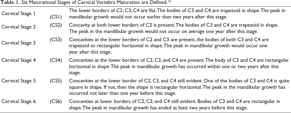
The CVM approach, which Baccetti et al., 2005 developed, was applied to determine skeletal maturity. Six separate maturation phases (CS1–CS6) build the CVM method, according to distinct structural features of cervical vertebrae 2, 3, and 4. The method was evaluated on two broad parameters: (a) presence or absence of concavity of lower border of cervical vertebrae 2, 3, and 4, (b) shape of body of cervical vertebrae 3 and 4. The preliminary step is to analyse the inferior border of the body, in order to determine whether the body is flat or concave (i.e., presence of notch). The second follow-up stage is to analyse the shape of cervical vertebrae 3 and 4 (CV3 and CV4). These morphological characteristics show the typical sequence, from progressing trapezoid to rectangular to square and to rectangular vertical as summarized in Table 1.
Six Maturational Stages of Cervical Vertebra Maturation are Defined. 13
Statistical Analysis
The statistical analysis was performed by using IBM SPSS software, version 22. The radiographs of lateral cephalograms were evaluated by two different examiners. To assess inter-examiner reliability, the Kappa statistic was used. Descriptive statistics were obtained by calculating the chronological ages for all six stages of cervical vertebrae. The Spearman rank order correlation coefficient was used to determine the relationship between chronological age and CVM stages separately for males and females. P value equal to or less than .05 was taken as statistically significant. In addition, sensitivity and accuracy analysis were performed in order to determine how variables affect each other under a set of assumptions. The age confidence interval of −95% and +95% was also assessed among all six CVM stages: CS1, CS2, CS3, CS4, CS5, and CS6. 12
Results
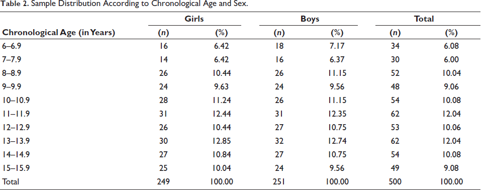
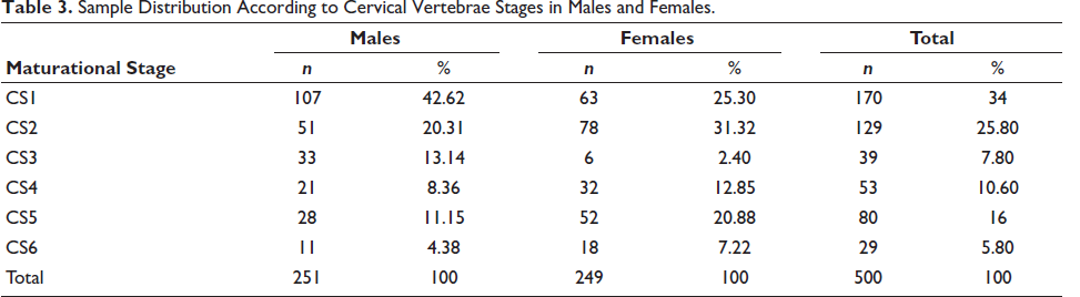
This present study is conducted on lateral cephalograms of 500 North Indian children, comprising 251 males and 249 females. Table 2 depicted the distribution of the entire sample according to age and sex. Table 3 showed the most frequent cervical stages among males and females. In females, the most frequent stage was CS2 (31.32%), followed by CS1 (25.30%) and CS5 (20.88%), followed by stages CS4 (12.85%), CS6 (7.22%) and CS3 (2.40%) however, among males the most frequently stage was CS1 (42.62%), followed by CS2 (20.31%) and CS3 (13.14%) followed by stages CS5 (11.15%), CS4 (8.36%), and CS6 (4.38%), respectively.
Sample Distribution According to Chronological Age and Sex.
Sample Distribution According to Cervical Vertebrae Stages in Males and Females.
The frequency distribution of each cervical vertebral maturation is shown separately for males and females in Table 4. In female subjects, the mean chronological age in CS1 was (8.62 + 0.9 years), CS2 was (14.09 + 0.4 years), CS3 was (13.12 + 2.7 years), CS4 was (14.80 + 1.6 years), CS5 was (11.72 + 0.9 years) and CS6 was (16.14 + 1.8 years). In males, CS1 was (8.72 + 1.6 years), CS2 was (14.09 + 0.4 years), CS3 was (13.21 + 0.07 years), CS4 was (14.80 + 1.4 years), CS5 was (12.54 + 1.1 years), and CS6 was (15.57 + 1.0 years).
Correlation Between Chronological Age and Cervical Vertebrae Stages Among Males and Females.
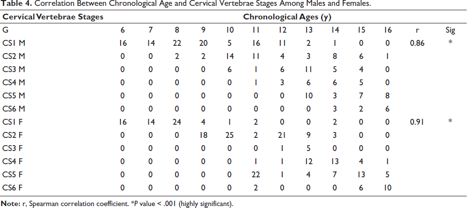
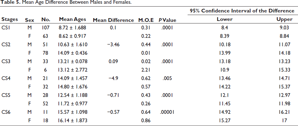
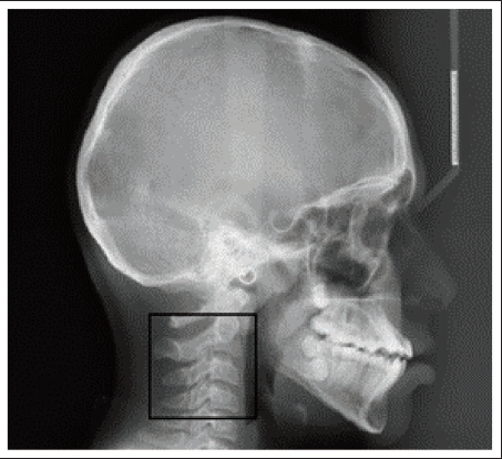
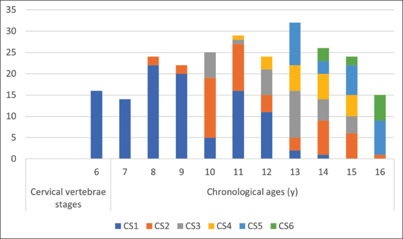
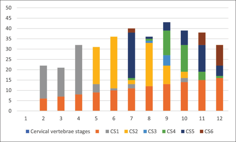
The digital panoramic view is illustrated in Figure 1. The sex dimorphism of the chronological age in each cervical vertebral maturation is shown in Figures 2 and 3. Table 5 represented the mean age of males and females according to cervical vertebral maturation stages. CS1 stage was most frequent in six to nine years, CS2 in 9–11 years, CS3 in 11–13 years, CS4 in 13–15 years, and CS6 was the most frequent in 15–16 years. The Spearman rank correlation between chronological age and cervical vertebral maturation stages was estimated for both the sexes (0.91 for males and 0.86 for females), and it was statistically significant at P value (<.001). The skeletal maturity stage: C2 was found to be more advanced among female subjects as compared to male subjects. CS5 and CS6 stages and mean chronological age of females in each cervical vertebral maturation stage was less than males, except CS6. The differences found in mean age in males and females were statistically significant in all CS stages. Inter-examiner agreement was found to be reliable and higher. The mean standard error of inter-examiner was 0.12 for females and 0.10 for males.
Mean Age Difference Between Males and Females.
Digital Panoramic View of Lateral Cephalogram of C2, C3, and C4.
Distribution of Chronological Age of Female Subjects According to Each Cervical Vertebral Stage.
Distribution of Chronological Age of Male Subjects According to Each Cervical Vertebral Stage.
Discussion
It has been observed from previous studies that children’s growth differs greatly, and thus, chronological age is a poor predictor of maturity. In recent years, the cervical vertebral maturation method has proven the validity and the ability to identify somatic maturation with respect to mandibular maturity, which is of great importance in dentofacial orthopedics. Several methods based on radiographic assessments, histological methods, physiological methods, and methods have been employed to determine skeletal maturity age among different population groups. 13
Baccetti et al. (2005) modified the original Hassel and Farman CVM method. This method was adopted in the present study because of its wide use in the current literature and demonstrated applicability among several population groups. 14 Sierra et al. found that the relationships between chronological age and cervical vertebral maturation assessment proved to have high correlation (0.58–0.79). 12 In the present study, high correlation was also found between chronological age and skeletal maturation method in both sexes (0.91 for females and 0.86 for males), respectively. The present study agreed with Al Hadlaq et al. (2007) (r value = 0.82 for males and 0.89 for females), respectively, but it is quite higher as compared to Sierra et al. (1987) and Ghulam et al. (2010) (0.69 for females and 0.52 for males). 15 The differences found can be attributed to different ethnic backgrounds, nutritional factors, environmental conditions, and research methodology associated with sample population size and sample distribution. Moreover, the growth potential may also vary according to different population groups, ethnic groups, and even among identical twins.
The high correlation values between chronological age and cervical vertebral maturation stages imply valuable clinical reliability in estimating skeletal maturity in orthodontics. In addition, the method is appropriate from the forensic point of view, for assessing the age of an individual whose birth record is not available.
The present study shows comparative stages of CVM stages in both sexes. It is apparent from the results that their mean chronological age increases as the cervical vertebrae show evident maturation in both groups. The skeletal development stage is more advanced among female subjects as compared to males. This finding was supported by previous studies conducted by Tanner et al. in 1992 and Hunter et al. in 1966. However, no evidence was found for the earliest CVM in either the sex in the study conducted by Montasser et al. among the Caucasian population and Thirupathi Reddy et al. among the South Indian population. 16
The value for different population was moderate/high due to the presence of errors occurring in most of the conducted studies, such as lack of systematic procedure performed for data collection, distinct age groups, and different methodology used for the analysis of cervical vertebrae growth pattern. These factors can infer the results of the research. 17 There are certain factors that might influence an individual’s body growth and maturation, such as genetic conditions, nutritional status, hormonal imbalance, hereditary disorders, and environmental influence. 18 Montasser et al. strongly recommended that racial background and sex differences should be considered when using the cervical vertebral maturation method. 16 However, this could not be a prime issue about the quality of the research. The selective inclusion criteria and detailed analysis of the radiograph leads to a useful method for age estimation. The study conducted by Heravi et al. showed different correlations of four different methods of cervical vertebrae and Hand wrist method formulated by Tanner-Whitehouse. 19 The parameters such as accuracy, correlation, as well as reproducibility of the proposed method can be influenced by the distinct analytical method. 20 Moreover, it is a good approach to study cervical vertebrae development phases by including qualitative analysis of morphology of cervical vertebrae, depth of concavity, ratios of height and width of CS 2, CS3, and CS4. 21 The outcome of the conclusion suggests that North Indian females show a maturation rate early as compared to males.
The appearance of definite concavity of the second vertebral body from the lateral cephalogram, evident the commencement of the rapid growth. The present study showed the concavity of C2 presents at a mean chronological age of 10.63 ± 1.610 in males and 14.09 ± 0.436 in females. The appearance of a visible concavity at the lower border of C3 identifies the stage immediately preceding the peak of growth (Baccetti et al., 2005). In the present study, it is noticed at 13.12 + 2.7 years in females and 13.21 + 0.07 years in males.
Conclusions
Skeletal maturity indicators play a significant role in an individual’s growth trajectory as compared to its chronological age. The lateral cephalograms were utilized in this study and proven to be a reliable radiological source of age estimation in the age group of 6–16 years. Hence, the CVM method could be applicable on the North Indian children population. To determine the precision in age estimation, CVM method could be better employed with other indices using Artificial Intelligence algorithms.
Footnotes
Acknowledgements
Authors are grateful to the Department of Oral Medicine, Radiology and Orthodontics, SGT University and Hospital, Gurugram, India, for providing the retrospective data (lateral cephalograms) and helping in analysing radiographs.
Author’s Contribution
RKK: Data collection, evaluated the radiographic staging, and original draft writing.
MA: Data interpretation, statistical analysis.
PPM: Supervision, validation, and writing-editing.
The authors read and approved the final manuscript.
Declaration of Conflicting Interest
The authors declared no potential conflicts of interest with respect to the research, authorship, and/or publication of this article.
Ethical Approval
The study was approved by the Ethical Research Committee of Amity University, Noida, Uttar Pradesh, India, and Ethical Clearance Certificate was obtained from the Institute having Reference Number: AUUP/IEC/2021/19 dated 25.05.2022.
Funding
The authors received no financial support for the research, authorship, and/or publication of this article.
Informed Consent
Not applicable.