
Abstract
Retinal organoid (RO) is the three-dimensional (3D) retinal culture derived from pluripotent or embryonic stem cells which recapitulates organ functions, which was a revolutionary milestone in stem cell technology. The purpose of this study is to explore the hotspots and future directions on ROs, as well as to better understand the fields of greatest research opportunities. Eligible publications related to RO from 2011 to 2022 were acquired from the Web of Science (WoS) Core Collection database. Bibliometric analysis was performed by using software including VOSviewer, CiteSpace, and ArcGIS. A total of 520 articles were included, and the number of annual publications showed a rapid increase with an average rate of 40.86%. The United States published the most articles (241/520, 46.35%) with highest total citation frequencies (5,344). University College London (UK) contributed the largest publication output (40/520, 7.69%) and received highest total citation frequencies. Investigative Ophthalmology & Visual Science was the most productive journal with 129 articles. Majlinda Lako contributed the most research with 32 articles, while Olivier Goureau has the strongest collaboration work. Research could be subdivided into four keyword clusters: “culture and differentiation,” “morphogenesis and modeling,” “gene therapy,” and “transplantation and visual restoration,” and evolution of keywords was identified. Last decade has witnessed the huge progress in the field of RO, which is a young and promising research area with extensive and in-depth studies. More attention should be paid to RO-related models and therapies based on specific retinal diseases, especially inherited retinopathies.
Introduction
The term organoid was first reported in 1946, which refers to the growth pattern of teratoma parallel to internal organs, or a morphological description of immature organ in the early stage of fetal development 1 . After continuous exploration of self-organization and organoid methodologies throughout more than a century, organoids are basically defined as a three-dimensional (3D) collection of cell types with organ specialty in vitro produced from stem cells or organ progenitors, and recapitulats self-organization through the process of cell sorting out as well as spatially constrained lineage commitment 2 .
Actually, the authentic origin of organoids could be traced back to 1907, when Wilson discovered the self-organization of dissociated sponge cells to form a whole regenerated organism 3 . Afterward, in the mid-20th century, researchers utilized single-cell suspensions from dissociated chick embryos to decipher their ability to reaggregate and recapitulate typical histogenesis in vitro culture4,5. Furthermore, the pattern of cell sorting out could be explained by Steinberg’s differential adhesion hypothesis (DAH) in the 1960s. Undoubtedly, the development of stem cell technology has injected new impetus into organoid research. It stemmed from the end of 20th century, and the most important events throughout these years including the establishment of mouse pluripotent cell lines in 19816,7, first human embryonic stem cell (ESC) line in 1998 8 , and first human-induced pluripotent stem cell (hiPSC) technology in 20079,10, opening up new avenues for the generation of different organoids (gut 11 , retinal 12 , brain 13 , kidney14,15, etc.) which has emerged as an immense breakthrough over the past decade.
The vertebrate retina is a complicated stratified structure endowed with various types of component cells to convert light energy into nerve impulses to form visual perception. During prenatal development of the eye, forebrain and bilateral neural folds derived from the neuroectoderm invaginate to form optic sulci, and optic sulci continues to sink deeper into the epidermal ectoderm and forms a chamber called optic vesicle (OV), and subsequently moves inward to form a double-layer cup-like structure, the optic cup, which develops into outer layer (pigment epithelium) and inner layer (neuroepithelium) of the retina.
In 2011, Yoshiki Sasai’s group established the first in vitro retinal organoid (RO) model by virtue of a 3D mouse ESC culture system, where the optic cup (retinal primordium) structure was formed spontaneously, and the interkinetic nuclear migration (INM) as well as differentiation were similar to those in vivo 12 . It was a revolutionary milestone in stem cell technology, for it promised a future access to generating specified and staged photoreceptors for transplantation therapy. Furthermore, they established optic-cup organoid model from human ESCs that was much larger than mouse ESC–derived one 16 . The morphology and cone differentiation were also different between two models in the developmental course of neural retina, presumably owing to species differences 16 . By optimizing the protocols generated by Sasai’s group, several groups have endeavored to perform functional studies by utilizing iPSC-derived organoids. For example, photoreceptors derived from hiPSC were observed to differentiate at an advanced level including detection of photosensitivity and outer-segment formation 17 . Researchers demonstrated the integrative and matured capability of ESC-derived photoreceptor cells to form outer segments (OSs) as well as synaptic connections after transplantation 18 . However, there is still a huge gap between conventional culture of RO and large-scale production for applications such as modeling, drug screening, and transplantation therapy, for the limitations lying in tedious culture cycles, significant heterogenicity under different conditions, as well as lack of vasculature and interplay connections between cell layers. This emphasizes the urgency of circumventing these bottlenecks for successful translation of RO.
During the decade that RO research has taken off, it is of great significance to obtain an overall landscape of the studies associated with ROs to explore the hotspots and future directions for translation of basic research to clinical setting, as well as to better understand the fields of greatest research opportunities. As the quantitative science of publications, bibliometrics are mostly utilized to measure the impact or value of research articles 19 . Nevertheless, to our knowledge, the quantitative study of RO-related research has not yet been reported. Herein, we undertook a bibliometric analysis of the literature on RO based on the Web of Science (WoS) database from 2011 to 2022. Moreover, we used ArcGIS, VOSviewer, and CiteSpace to visualize the topics related to RO in the form of bibliometric coupling and co-occurrence maps.
In this context, the purposes of this study are
To evaluate the research hotspots and publication trends in the field of RO by bibliometric analysis.
To reveal the collaborative patterns between different countries/regions, institutions, journals, and authors on RO-related research.
To identify and analyze the most cited keywords with the strongest citation bursts.
To navigate the areas of greatest potential and opportunity for research.
Materials and Methods
Ethics Statement
This research was built upon published papers and books which did not involve human or animal experiments. As a result, this study did not require ethical approval.
Data Source and Search Strategy
We selected eligible articles related to RO from the WoS Core Collection, which is considered as the most authoritative, reliable, and comprehensive database for scientific exploration as well as appraisal 20 . Detailed publication information involves literature, authors, publication time, journals, countries/regions, and so on. In view of fact that in the early studies, researchers did not systematically define ROs, but describe it through its morphological identity (e.g., 3D retinal tissue induced by ESC or PSC), we optimized the retrieval formula to cut down the missed articles relevant to our topic, which was designed as follows: TS (Topic) = (((“pluripotent stem cells” OR “embryonic stem cells”) AND (“retina” OR “retinal”) AND “three-dimensional”) OR (“retinal organoids”) OR ((“organoids” OR “organoid”) AND “retina”) OR (“retinal organoid”)). The scope of document type includes original article, review article, and proceeding. The timespan was from January 1, 2011 to December 31, 2022. The download date was February 18, 2023. The language was English. In total, 573 valid literature in the RO research were acquired. Detailed process of identification and screening is shown in Fig. 1.

Flow diagram of the retrieval process. WoS: Web of Science.
Data Collection
We extracted all the records of correlative publications, including titles, author names, keywords, abstracts, publication time, countries and regions, journals, and so on, from Science Citation Index (SCI) Expanded database, and then inputted them into VOSviewer version 1.6.18 for subsequent analysis and presentation. The inclusion criteria were displayed as follows: (1) the papers focused on the topic of RO; (2) the document types are Original Article, Review Article, and Proceeding; and (3) the papers must be written in English. And the exclusion criteria were shown as follows: (1) the papers are not related to the research of RO, and (2) the papers do not meet the expected document types. Finally, 520 papers were selected for the bibliometric analysis.
Bibliometric Analysis
Bibliometric analysis functions as a statistic tool to present an overview of research area, which is underpinned by quantitative analysis of a myriad of literature 21 . Data including authors, article titles, sources, document types, keywords, abstract, and so on were extracted into Microsoft Excel 2019 for data processing and analysis. Relative research interest (RRI) is an index defined as the proportion of publication counts in a certain field to all-field publication counts every year 22 . The Hirsch index (H-index) was a bibliometric indicator of the scientific impact or academic productivity of individuals, meaning that a researcher has published h number of articles and each of them has been cited at least h times 23 .
Visualized Analysis
As a powerful and user-friendly tool, VOSviewer exerts its advantages in constructing and displaying bibliometric maps to better undertake co-occurrence and co-citation analysis 24 . In our study, VOSviewer was utilized to construct networks between journals, authors, countries/regions, and publications, based on co-citations and co-authorship relationships, co-occurrence of keywords, and bibliometric couplings. CiteSpace, developed by Professor Chaomei Chen (College of Information Science and Technology, Drexel University, Philadelphia, USA) 25 , is a potent visualization tool for identifying burst keywords and anticipating the possible research trends and frontiers in the foreseeable future, which is also used in our study. Besides, ArcGIS was utilized to show the geospatial distribution of total publications on RO.
Results
Global Publications
According to the paradigm of retrieval process, 520 articles were selected for the analysis, including 307 original articles, 136 proceedings, and 77 review articles. From 2011 to 2022, the number of annual publications rose sharply from 2017 (n = 24, 4.62%) to 2018 (n = 56, 10.77%), and reached its peak in 2022 (n = 132, 25.14%) (Fig. 2A). Furthermore, the fitting curve of global trends in RO was Y = 11.972 × X − 24098 (R2 = 0.8793), indicating that the number of global publications in the future might increase at a relatively stable rate (Fig. 2B). RRI in the RO field was of sustained growth, and increased rapidly between 2017 and 2018. To explore the most impactful articles and related research interest throughout the years, Table 1 summarizes the top 10 most cited articles over these years, of which there were 9 original articles and 1 review article.

Global publications. (A) Trends of global publications and relative research interest. (B) Fitting curve of global publications.
Top 10 Most Cited Articles Published From 2011 to 2022.
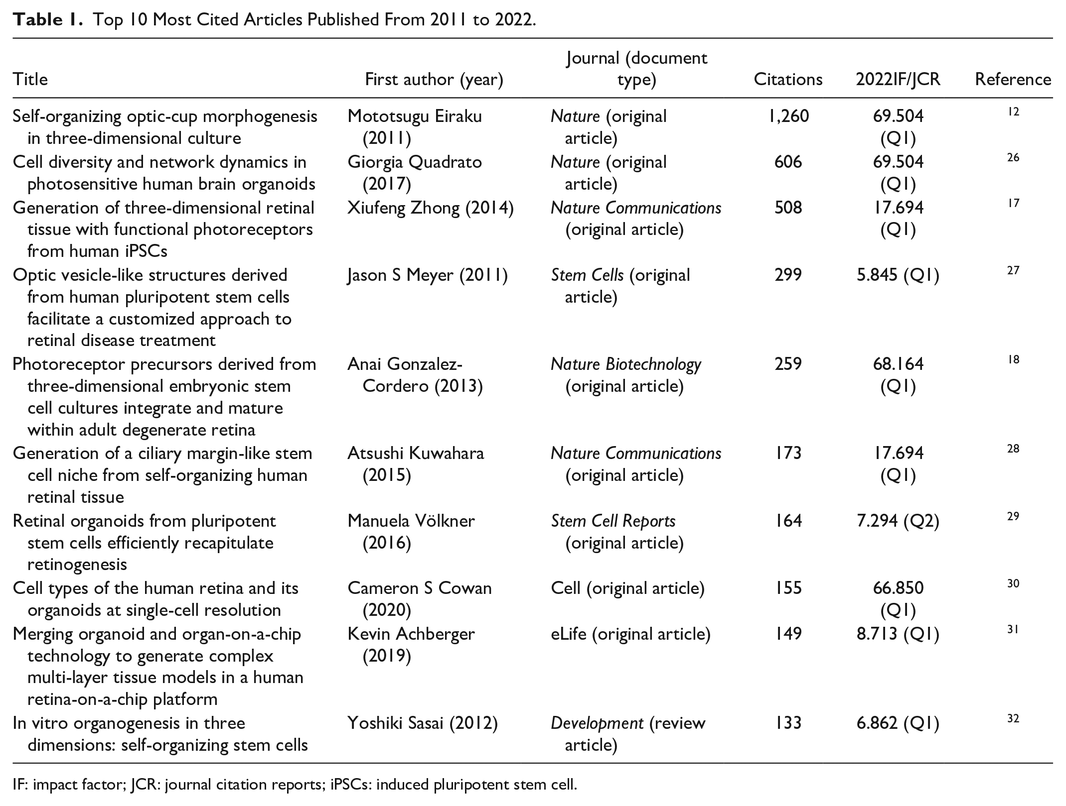
IF: impact factor; JCR: journal citation reports; iPSCs: induced pluripotent stem cell.
Publication Analysis of Countries/Regions and Institutions
According to the results of the WoS database, we counted the documents, citations/average citations and H-index of different countries/regions, and institutions to analyze the global academic contributions to the research of RO. There are a total of 35 countries/regions, 640 institutions contributing to the publications on the topic, seen in Fig. 3. We listed the top 10 countries/regions ranked by the most publication counts, which accounts for 94.04% of all included articles (Table 2). The United States published the most articles (241/520, 46.35%) with highest total citation frequencies (5,344). The United Kingdom and China were the second leading countries/regions in document counts (85/520, 16.35%), followed by Germany and Japan. Of the top 10 countries/regions with highest total publications, publications from Japan showed highest average citation frequencies (55.83), followed by Spain (31.26), France (25.73), Switzerland (25), and Germany (22.51). In terms of H-index, the relative publications of the United States had the highest H-index (38), followed by the United Kingdom (23), China (19), Germany (16), and Japan (16).

Geographical distribution of publications on retinal organoids from 2011 to 2022.
Top 10 Countries/Regions and Institutions.

We also analyzed the publication patterns of different institutions across the world. Overall, in the top 10 institutions ranked by the total document counts, University College London (UK) contributed most articles (40/520, 7.69%) with highest total citation frequencies (1,225), and Newcastle University (UK) was the second leading institution (37/520, 7.12%) with 672 citations, followed by Sun Yat-Sen University (China) (31/520, 5.96%), Johns Hopkins University (USA) (29/520, 5.58%), and National Eye Institute (USA) (28/520, 5.38%). In regard to average citation analysis, Harvard Medical School (USA) had the highest average citation frequencies (48.73), followed by Johns Hopkins University (USA) (41.79), Technische Universität Dresden (Germany) (36.8), University College London (UK) (30.625), and National Eye Institute (USA) (29.04). Besides, the relative publications from University College London (UK) had the highest H-index (17), followed by National Eye Institute (USA) (15), Newcastle University (UK) (12), and Johns Hopkins University (USA) (10).
Publication Analysis of Journals, Authors, and Research Areas
We listed top 20 productive journals ranked by number of total publications which accounts for 63.46% of all articles in this field (Table 3). Investigative Ophthalmology & Visual Science published the most with 129 articles [impact factor (IF) = 4.925, Q1], followed by Stem Cell Reports (26 articles, IF = 7.294, Q2), Stem Cells (23 articles, IF = 5.845, Q1), International Journal of Molecular Sciences (15 articles, IF = 6.208, Q1), and Scientific Reports (14 articles, IF = 4.996, Q2). The average citations from Nature Communications ranked first with 124.86 citations per article, followed by Stem Cells (52.04), Development (49.875), Progress in Retinal and Eye Research (35.5), and Stem Cell Reports (33.92). The relative publications from Stem Cells, Stem Cell Reports, and Investigative Ophthalmology & Visual Science had highest H-index, with 15, 14, and 11, respectively.
Top 20 Most Productive Journals Ranked by Total Publication Counts.

IF: impact factor; JCR: journal citation reports.
The list of top 20 authors ranked by total publications was shown in Fig. 4A. Majlinda Lako contributed the most research with 32 articles, followed by Anand Swaroop with 22 articles, Birthe Dorgau with 18 articles, Joseph Collin with 17 articles, and Magdalene J Seiler with 17 articles. The average citations of the authors were also shown in this figure, where David M Gamm ranked first (68.25), Jane C Sowden ranked second (54.70), and Anai Gonzalez-Cordero ranked third (45.25).
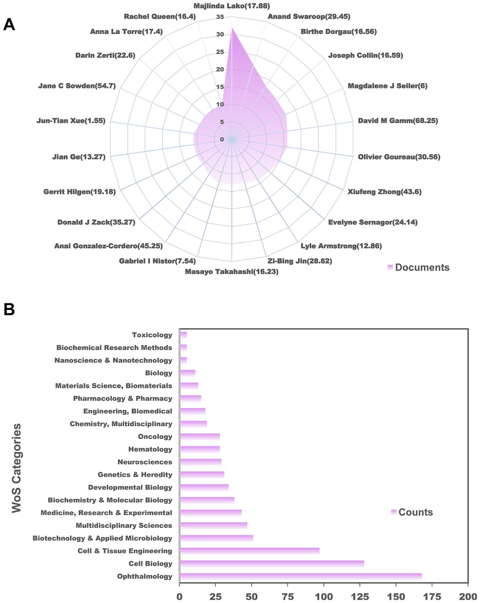
Publication of authors and research areas. (A) The top 20 authors with most publications. (B) The top 20 research areas of retinal organoid research.
Fig. 4B shows the top 20 research categories belonging to ROs during 2011 to 2022. Of all the included documents, ophthalmology (168, 32.31%), cell biology (128, 24.62%), cell and tissue engineering (97, 18.65%) are the most frequently studied areas.
Bibliographic Coupling Analysis of Authors, Institutions, Countries/Regions, and Journals
Fig. 5A shows the bibliographic coupling analysis of the 2,430 authors. Total link strength (TLS), which represents the inter-collaboration strength of a given researcher with other researchers, was calculated. VOSviewer showed that the productive authors with largest TLS were Olivier Goureau, Majlinda Lako, David M Gamm, Anand Swaroop, Igor O Nasonkin whose TLS equaled to 47,865, 42,817, 39,034, 36,564, and 29,743 times, respectively.
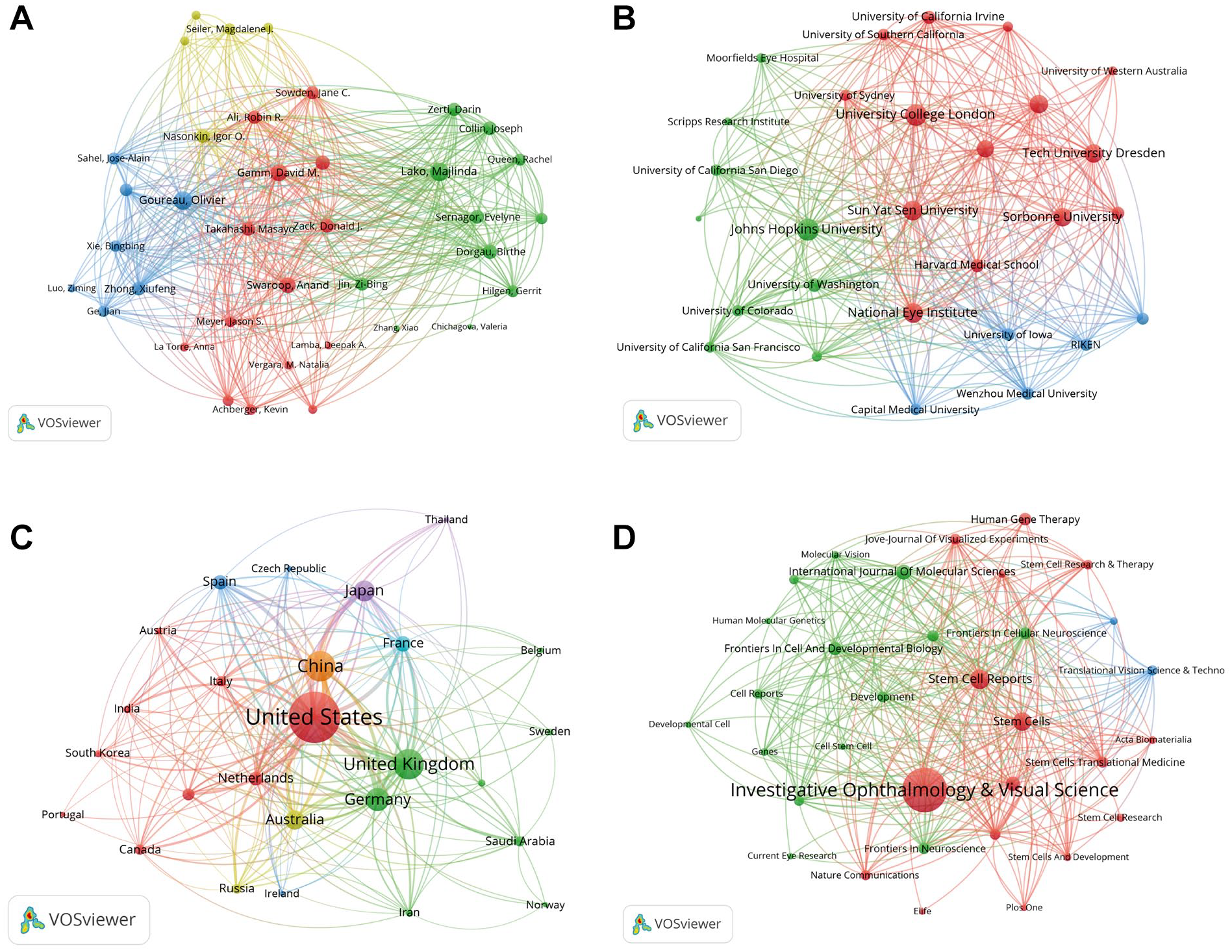
Bibliographic coupling analysis of authors, institutions, countries/regions, and journals. (A) Mapping of the 2,430 authors. (B) Mapping of the 640 institutions. (C) Mapping of the 35 countries/regions. (D) Mapping of the 149 journals. The circle size represents the publication count; the link between different points represents the collaboration between authors/institutions/countries/regions/journals.
Publications from 640 institutions were identified and analyzed by VOSviewer. We screened documents co-authored by more than 25 organizations and defined the minimum number of documents per organization as 5. Fig. 5B showed the top 5 institutions with strongest TLS, Johns Hopkins University (USA), University College London (UK), National Eye Institute (USA), Sun Yat-Sen University (China), and Newcastle University (UK), whose TLS equaled to 31,331, 31,007, 26,444, 25,493, 22,590 times, respectively.
We also analyzed the bibliometric coupling of 38 countries/regions via VOSviewer. Fig. 5C presents the top 5 countries/regions with largest TLS: the United States (164,592), China (80,773), the United Kingdom (67,389), Germany (58,293), and Japan (41,386).
We also analyzed the collaborative relationship between 149 journals which were involved in our study. As shown in Fig. 5D, Stem Cells (IF = 5.845, Q1), Progress in Retinal and Eye Research (IF = 19.704, Q1), Stem Cell Reports (IF = 7.294, Q2), International Journal of Molecular Sciences (IF = 6.208, Q1), and Scientific Reports (IF = 4.996, Q2) were the top 5 journals with largest TLS equaling to 24,129, 19,686, 18,799, 17,145, and 16,217, respectively.
Analysis of Keywords
To gain the insight of the several basic components of research hotspots, we screened 91 keywords from 1,665 keywords of all documents included (the minimum number of occurrences per word was defined as 7) and visualized the co-occurred keywords by VOSviewer which were mainly subdivided into four clusters: culture and differentiation (cluster 1, blue), morphogenesis and modeling (cluster 2, green), gene therapy (cluster 3, yellow), and transplantation and visual restoration (cluster 4, red) (Fig. 6A). These clusters indicated the most popular directions of researches.

(A) The visualization network mapping of the co-occurrence analysis of keywords. The keywords of the research fields were mainly subdivided into four clusters: culture and differentiation (blue), morphogenesis and modeling (green), gene therapy (yellow), and transplantation and visual restoration (red). (B) Visualization of distribution of keywords. Points in blue and yellow color represent an earlier or later appearance, respectively. (C) The top 15 keywords with strongest citation bursts. Burst detection years were shown in red squares. Keywords emerging in recent years would indicate the potential research directions in the future.
Furthermore, we also visualized the time distribution of these co-occurred keywords, which were displayed with distinct colors according to the time order in which these keywords appeared (Fig. 6B). The trends of the main scopes have not changed significantly with time.
We identified the keywords with strong citation bursts which served as an indicator to present and direct the research trend. As shown in Fig. 6C, we generated a keyword burst map including the burst strength and duration of the top 15 keywords via CiteSpace. Previous studies playing close attention to “directed differentiation” (began in 2011), “embryonic stem cell” (began in 2011), and “optic vesicle” (began in 2012). The burst keywords emerging in recent years, such as “proliferation” (began in 2019), would guide researchers to the potential research directions in the future.
Discussion
Research Trends of ROs
Bibliometric analysis and visualization can illustrate the current research trends as well as provide novel insight into future directions 33 . Last decade has witnessed a steady increase in the number of publications, which grew from 3 to 130 (from 2011 to 2022), and the overall growth rate reached 40.86%, indicating RO as an exciting and potential research field. Furthermore, we can predict the future publications based on the fitting curve of global trends in RO (Fig. 2B). More high-quality articles are expected to be published in the foreseeable future.
Besides, Table 1 shows the top 10 most cited articles in RO research. The earlier published article was in 2011, when Yoshiki Sasai’s group established the first in vitro RO model through a 3D mouse ESC culture system as previously mentioned 12 . After the most impactful study provided an immense breakthrough in 2011, various of RO-related high-quality researches have sprung up, whose common features were as follows: (1) the findings have been published in high-impact journals (e.g., Nature, Cell, Nature Biotechnology); (2) innovative strategies in generation and development of ROs, as well as integration of research progress in other fields (e.g., tissue engineering); (3) the bridge between basic research of ROs and clinical translation. These articles laid the groundwork for subsequent studies and presented necessary insights to future publications based on RO.
Global Publications and Collaboration Pattern
In total, the 520 publications were published in 38 countries and regions. In Table 2, with respect to these evaluation indicators (e.g., document counts, total citations, and H-index), the publication pattern illustrated that the United States made the biggest contribution in this area, playing an indispensable role in RO research. China and the United Kingdom had equal number of publications, ranking second and third, respectively, but the average citation times and H-index of documents published in China were slightly lower than that of the United Kingdom. Presumably, the main reason lied in the slight discrepancy between the quality and quantity of literature in China. Although Japan published the fifth largest documents, it ranked second in total citations and took the lead according to average citation, which may be due to several high-impact and high-cited articles from Japan12,28,34,35.
An increasing number of RO-related documents have been published from the leading institutions of RO research, of which the top 3 were University College London (UK), Newcastle University (UK), and Sun Yat-Sen University (China) (Table 2). Besides, University College London (UK) was the institution with largest total citations and highest H-index. The average citations of documents published in Harvard Medical School (USA) ranked first, which showed high-quality papers from the institution attracted more interest and attention from researchers 26 . In this study, Quadrato et al. 26 described the prolonged development (over 9 months) of human brain organoids into diverse cell types including the retina, and neuronal activity could be modulated by light stimulation of photosensitive cells, which demonstrated the possibility of network activity modulation by using physiological sensory mechanisms.
We found that more articles were published in Investigative Ophthalmology & Visual Science, Stem Cell Reports, and Stem Cells. Of the top 20 journals with the most publications, Stem Cells was the journal with largest total citations and highest H-index (Table 3). Nature communications was the leading journal with largest average citations and published seven articles related to RO among which one article received 508 citations from 2011 to 2022 17 . We also listed the top 20 authors with largest number of documents, among which Majlinda Lako was the most productive authors, and David M Gamm was the leading author with highest citation times as well as average citation times (Fig. 4A). As for the research categories, apart for “ophthalmology,” the undoubted core categories in our research, “cell biology” and “cell & tissue engineering,” were the hotter research categories (Fig. 4B). Future publications are expected to appear at the top of the journal list, and future literature are more likely to be distributed in subareas such as cell biology and cell and tissue engineering. The authors in the list are likely to contribute to more interesting, valuable, and innovative researches in the foreseeable future.
In our study, the relationship among 2,430 authors, 640 institutions, 38 countries/regions, and 149 journals were established through bibliographic coupling analysis, indicating that two works co-citing the third work in their bibliography. VOSviewer showed that Olivier Goureau, Majlinda Lako, David M Gamm were the leading authors in this field. Besides, Johns Hopkins University (USA), University College London (UK), National Eye Institute (USA) made the biggest contributions to this area. The United States, China, and the United Kingdom were the top 3 countries/regions with largest TLS in this field. Besides, Stem Cells, Progress in Retinal and Eye Research, and Stem Cell Reports were the most relevant journals. These data showed the aforementioned authors, institutions, countries/regions, and journals had the strongest collaboration work.
Keyword Analysis of ROs
Generally, the frequently appeared keywords reflect the research hotpots of RO. By co-occurrence analysis of keywords, we identified dominant keywords and divided the relevant literature into four basic components which meant the different subareas. Based on the retrieval formula, the most frequent keywords included “pluripotent stem cells,” “retinal organoids,” “neural retina,” and “induced pluripotent stem cells.” Besides, some keywords relating to retinal structure such as “retinal pigment epithelium” and “photoreceptors” were the dominant keywords in the literature. The impairment or loss-of-function of photoreceptors or supportive retinal pigment epithelium (RPE) is the main cause of permanent loss of vision, such as age-related macular degeneration (AMD), and hereditary retinopathies, such as retinitis pigmentosa (RP) 36 . The introduction of RO technology provided hope for unlimited source of photoreceptor cell transplantation. Furthermore, keywords associated with processes of RO technology, such as “differentiation” and “generation,” and RO-based therapy, such as “transplantation” and “expression,” also frequently appeared in our literature. Since the crucial protocol of in vitro optic-cup generation was reported by the Sasai’s group 12 , various research groups have endeavored to utilize 3D RO technology for the differentiation of retina components and generation of photoreceptor precursor cells or retinal sheets17,18,37,38. Besides, gene correction is a cutting-edge approach combined with disease modeling in the application of RO technology. Deng et al. 39 generated iPSCs from RP patients with RP GTPase regulator (RPGR) mutations, and then differentiated them into RPE cells and ROs, and showed significant developmental defects were observed. Furthermore, gene correction of the RPGR mutations by CRISPR/Cas9 contributed to rescue of photoreceptor development, gene expression, electrophysiological level, and ciliopathy.
The identified keywords were previously subdivided into four clusters, including “culture and differentiation,” “morphogenesis and modeling,” “gene therapy,” and “transplantation and visual restoration.” These results exhibited the most popular fields of RO centered on the four clusters, which vividly presented the future directions. At the center of this co-occurrence map, keywords such as “differentiation,” “gene expression,” and “mouse model” have relatively large weight. Future high-quality studies of RO associated with these four directions are still demanded. The colors of points in the overlay visualization map ranged gradually from blue to yellow, set by the sequential order of appearance (mainly from 2018 to 2021). According to the map, the topics of early publications mainly included process of RO production such as “mouse model,” “differentiation,” and “generation,” and gradually transited to retinal diseases including “age-related macular degeneration,” “inherited retinal diseases,” and “retinoblastoma.” Burst keywords reflected the research hotpots on the timeline, and the transformation from RO generation (e.g., directed differentiation, ESC) to vision recovery of retinal diseases [e.g., Leber congenital amaurosis (LCA), restoration].
Modeling of Retinal Diseases
Recently, ROs modeling the retinal disease have been established to decipher the molecular mechanism of diseases, drug screening and evaluate the efficiency of gene therapy in preclinical trials. As an irreversible and hereditary retinopathy, the exact pathogenic mechanism of RP is still elusive. Researchers have established RP models by patient iPSC–derived ROs with mutations in the genes including RPGR 39 , pre-mRNA processing factors (PRPFs) 40 , nuclear receptor subfamily 2, group E, member 3 (NR2E3), 41 and so on, recapitulating defects in retinal development. Zi-Bing Jin’s group also established RP model with stable phenotype, which was the first RO modeling late-onset retinal degeneration 42 . Besides, researchers have established in vitro models of LCA in ROs, and defects in photoreceptor maturation and downregulated gene expression were observed43,44. X-linked juvenile retinoschisis (XLRS) is also an inherited retinal disease linked with genetic alterations in the RS1 gene, characterized by splitting of different retinal layers and mild-to-severe visual impairment at a young age. Researchers established RS1 mutant RO derived from XLRS patients, and then utilized CRISPR-Cas9 gene editing to correct the mutation and rescued the phenotype of XLRS successfully 45 . Retinoblastoma (Rb) is a rare and life-threatening pediatric malignancy with global survival rate less than 30%, linked to the biallelic inactivation of RB1 gene 46 . Liu et al. 47 developed Rb organoid differentiated from genetically engineered hESCs with biallelic RB1 mutation, which recapitulated the tumorigenesis, genetic, and epigenetic features of Rb. In another study based on Rb, ROs were established, separated, and injected into the eyes of immunocompromised mice to support tumorigenesis. Organoid-derived Rb recapitulated the molecular, cellular, genetic, and epigenetic properties of human Rb, which paved a new avenue for combined therapeutic strategies for individual patients 48 .
As an unlimited source of stimulating native structure and presenting cellular interactions, ROs derived from hiPSCs hold significant promise and potential for accelerating drug discovery and toxicity screening process. Formidable obstacles should be circumvented, such as lack of quantitative assays for 3D retinal models due to complicated cell types self-organized in specific spatial arrangements 49 . To satisfy the demand, researchers developed a screening platform based on precise quantification of fluorescent reporters, which met the speed, sensitivity, and reproducibility parameters. Subsequently, they demonstrated the capacity of the system to monitor mitochondrial health in photoreceptors in longitudinal studies, which served as the proof-of-concept drug testing 50 . Advances in high-throughput imaging technology have allowed for high-content screening of 3D culture such as organoids 51 . This technology has been utilized in RO culture, which is expected to undertake pharmacological and toxicological researches 52 .
Nowadays, RO models, in combination with newly developing single-cell RNA sequencing technologies, are expected to break through the bottleneck of traditional retinal disease model and facilitate drug research and development, precision medicine, as well as regenerative medicine. For example, in 2020, a study has developed light-sensitive human ROs with highly complex anatomical and functional structure and utilized single-cell sequencing to demonstrate that (1) the rate at which RO transcriptomes reached its stable and mature state in vitro was similar to human retina in vivo; (2) the RO transcriptomes converged to adult human peripheral retinal transcriptomes; and (3) the exploration of genetic disease maps corroborated retinal diseases possessed cell-type specialty, which can also be seen in organoids 17 . These promising results present necessary insights of modeling retinal disease, deciphering cellular targets and targeted therapeutic strategies in ROs in the foreseeable future.
Transplantation and Gene Therapy
Owing to the advantages of convenience, stability, unlimitedness, and similarity in stratified structure and cellular connections, RO has been a potent resource for transplantation as well as gene therapy 53 .
Generally, RO-based transplantation can be divided into two approaches: transplantation of retinal sheets (tissue) and transplantation of single-cell suspensions. The former refers transplantation of tissue dissociated from the fetal retina or developed optic cup to preserve characteristic stratified structures and neural circuits 54 . Takahashi’s group successfully transplanted retina sheets derived from ROs into the end-stage retina-degeneration mice, which resulted in the maturation of photoreceptors to fully form OS and establish host-graft synaptic connections, displayed light-responsive identity, and recovered visual function to some extent55,56. After transplanted into retina-degeneration mice, RO sheets were observed to differentiate, integrate, and enhance visual function which was assessed by optokinetic response (OKT) and electrophysiologic recordings in the superior colliculus 57 .
Nonetheless, single-cell transplantation has been much more explored than sheet transplantation for advantages as follows: accurate quantity of transplanted cells, better host–graft connections, and reduced surgical invasion 58 . RPE, photoreceptors, and retinal progenitor cells (RPCs) have been utilized in preclinical trials 59 , and purification by specific surface biomarkers of single cells was thoroughly reviewed by Zhang et al. 54 Several studies demonstrated that cell sorting approach contributed to higher integration levels and capability of survival and maturation proximal to the host retina60,61, and it can improve the microenvironment of the degenerative host retina by modulating microglia activation 62 , which is a crucial issue on track of better survival, differentiation, and integration.
ROs provide a great opportunity to explore and develop therapeutic strategies for degenerative retinal dystrophies, which may lead to irreversible loss of vision 63 . Viral vectors based on adeno-associated virus (AAV) serve as a safe and efficient tool for gene delivery to ROs, and recombinant AAV (rAAV) is one of the most frequently used viral vectors. In vitro ROs derived from patients can significantly improve the efficacy of transduction in place of animal models63,64. As a human promoter, the gene augmentation of cone–rod homeobox (CRX) mediated by an AAV vector, which accurately targeted a distinct apical lamina within the photoreceptor layer, can partially restore the expression of cone and rod genes, and phototransduction-related genes 44 .
Besides, the application of CRISPR/Cas9-mediated genome editing technology has promoted the establishment of retinopathy models including RP 65 , glaucoma 66 , and Rb 67 , as well as the rescue of phenotypes in retinopathy models39,45,68, promising a therapeutic strategy based on gene editing to treat retinal dystrophies.
Organoid on a Chip
There are significant hurdles in the exploration of human retinal development, visual function, or response to therapy, such as absence of critical cell types, vascularization, and physiological connections between matured photoreceptors and RPE 31 . Same as transformative technology, the microphysiological systems (MPS) or organ-on-chips have evolved into a great alternative of conventional cell culture with vasculature and microfluidic perfusion, recapitulating in vitro microstructure and functions of living human organs. In view of this property, organ-on-chips technology can be excellently applied to model blood–retina barriers for architecture of multilayered cells and vasculature-like perfusion 69 . Achberger et al. first introduced a retina-on-a-chip (RoC) by combining ROs and RPE both derived from hiPSCs. This microphysiological platform presented optimized formation and preservation of inner segment and OS, the interaction between photoreceptors and RPE, and precisely modulated sub-RPE choroidal-like perfusion 31 . This study presented necessary insights to exploring nervous innervation, integration of blood–retina barriers, and establishment of specific retinopathies.
Strengths and Limitations
To gain an overview of publications on RO from 2011 to 2022, we extracted all documents from SCI Expanded of WoS for acquiring comprehensive, reliable, and objective statistics. Subsequently, we undertook bibliometric and visualized analysis by virtue of ArcGIS, VOSviewer, and CiteSpace; analyzed the quality of publication; and discussed the research hotpots as well as future directions. Undeniably, there are some limitations existing in our study. First, publications were only derived from SCI Expanded of WoS database, so we may miss some valuable and relevant articles due to database bias. Second, the retrieval formula may not comprehensively include all relevant documents. Third, due to the frequent appearance of many keywords belonging to any deeper subdomains, a part of keywords presented in the figures make no sense. Finally, given the absence of uniform standards for parameters, results of cluster analysis can be slightly distinct with regard to different settings.
Conclusion
This study displayed a comprehensive overview of global publications on RO which is nowadays the hotpot of stem cell medicine. We also discussed the current applications of RO at the end of this article. It is a young and promising research area with many extensive and in-depth studies, and the next directions should circumvent the hurdles of RO, such as defects in vasculature and interplay of different cell types. More attention should be paid to models and therapies based on specific retinal diseases, especially inherited retinopathies in the field of RO.
Footnotes
Author Contributions
Design of the work: WS, AS, KJ, and JY. Collection and screening of the data for the work: WS, AS, WZ, LL, AG, KJ, and JY. Analysis of the data: AS. Drafting the work: WS. Revising of the manuscript: WZ, LL, AG, KJ, and JY. All authors contributed to the article and approved the submitted version.
Data Availability
The data sets used and/or analyzed during the current study are available from the corresponding author on reasonable request.
Ethical Approval
This study was approved by our institutional review board.
Statement of Human and Animal Rights
This article does not contain any studies with human or animal subjects.
Statement of Informed Consent
There are no human subjects in this article and informed consent is not applicable.
Declaration of Conflicting Interests
The author(s) declared no potential conflicts of interest with respect to the research, authorship, and/or publication of this article.
Funding
The author(s) disclosed receipt of the following financial support for the research, authorship, and/or publication of this article: This work has been financially supported by National Key Research and Development Program of China (grant number 2019YFC0118400), Natural Science Foundation of China (grant number 82201195), Natural Science Foundation of Zhejiang Province (grant number LQ21H120002), Medical and Health Science and Technology Program of Zhejiang Province (grant number 2021RC064), and Clinical Medical Research Center for Eye Diseases of Zhejiang Province (grant number 2021E50007). The funding body played no role in the design of the study and collection, analysis, and interpretation of data and in writing the manuscript.