
Abstract
Background
Diabetes is a serious global health concern which severely affected public health as well as socio-economic growth worldwide. Scutellarin (SCU), a bioactive flavonoid, is known for its efficacious action against a range of ailments including cardiovascular problems. The present study was conducted to find out possible protective effect and its associated mechanisms of SCU on experimental type 2 diabetes-induced cardiac injury.
Methods
Type 2 diabetes was induced by treating animals with high fat diet for 4 weeks and a single intraperitoneal dose (35 mg/kg body weight) of streptozotocin and diabetic animals received SCU (10 or 20 mg/kg/day) for 6 weeks.
Results
Scutellarin attenuated type 2 diabetes-induced hyperglycemia, bodyweight loss, hyperlipidaemia, cardiac functional damage with histopathological alterations and fibrosis. Scutellarin treatment to type 2 diabetic mice ameliorated oxidative stress, inflammatory status and apoptosis in heart. Furthermore, the underlying mechanisms for such mitigation of oxidative stress, inflammation and apoptosis in heart involved modulation of Nrf2/Keap1 pathway, TLR4/MyD88/NF-κB mediated inflammatory pathway and intrinsic (mitochondrial) apoptosis pathway, respectively.
Conclusions
The current findings suggest that SCU is effective in protecting type 2 diabetes-induced cardiac injury by attenuating oxidative stress and inflammatory responses and apoptosis, and it is also worth considering the efficacious potential of SCU to treat diabetic cardiomyopathy patients.
Introduction
Diabetes is a major global health concern which severely affected public health as well as socio-economic growth worldwide. As per estimates of International Diabetes Federation, the diabetic cases were 451 million in 2017 with unimaginable predicted rise to 693 million by 2045. 1 Diabetes affects multiple organs and tissues and cause complications including cardiovascular diseases (CVDs). 2 Cardiovascular disease is a chronic disease with the highest morbidity and mortality (costing 17.9 million deaths every year) across the globe. Even though the aetiology for this occurrence is not fully understood, evidence suggests that diabetes has been considered as prime factor for CVDs. 3
Diabetes induces pathological changes in myocardium at morphological, biochemical and molecular level and eventually leads to serious cardiac events.4,5 Heart being an active organ experiences severe stress during hyperglycaemic condition and alters energy preferences in cardiomyocytes thereby elevates free radicals. Hyperglycemia reduced antioxidant defences along with elevated levels of free radical’s/lipid peroxidation 5 making oxidative stress in cardiomyocytes, which may remodel cardiac structure, a hallmark of cardiac damage. 6 At this juncture, it is worth mentioning to consider the critic role of Nrf2 in the protection of cardiomyocytes from oxidative injury. 7 Previous findings cited the implication of oxidative stress in instigation of various signalling pathways of inflammation and generates inflammatory cytokines, which negatively affects cardiac function.8,9 Among the signalling pathways of inflammation, Toll-Like Receptors (TLRs) are of great importance due to their regulation of immune signalling in cardiomyocytes. 10 Immune signalling converges in activation of nuclear factor κB (NF-κB), which plays an important role in the regulation of immune reaction. 11 Scientific literature also revealed hyperglycemia-mediated induction of oxidative stress, which inextricably work with inflammation to provoke apoptosis.8-10
Despite the availability of conventional medicines to regulate blood glucose levels, they are ineffective in protecting against diabetes-related consequences such as cardiac damage.12,13 Supplementation of phytochemicals has gained significance due to their safety and/or non-toxic nature 14 and protective principles in the form of a range of biological activities. Scutellarin (SCU) (5,6,7,4′-tetrahydroxyflavone-7-O-β-glucuronide), the main component (≥90%) in the extract of the herb Erigeron breviscapus, which has been in traditional Chinese medicine use for decades mainly for effective actions against brain and heart related ailments. 15 The therapeutic formulations of SCU were in the form of tablets, capsules and injectable with concentrations more than 90% and 98% in oral and injectables, respectively. 11 Clinical data and experimental data also revealed the efficacy of SCU in the treatment of range of diseases and disorders including cardiovascular disease, cerebrovascular disease and diabetic complications. 11 The medical claim that was attributed to this extract was antioxidant activity based on evaluation of many assays. 16 In recent years, the focus has been shifted towards finding the efficacy and mechanism of action of SCU. Scutellarin is well known for its protective potential against neurotoxicity, 17 diabetic nephropathy,18,19 diabetic retinopathy,20,21 diabetes-induced testicular damage 22 and diabetes-induced liver injury 23 through its antioxidant 24 and anti-inflammatory 25 actions. Previously, studies reported that SCU protects doxorubicin-induced acute cardiotoxicity, 26 isopranaline-induced myocardial infarction 27 and ischaemia–reperfusion (I/R)-induced myocardial injury. 25 Also, SCU was proved to be effective by alleviating interstitial fibrosis and cardiac dysfunction in infarct rats. 28 Furthermore, preclinical safety evaluation of SCU by acute and sub-acute studies revealed minimal toxicity or non-toxicity in rodents. 29
By considering diabetes-induced cardiac complications and SCU safety and efficacy potential, the present study is planned and executed to explore possible protective effects and mechanisms of SCU against cardiac injury in well-established experimental type 2 diabetes (high fat diet/STZ) mice model. The results are of value in potential implication of SCU on cardiac injury with the aim to improve cardiac function in type 2 diabetic condition.
Methodology
Drugs and chemicals
Scutellarin and streptozotocin (STZ) were selected as the test chemicals for the present study and purchased from Sigma Chemical Company (St. Louis, MA, USA) and other chemicals (highest grade) were bought from local commercial suppliers.
Animals
Adult healthy male Swiss mice (22 ± 03 g) were procured and allowed to acclimatize for 7 days before being used for the present study. Animals were submitted to ophthalmological and detailed clinical examination during acclimatization. Also, growth of animals was monitored regularly and the animals displaying poor growth were not included in the study. The mice were housed in polypropylene cages by using sterilised corn cob for bedding purpose and allowed ad libitum access to reverse osmosis autoclaved water and standard rodent feed. The rats were maintained in an automatic controlled well-controlled laboratory conditions (temperature 23 ± 2°C; 12-h light and 12-h dark cycle; humidity 50 ± 10%). The animal experiment procedure was reviewed and approved by a panel of Institutional Animal Ethical Committee members.
Induction of experimental type 2 diabetes
To induce experimental type 2 diabetes in mice, animals were supplied with ad libitum access to high fat diet (58% energy from fat) for 4 weeks and given a single intraperitoneal injection of STZ (35 mg/kg body weight) in a citrate buffer of pH 4.5. 30 Later 1 week of STZ injection, animals exhibiting hyperglycaemia, that is, fasting blood glucose level > 12.5 mmol/L, were considered as type 2 diabetic animals and further used for the research. 31
Experimental design
One-week after adaptation period, a total of 40 animals were randomly categorised into four groups (n = 10) Group 1: Normal control group-received vehicle. Group 2: Type 2 diabetes group. Group 3: Diabetic mice received 10 mg/kg body weight of SCU. Group 4: Diabetic mice received 20 mg/kg body weight of SCU.
Scutellarin was dissolved in 5% DMSO 27 and distilled water (1:9) and treatment for 6 weeks via orally with oral gavage tube. The dosage of SCU was based on earlier study.3,28 The study protocol was approved by the Ethics Committee of First Affiliated Hospital of Heilongjiang University of Traditional Chinese Medicine (No. DXBY2020).
Determination of body weight and blood glucose levels
Weekly body weights were measured using calibrated weighing balance, and fasting blood glucose levels were monitored once in a week using a glucometer (Accu-Check, Roche, Germany).
Biochemical analysis
At the end of the study, blood samples from the control and experimental mice were withdrawn via intraocular puncture using mild ether anaesthesia. The blood samples were allowed to clot at room temperature for at least 40 min, and sera were separated by centrifuging the samples at 3500 r/min for 15 min. The serum concentrations of total cholesterol (TC), triglycerides (TG), high density lipoprotein (HDL) and low density lipoprotein (LDL) were analysed using automatic Beckman Coulter analyzer.
Analyses of serum concentrations of CK-MB, Troponin and BNP were based on ELISA kit method. Hearts were quickly separated after euthanizing animals, blotted free of blood, weighed wet to the nearest milligram by using an electronic balance and further used for histopathological, biochemical and molecular analyses. Left cardiac portion was used for biochemical and molecular analyses, while right cardiac portion was fixed in 10% neutral formalin saline for histopathological studies.
Histopathology
For histological study, hearts fixed in neutral buffer saline were taken out and trimmed off excessive tissue, processed in tissue processor with increasing series of alcohol washes, embedded in paraffin blocks, cut into 5 μm thick sections using microtome, and sections were placed on adhesive slides and stained with Periodic acid-Schiff (PAS) and Masson’s trichrome stains. Then, the sections were examined under Olympus phase contrast microscope (Tokyo, Japan), and digital images were captured by microscope camera (Nikon Coolpix 5400, Nikon, Japan).
Immunohistochemistry and immunofluorescence
To perform immunohistochemistry, heart sections from all the animals were deparaffinized, rehydration, antigen retrieved, blocked the endogenous peroxidase activity with 0.3% H2O2 and incubated for overnight at 4°C with primary antibodies such as Nrf2 (NBP1-32822; Novus Biologicals, Littleton, CO, USA), Keap1 (sc-514914; Santa Cruz Biotechnology, CA, USA), IKKβ (NB100-56509; Novus Biologicals, Littleton, CO, USA), NF-κB (sc-8008; Santa Cruz Biotechnology, CA, USA), TNF-α (ab109322; Abcam, Cambridge, MA, USA), BCL-2 (NB100-56098; Novus Biologicals, Littleton, CO, USA) and Bax (sc-7480; Santa Cruz Biotechnology, CA, USA) at a dilution of 1:200 in 5% bovine serum albumin. After three PBS washes, the sections were incubated with appropriate secondary antibody for 1 h and were stained and counterstained with 3'-Diaminobenzidine and haematoxylin, respectively. Also, negative control was also kept to ensure antibody specificity by using normal rabbit serum instead of primary antibody to confirm the specificity.
The double immunofluorescence was performed according to the method as previously described.33,34 The deparaffinised, rehydrated and antigen retrieved sections were blocked with 2.5% normal horse serum provided in the kit (Vector Laboratories, Burlingame, CA, USA) for 30 min at room temperature. The slides were incubated with first primary antibodies like caspase-3 (NB100-56708; Novus Biologicals, Littleton, CO, USA) at ratio of 1:1000 for 2 h at room temperature, then the slides were washed with PBS for three times. The slides were incubated with secondary antibody, caspase-9 (NB100-56118; Novus Biologicals, Littleton, CO, USA) at ratio of 1:1000 for 2 h at room temperature. After washing 3 times with PBS, the slides were incubated in DyLight® 594 Anti-Rabbit (red), DyLight® 488 Anti-Mouse (green) secondary antibodies (VectaFluor™ Duet Immunofluorescence Double Labeling Kit; DK-8828; Vector Laboratories, Burlingame, CA, USA) for 2 h at room temperature. Then, the sections were mounted with Mounting Medium with DAPI (Santa Cruz Biotechnology, CA, USA). The slides were viewed under confocal microscope (Leica TCS SP5 II, Wetzlar, Germany).
Oxidative and anti-oxidative status in the heart
Oxidative stress parameters such as lipid peroxidation products (level of thiobarbuturic acid reactive substances as a measure of malondialdehyde-MDA levels) and protein carbonyls and antioxidant enzyme activities such as superoxide dismutase (SOD; E-BC-K020-M), catalase (CAT; E-BC-K031-M), glutathione peroxidase (GPx; E-BC-K096-M), glutathione S-transferase (GST; E-BC-K278-S) in 10% heart tissue homogenates samples were measured with commercially available kits (Elabscience Biotechnology, Wuhan, China) as per supplier kit protocol. Protein content in the enzyme source was estimated by Bradford protein assay kit (Sigma Company, St. Louis, MO, USA).
Quantitative reverse transcription polymerase chain reaction analysis

Statistical analysis
Results were expressed as mean ± SD of 10 determinants. The difference between the groups were analysed using one-way analysis of variance (ANOVA) followed by Tukey’s post hoc test in SPSS version 21 (SPSS, Chicago, IL). Statistical significance was considered when p < 0.05.
Results
Effect of SCU on bodyweight, blood glucose levels and serum lipid profile in high fat diet and low dose of STZ-induced diabetic mice
Figure 1(a) shows the weekly body weights in control and experimental animals. Significantly higher (p < 0.05) initial body weights of HFD-fed mice were observed when compared to normal diet fed mice. After induction of diabetes, significant body weight loss was seen in diabetic mice in comparison to control mice, while significant decrease in body weight losses was observed in 10 (p < 0.05) or 20 (p < 0.05) mg SCU/kg body weight treated mice when compared to diabetic mice. Effect of Scutellarin on body weight, heart weight, serum lipid profile and serum cardiac markers in high fat diet and low dose of STZ-induced diabetic mice. (A) Bodyweight; (B) blood glucose levels; (C) total cholesterol (TC); (D) triglyceride (TG); (E) high density lipoprotein (HDL); (F) low density lipoprotein (LDL); (G) troponin; (H) creatine kinase-MB (CK-MB); (I) brain natriuretic peptide (BNP). NC: normal control mice; DM: diabetes mellitus; DM+10 Scutellarin: diabetic mice treated with 10 mg/kg/bw of Scutellarin; DM+20 Scutellarin: diabetic mice treated with 20 mg/kg/bw of Scutellarin. Data are mean ± SD of 10 mice. Bars or line with different superscripts differ significantly at p < 0.05.
The results of this study revealed diabetic mice experienced hyperglycaemia through significant (p < 0.05) rise in FBG levels compared to normal controls. However, FBG levels in diabetic animals treated with SCU (10 or 20 mg/kg bw) for 6 weeks showed significant (p < 0.05) improvement in the form of decreased levels of glucose levels when compared to diabetic controls (Figure 1(b)).
Lipid metabolic alterations in diabetic mice were evidenced from the study through significant increase in serum TC (p < 0.05) (Figure 1(c)), TG (p < 0.05) (Figure 1(d)), LDL (p < 0.05) (Figure 1(f)), and significant reduction in levels of serum HDL (p < 0.05) (Figure 1(e)) with respect to control mice. Whereas, treatment with SCU 10 or 20 mg/kg bw to diabetic mice for 6 weeks significantly (p < 0.05) curbed the dyslipidaemia in diabetic mice.
Effect of SCU on serum cardiac markers in high fat diet and low dose of STZ-induced diabetic mice
To know the protective effect of SCU against diabetes-mediated cardiac injury, we analysed the levels of serum cardiac markers such as Troponin (Figure 1(g)), CK-MB (Figure 1(h)) and BNP (Figure 1(i)) levels. The findings revealed that diabetic mice exhibited cardiomyocyte-injury by significantly (p < 0.05) elevating CK-MB, Troponin and BNP levels in diabetic mice compared to normal controls. Conversely, SCU (10 or 20 mg/kg bw) administration to diabetic mice for 42 days showed significant (p < 0.05) improvement in cardiac function by decreasing CK-MB, Troponin, and BNP levels when compared to only diabetic control group without SCU treatment.
Effect of SCU on histopathology of heart in high fat diet and low dose of STZ-induced diabetic mice
Histological analyses of PAS (Figure 2(a)) and Masson-trichrome (Figure 2(b)) stained sections confirmed the cardio-protective effect of SCU in type 2 diabetes-induced myocardial injury. Control mice showed usual morphological features of heart with normal myocardial fibres, while the diabetic heart sections showed extensive damage in myocardium through presence of necrotic myocytes, vacuolation and infiltration of inflammatory cells with interstitial fibrosis. However, SCU (10 or 20 mg/kg bw) treatment for 6 weeks ameliorated diabetes-induced histopathological alterations and myocardial fibrosis in heart. Effect of Scutellarin on histopathological changes in high fat diet and low dose of STZ-induced diabetic mice. (A) Periodic acid-Schiff (PAS); (B) Masson trichrome. NC: normal control mice; DM: diabetes mellitus; DM+10 Scutellarin: diabetic mice treated with 10 mg/kg/bw of Scutellarin; DM+20 Scutellarin: diabetic mice treated with 20 mg/kg/bw of Scutellarin. Scale bar: 100 µm. Magnification: 40X.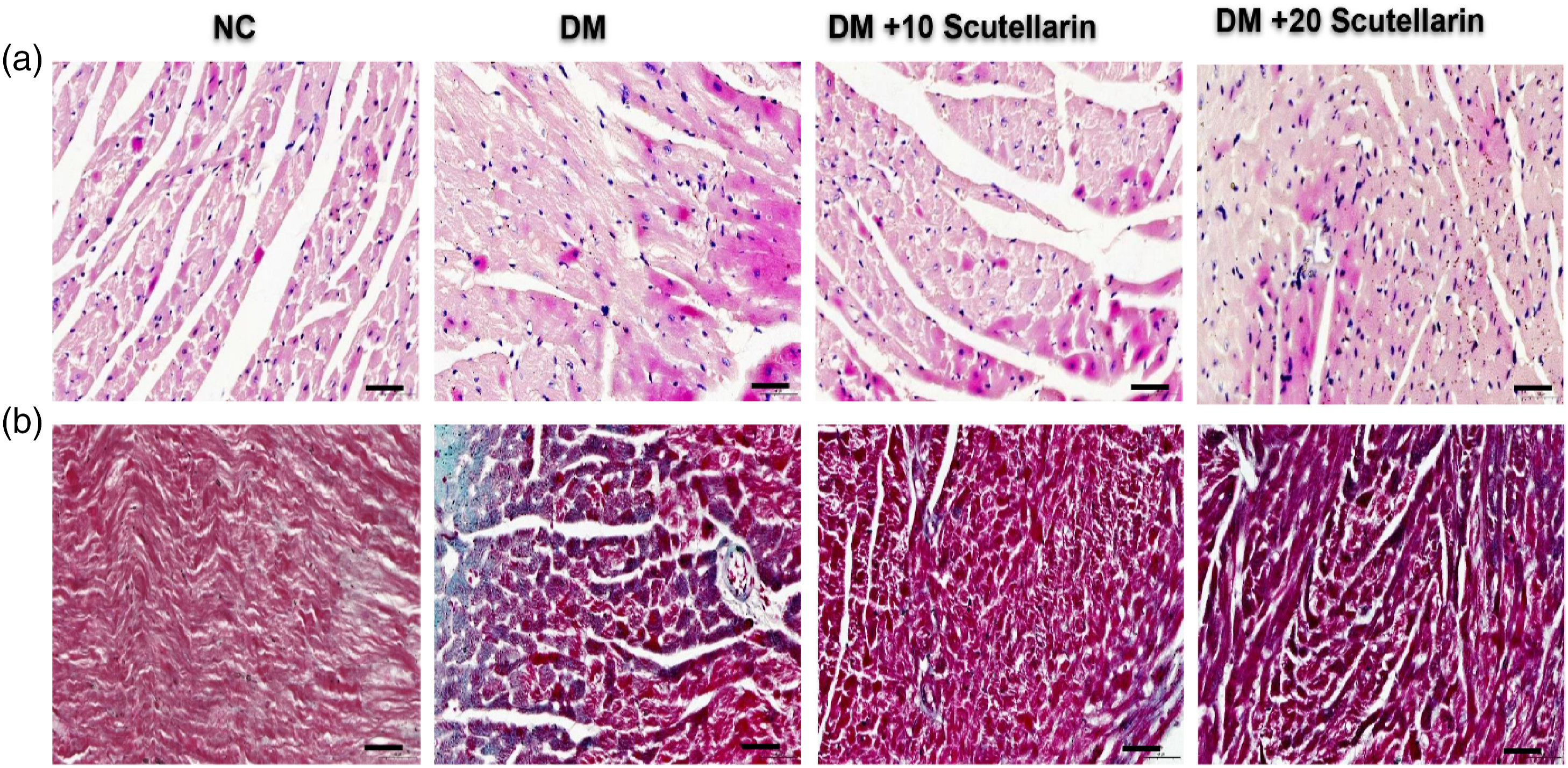
Effect of SCU on oxidative status in high fat diet and low dose of STZ-induced diabetic mice
Herein, diabetic mice exhibited oxidative stress in hearts through significant (p < 0.05) elevation in MDA (Figure 3(f)) and protein carbonyl (Figure 3(e)) levels along with significant (p < 0.05) reduction in antioxidant defence enzyme activities such as SOD (p < 0.05) (Figure 3(a)), CAT (Figure 3(b)), GPx (Figure 3(c)) and GST (Figure 3(d)) compared to respective levels and enzyme activities in hearts of control mice. Nevertheless, these alterations were attenuated after treating diabetic animals with SCU at doses 10 or 20 mg/kg (p < 0.05) for 42 days. Effect of Scutellarin on oxidative stress and antioxidant enzyme levels in cardiac tissue of high fat diet and low dose of STZ-induced diabetic mice. (A) SOD; (B) CAT; (C) GPx; (D) GST; (E) protein carbonyl; (F) Lipid peroxidation products malondialdehyde (MDA). NC: normal control mice; DM: diabetes mellitus; DM+10 Scutellarin: diabetic mice treated with 10 mg/kg/bw of Scutellarin; DM+20 Scutellarin: diabetic mice treated with 20 mg/kg/bw of Scutellarin. Each bar is mean ± standard deviation of 10 mice. Bars with different superscripts differ significantly at p < 0.05.
To know the molecular mechanisms behind antioxidant property of SCU, a battery of genes and protein distribution levels were estimated by RT-PCR and immunohistochemistry, respectively. The results revealed downregulated mRNA expression of Nrf2 (Figure 4(a)), Nqo-1 (Figure 4(c)), and Ho-1 (Figure 4(d)) with upregulated Keap1 (Figure 4(b)) mRNA expression in diabetic hearts of mice compared to same expression in normal healthy mice. While, diabetes-induced and SCU treatment for 42 days significant ameliorated the alterations observed in antioxidant genes of only diabetic mice hearts. Effect of Scutellarin on relative mRNA expression of antioxidant enzymes in high fat diet and low dose of STZ-induced diabetic mice. Relative mRNA expression of (A) Nrf2; (B) Keap-1; (C) Nqo1; (D) and Ho-1. (E) Immunostaining of NRF2 and (F) Keap1. NC: normal control mice; DM: diabetes mellitus; DM+10 Scutellarin: diabetic mice treated with 10 mg/kg/bw of Scutellarin; DM+20 Scutellarin: diabetic mice treated with 20 mg/kg/bw of Scutellarin. Each bar is mean ± SD of 10 mice. Bars with different superscripts differ significantly at p < 0.05. Scale bar: 100 µm. Magnification: 40X.
Furthermore, immunohistochemistry studies supported the results of RT-PCR through reduced Nrf2 protein distribution (Figure 4(e)), and elevated Keap-1 protein distribution (Figure 4(f)) in the heart of diabetic mice. Whereas, 10 or 20 mg/kg bw SCU treatment for 42 days has resulted in an elevated protein distribution of Nrf2 protein with lessened Keap-1 protein distribution.
Effect of SCU on inflammatory markers in cardiac tissue of high fat diet and low dose of STZ-induced diabetic mice
The relative mRNA expression levels of inflammatory markers such as Tlr4 (Figure 5(a)), Myd88 (Figure 5(b)), Nf-κb 1 (Figure 5(c)), Il 6 (Figure 5(e)) and TNf-α (Figure 5(f)) were upregulated, and mRNA expression level of IkBβ (Figure 5(d)) was decreased in hearts of HFD/STZ-induced mice when compared with normal control animals. Conversely, diabetes-induced and SCU (10 or 20 mg/kg bw) treated mice showed significant (p < 0.05) alleviation in expression levels of the selected inflammatory markers in hearts. Effect of Scutellarin on inflammatory markers in cardiac tissue of high fat diet and low dose of STZ-induced diabetic mice. mRNA expression of (A) Tlr4, (B) Myd88; (C) Nf-kb1; (D) Ikbβ; (E) Il-6; and (F) TNF-α. NC: normal control mice; DM: diabetes mellitus; DM+10 Scutellarin: diabetic mice treated with 10 mg/kg/bw of Scutellarin; DM+20 Scutellarin: diabetic mice treated with 20 mg/kg/bw of Scutellarin. Each bar is mean ± SD of 10 mice. Bars with different superscripts differ significantly at p < 0.05.
Additionally, protein distribution levels of NF-κB (Figure 6(a)) and TNF α (Figure 6(c)) were increased, and protein distribution of IKKβ (Figure 6(b)) was decreased in immunohistochemistry sections of diabetic heart when compared to normal control mice. Conversely, SCU (10 or 20 mg/kg bw) treatment resulted in marked improvement in inflammatory protein distribution profile. Effect of Scutellarin on inflammatory markers in cardiac tissue of high fat diet and low dose of STZ-induced diabetic mice. Immunostaining of (A) NFKβ; (B) IkBβ; and (C)TNF-α. NC: normal control mice; DM: diabetes mellitus; DM+10 Scutellarin: diabetic mice treated with 10 mg/kg/bw of Scutellarin; DM+20 Scutellarin: Diabetic mice treated with 20 mg/kg/bw of Scutellarin. Scale bar: 100 µm. Magnification: 40X.
Effect of SCU on apoptosis markers in cardiac tissue of high fat diet and low dose of STZ-induced diabetic mice
Apoptotic profile was revealed by RT-PCR and immunohistochemistry experiments. There was an upregulation in gene expression of Bax (Figure 7(a)), Cyt-c (Figure 7(c)), Caspase-9 (Figure 7(d)), Caspase-3 (Figure 7(e)) and Parp 1 (Figure 7(f)) and downregulation in gene expression of bcl-2 (Figure 7(b)) in cardiac tissue of hyperglycaemic mice in comparison with non-diabetic mice. While, either 10 or 20 mg/kg SCU treatment to diabetic mice has shown significant improvement in apoptotic profile. Effect of Scutellarin on apoptosis markers in cardiac tissue of high fat diet and low dose of STZ-induced diabetic mice. Relative mRNA expression of (A) Bax; (B) Bcl-2; (C) Cytc; (D) Caspase-9; (E) Caspase-3; and (F) Parp 1; NC: normal control mice; DM: diabetes mellitus; DM+10 Scutellarin: diabetic mice treated with 10 mg/kg/bw of Scutellarin; DM+20 Scutellarin: diabetic mice treated with 20 mg/kg/bw of scutellarin. Each bar is mean ± standard deviation of six mice. Bars with different superscripts differ significantly at p < 0.05.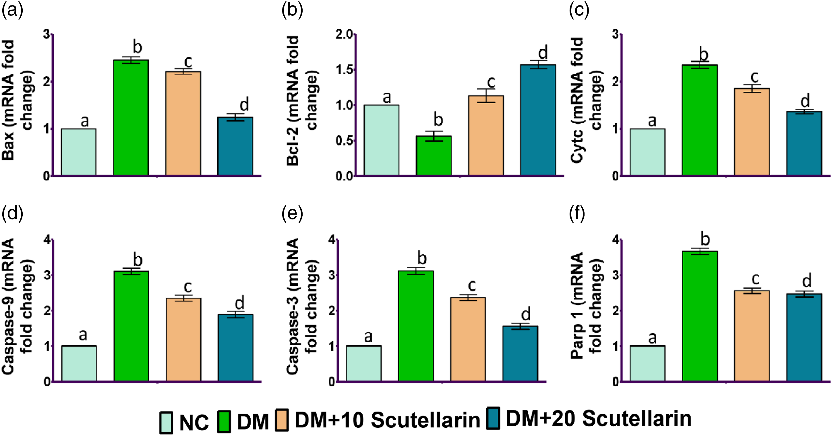
In addition, protein distribution level of apoptotic markers, that is, Bax (Figure 8(a)), was increased with reduction in protein distribution level of Bcl2 (Figure 8(b)) in cardiac tissue of diabetic mice. Whereas, treatment with either 10 or 20 mg/kg SCU resulted in marked improvement in apoptotic status. Effect of Scutellarin on apoptosis markers in cardiac tissue of high fat diet and low dose of STZ-induced diabetic mice
Moreover, immunofluorescence results also confirm that higher caspase-3 (green signals) and caspase-9 (red signals) (Figure 9) distribution was observed in cardiac tissue of diabetic mice. However, diabetic mice treatment with either 10 or 20 mg/kg SCU showed lower distribution of caspase-3 and caspase-9 when compared with diabetic mice. Effect of Scutellarin on apoptosis markers in cardiac tissue of high fat diet and low dose of STZ-induced diabetic mice. Immunostaining of (A) Caspase-3 (green signals) (B) Caspase-9 (red signals). NC: normal control mice; DM: diabetes mellitus; DM+10 Scutellarin: diabetic mice treated with 10 mg/kg/bw of Scutellarin; DM+20 Scutellarin: diabetic mice treated with 20 mg/kg/bw of Scutellarin. Scale bar: 50 µm. Magnification: 40X.
Discussion
Diabetes-mediated cardiac injury has been considered as a main cause of heart failure and death in diabetic patients. Epidemiological studies have shown that diabetic patients are exposed to 2- to 5-fold increased risk of developing cardiac abnormalities compared with non-diabetics. On the other hand, phenolic compounds possess cardio-protective potential through its modulatory effects on oxidative stress and inflammatory responses involved in CVDs. Scutellarin , a well-known polyphenolic flavonoid, is the major effective ingredient of breviscapine, which is extensively being used in the treatment of various ailments including CVDs.35,36 However, the effect of SCU against type 2 diabetes-induced cardiac complications is still not yet unveiled. Taking this into consideration, the present study is designed to elucidate the possible role and mechanisms of SCU in experimentally (HFD/STZ)-induced type 2 diabetes rat model. The results of this study revealed SCU attenuates type 2 diabetes-induced cardiac complications through its modifying effects on oxidative stress (Nrf2/Keap1/ARE pathway), inflammation (TLR4/MyD88/NF-κB pathway) and apoptosis (mitochondrial-dependent pathway). These findings indicate that SCU could be used as an alternative therapy to treat and/or prevent type 2 diabetes-induced cardiac complications.
The results of present study revealed that weekly body weights of type 2 diabetic mice were significantly reduced in comparison with counterpart non-diabetic mice suggesting burning up of proteins and fatty acids as an energy source because of unavailability of carbohydrates for energy needs. The decreased body weights in diabetic animals were already reported in earlier studies.8,37 While, treatment of diabetic animals with SCU (10 and 20 mg/kg body weight) for 6 weeks has resulted in significant increase in body weights indicating consumption of carbohydrates for energy needs. The maintenance of high levels of blood glucose is a characteristic feature of type 2 diabetic animals.19,38 Similarly, the present findings have revealed presence of high levels of blood glucose levels throughout the study in diabetic animals when compared to that of control animals. However, significant glycaemic control was observed in 10 and 20 mg SCU/kg bodyweight treated mice in comparison with untreated diabetic mice demonstrating the hypoglycaemic efficacy of SCU. 19
In type 2 diabetics, the prevalence of hyperlipidaemia is reported (Matsuzaka and Shimano, 2020), which later leads to CVDs. 39 Under normal biological condition, insulin stimulates lipoprotein lipase to make fatty acids and glycerol from TG, the formed TG are stored as fuel or can serve as energy source.39,40 Earlier, it was reported that insulin resistance or its deficiency, which generally happens during diabetic condition, can inactivate lipoprotein lipase and thereby shoots up triglyceride levels. 41 Low density lipoproteins transport cholesterol from liver to other tissues of body, 42 and HDLs transport endogenous cholesterol from other tissues to liver for metabolization and excretion. 43 In the present investigation, alterations in lipid metabolism of diabetic mice are evidenced by significant increase in serum TC, LDL, and TG, and significant decrease in the levels of HDL when compared with non-diabetic mice. Considering the fact that hyperlipidaemia increases the risk of diabetes-mediated cardiovascular complications, control of dyslipidaemia is a key to protect cardiovascular health . 44 Earlier, it was scientifically proved that polyphenols are effective in lowering serum lipids and thereby prevent the development of cardiac diseases and its complications.45,46 However, in this study, administration of 10 or 20 mg SCU/kg bw for 6 weeks curbed the lipid metabolic alterations at varying degrees (dose-dependent increase in LDL) in diabetic mice, which is an indication of hypolipidemic action of SCU. The results are well supported by observations of Liu et al. 47 who reported alleviation of dyslipidaemia by decreasing TG and TC levels and enhancing high density lipoprotein levels in db/db mice after treating animals with SCU.
Evaluation of cardiac functional biomarkers has been considered as a powerful tool to find out cardiac injury and institute therapy to myocardial damage. Analysis of serum cardiac injury biomarkers such as CK-MB, troponin and BNP is of utmost important as myocardial damage leaks these biomarkers into bloodstream. 48 In the present study, myocardial damage in diabetic mice is evidenced from increased levels of CK-MB, troponin and BNP in serum. Similar increase in serum levels of CK-MB, troponin and BNP in serum was earlier reported in diabetic animals. 49 Alleviation of myocardial damage by means of significant decrease in serum levels of CK-MB, troponin and BNP was observed in diabetic mice treated with SCU (10 and 20 mg/kg bw) in a dose-dependent manner compared to only diabetic mice demonstrating cardio-protective potential of SCU. Considering the role of lipid metabolic alterations in aggravating diabetes heart injury, 50 the cardio-protective potential of SCU could be attributed to its anti-hyperlipidaemia effect. Earlier, it was also reported that breviscapine treatment results in reversal of cardiac dysfunction in diabetic cardiomyopathy rats. 51
A large number of scientific evidences pointed out that oxidative stress phenomenon plays a significant role in the development of type 2 diabetes-induced complications including cardiac complications.52,53 Oxidative stress occurs when there is an overproduction of pro-oxidants relative to antioxidant defences. 54 In hyperglycaemic condition, an increased mitochondrial glucose oxidation results in release of large amount of reactive oxygen species into cytoplasm thereby ROS outweighs the antioxidant defences, and the resulting oxidative stress can negatively impact the cardiomyocytes. 55 In this study, increased levels of MDA and protein carbonyls and decreased activities of antioxidant enzymes such as SOD, CAT, GPx and GST were observed indicating elevated oxidative stress in cardiac tissues of diabetic mice. The findings are in consonance with the earlier study of Althunibat et al. 5 , who reported elevated levels of MDA and protein carbonyls with reduced activities of antioxidant defences in diabetic heart. Conversely, diabetic mice treated with SCU (10 or 20 mg/kg body weight) showed attenuation in oxidative stress and boosting of antioxidant defence system in the form of dose-dependent decrease in levels of MDA and protein carbonyls and increase in SOD, CAT, GPx and GST enzyme activities when compared to only diabetic animals without SCU treatment. In agreement with this, SCU ameliorated ISO-induced myocardial infarction through marked improvement in antioxidant defence system and reduction in oxidative status. 27
Additionally, we suspected activation of other cytoprotective proteins might be helping in antioxidant potential of SCU. In this context, Nuclear factor erythroid 2-related factor 2 (Nrf2), a ubiquitous transcriptional factor present in various organs and tissues including heart 56 which is involved in the regulation of various antioxidant defences to protect against oxidative damage.57,58 During normal biological conditions, Nrf2 is tightly coupled by Keap1 (Kelch-like ECH-associated protein 1) in the cytoplasm, which facilitates its proteasomal degradation. 59 Under stimulated conditions, Nrf2 uncouples from Keap-1 and translocate to nucleus to activate various genes to protect cells against oxidative stress.60,61 Of note, knockout of Nrf2 (Nrf2−/−) did not reveal any structural and functional abnormalities in unstressed heart; however, Nrf2−/− mice had significant oxidative stress-induced cardiac hypertrophy, myocardial fibrosis and apoptosis, overt heart failure in stressed condition. 29 In the current study, diabetic stress in cardiac tissue resulted in significant downregulation of Nrf2 (mRNA and protein), Nqo-1 (mRNA) and Ho-1 (mRNA) genes and upregulation of Keap1 (mRNA and protein). The present findings are in line with earlier reports of Wen et al. 62 showing upregulation in mRNA expression levels of Nrf2, NQO1 and HO-1 and protein expression level of Nrf2 along with downregulated Keap1 mRNA expression level in different tissues of experimentally induced type 2 diabetes rat model. Interestingly, the current study results also revealed dose-dependent upregulation in gene (Nrf2, Nqo-1 and Ho-1) and protein (Nrf2) expression with downregulation in Keap-1 mRNA and protein expression in diabetic mice treated with 10 or 20 mg SCU/kg body weight in comparison with only diabetic mice. In accordance, SCU showed reno-protective effects in db/db mice through modulation of Nrf2/HO-1 signalling pathway. 19 These findings suggest that SCU can attenuate diabetes-induced cardiac injury through attenuation of oxidative stress and boosting of antioxidant defence system.
Several studies have claimed a strong correlation between oxidative stress and inflammation, which have been considered as critical mediators in the pathogenesis of cardiac injury in diabetes5,63,65, Among the various inflammatory pathways, TLR-mediated NF-κB pathway plays a pivotal role in the regulation of inflammatory responses.64,65 Upon activation, TLR signalling is carried through myeloid differentiation primary-response protein 88 (MyD88) and inhibitor-kB (IkB) kinase, which degrades inhibitor of NF-kB, that is, Inhibitor-κB (IκB), and thereby translocates NF-κB into nucleus to activate various inflammatory cytokines including IL-6 and TNF-α. 11 In this study, the relative mRNA expression and protein distribution of inflammatory mediators (mRNA expression: Tlr4, Myd88 and NF-κB; protein distribution: NF-κB) and inflammatory cytokines (mRNA expression: IL-6 and TNF- α; protein distribution: TNF- α) were elevated with reduced mRNA expression and protein distribution of inflammatory inhibitor, that is, IkB in cardiac tissue of type 2 diabetic mice. Previously, studies also demonstrated the involvement of TLR4/MYD88/NF-κB signaling pathway in provoking inflammation and cardiac injury to promote inflammation and cardiac injury in diabetic animals.66,67 In contrast, treatment of diabetic mice SCU (10 or 20 mg/kg bw) for 42 days resulted in significant and dose-dependent attenuation in mRNA expression and protein distribution levels of selected inflammatory markers 32 . Supporting to this data, earlier Huang et al. 27 reported the anti-inflammatory effect of SCU through its inhibition of NF-κB translocation from cytoplasm to nucleus and by reducing inflammatory cytokine levels. These results indicate the anti-inflammatory action of SCU through its suppression of TLR4/MyD88/NF-κB pathway, thereby limiting the inflammatory responses. 68
A plethora of scientific evidences revealed that several mechanisms including oxidative stress and inflammation instigate apoptosis.5,69 The activation of apoptosis involves sequence of events such as promotion of pro-apoptotic factors (Bax), deactivation of anti-apoptotic proteins (Bcl2), destabilisation of mitochondrial membrane, leakage of cytochrome c (cyt c) and further activation of caspase-9, caspase-3 and Parp1, which eventually end up in cell by activating caspase-3-activated DNase-mediated DNA fragmentation.70,71 Type 2 diabetic mice in this study showed upregulated mRNA expression of apoptotic factors such as Bax, cyt c, caspase-9, caspase-3 and Parp1 with downregulated expression of Bcl2 in cardiac tissue. Gene expression studies were also reflected in immunohistochemistry studies through an increased protein distribution of caspase-3 and decreased protein distribution of Bcl2 in cardiac tissue of diabetic mice. These results indicate that type 2 diabetic condition induced oxidative stress and inflammation and thereby activates apoptosis in diabetic heart (Figure 10). However, studies are further warranted to know the interaction of oxidative stress and inflammation in the induction of apoptosis by blocking oxidative stress or inflammation individually and studying the apoptotic status under diabetic mellitus. The observations in the present study are in agreement with earlier findings, which reported increased gene and protein expressions of pro-apoptotic factor (Bax), caspase-9 and 3, and decreased gene and protein expression of anti-apoptotic factor (Bcl2) in experimentally induced type 2 diabetic heart of mice.67,72 Schematic representation of the mechanisms by which Scutellarin reduces cardiac oxidative stress, inflammation and thereby apoptosis. Type 2 diabetes downregulates Nrf2, which affects antioxidant defences in the heart. On the other hand, hyperglycemia directly (Tlr4 mediated) and indirectly (oxidative stress) elicits NF-kB activation, which thereby triggers inflammatory cytokines. Oxidative stress and inflammation form a viscous cycle and culminates in induction of apoptosis. Scutellarin directly by containing hyperglycemia and indirectly by reducing oxidative stress and inflammation reduces apoptosis. Red and green arrows indicate effects mediated by type 2 diabetes and type 2 diabetes + Scutellarin, respectively. Upward and downward arrows indicate upregulation and downregulation, respectively.
Previously, suppression of oxidative stress by N-acetylcysteine (a free radical scavenger) treatment resulted in partial reversion of PA-induced H9C2 cell apoptosis showing oxidative stress was an inducer in PA-induced cell apoptosis. 73 Similarly, SCU dose (10 or 20 mg/kg body weight) dependently downregulated Bax, cyt-c, caspase-9, caspase-3 and parp-1 and increased Bcl2 in diabetic heart indicating anti-apoptotic effect, which is at least in part due to antioxidant effect of SCU. Previously studies reported the anti-apoptotic protective effect of SCU against I/R (In vitro)- and ISO (In vivo)-induced myocardial injury.25,27
Additionally, supporting biochemical and molecular alterations were observed in this study, the diabetic heart sections showed extensive damage in the myocardium by means of necrosis, vacuolation and infiltration of inflammatory cells. Additionally, interstitial fibrosis was observed in cardiac muscle fibres of HFD/STZ induced mice. The observed histopathological changes are supported by earlier studies reporting histological and fibrotic changes in diabetic heart.8,46 While, SCU treatment minimised pathological changes in myocardium and attenuated myocardial fibrosis in diabetic animals. Earlier reports also observed amelioration of histopathological alterations and fibrosis after treating diabetic animals with SUC.27,28,74 The observed effects might be due to its antioxidant, anti-inflammatory and anti-apoptotic potential of SCU.
In conclusion, type 2 diabetes (HFD/STZ) causes cardiac damage through altering metabolic parameters, lipid profile, cardiac functional parameters, oxidative and anti-oxidative state, inflammatory mediators and thus the apoptotic profile. Moreover, treatment of type 2 diabetic mice with SCU at dose levels of 10 or 20 mg/kg body weight significantly attenuated metabolic, lipidemic and cardiac functionality markers. Furthermore, the perturbations occurred in oxidative stress, inflammation and apoptosis in type 2 diabetic heart were effectively attenuated by SCU due to activation of Nrf2/Keap1/ARE pathway and suppression of TLR/MYD88/NF-kB pathway and mitochondrial apoptotic pathway, respectively. The limitation of this study is lack of pharmacokinetics data; however, drug development studies involving drug complexes with improved drug delivery systems are warranted to improve pharmacokinetics and efficacy of SCU. Furthermore, clinically relevant translational studies with sufficient patient enrolment are further warranted to further validate the efficacy of SCU for the treatment and/or prevention of diabetes-induced cardiac injury.
Footnotes
Acknowledgements
The authors would like to thank all the research staff of Xuzhou Medical University, the Second People’s Hospital in Kashgar, the First People’s Hospital in Kashgar and Heilongjiang University of Traditional Chinese Medicine who worked on this research.
Authors contributions
YH, AM and RQ designed that study and critical revision of the manuscript for important intellectual content. YH, AM, RC, Abudukadier Mijiti and ZG carried out the laboratory analysis. ZG, MA and ZW analysed the data and interpretation. YH and AM wrote the first draft of the manuscript. RQ edited the final manuscript and funding. All authors contributed to the final version of the manuscript. All authors read and approved the final manuscript.
Declaration of conflicting interests
The author(s) declared no potential conflicts of interest with respect to the research, authorship, and/or publication of this article.
Funding
The author(s) disclosed receipt of the following financial support for the research, authorship, and/or publication of this article: This research was supported by grants First Affiliated Hospital of Heilongjiang University of Traditional Chinese Medicine (Grant No. 24587).
Ethics approval and consent to participate
The study was approved by the Ethics Committee of First Affiliated Hospital of Heilongjiang University of Traditional Chinese Medicine (No. DXBY2020).