
Abstract
Pleckstrin homology-like domain, family A, member 1 (PHLDA1) is a multifunctional protein that plays a role in diverse pathological conditions. However, whether PHLDA1 participates in cerebral ischemia-reperfusion injury has not been reported. The goals of the present work were to assess the possible relationship between PHLDA1 and cerebral ischemia-reperfusion injury. Hippocampal neurons were subjected to oxygen-glucose deprivation/reoxygenation (OGD/R) to simulate cerebral ischemia-reperfusion injury in vitro, which led to significant increases in the expression of PHLDA1. Cellular functional studies showed that the knockdown of PHLDA1 produced a protective role in OGD/R-injured neurons via the down-regulation of neuronal apoptosis, oxidative stress and proinflammatory cytokine release. On the contrary, the overexpression of PHLDA1 rendered neurons more vulnerable to OGD/R injury. In-depth research revealed that the inhibition of PHLDA1 resulted in the enhancement of nuclear factor erythroid 2 like 2 (Nrf2) signaling in OGD/R-injured neurons. The reactivation of glycogen synthase kinase 3β (GSK-3β) abolished the PHLDA1-inhibition-mediated activation of Nrf2 signaling. Moreover, the restraint of Nrf2 signaling diminished the PHLDA1-knockdown-induced neuroprotective effects in OGD/R-injured neurons. In summary, the data of our work show that the loss of PHLDA1 protects against OGD/R injury via potentiating Nrf2 signaling via the regulation of GSK-3β. This work underscores a potential role of PHLDA1 in cerebral ischemia-reperfusion injury and proposes PHLDA1 as an attractive target for the development of neuroprotective therapy.
Keywords
Introduction
Ischemic stroke is a life-threatening neurological disease that is a vital cause of acquired adult disability and death globally. 1 Embolism and thrombosis in cerebrovascular vessels result in a disruption of blood supply to the brain, which leads to ischemic injury. 2 The major treatment approach for ischemic stroke is recovery of the blood supply; however, reperfusion of the blood supply causes secondary cerebral tissue injury, a clinical phenomenon known as cerebral ischemia-reperfusion injury. 3 Cerebral ischemia-reperfusion injury causes a severe social and economic burden; however, there is still a shortage of effective treatment options. A variety of pathological events, including neuronal apoptosis, oxidative stress and inflammatory response, occur during the progression of cerebral ischemia-reperfusion injury. 4 However, the mechanisms responsible have not been well elucidated. Therefore, it is imperative to identify new and key genes that contribute to the regulation of this pathological condition, which will provide a fundamental theory for exploiting effective targeted therapies for cerebral ischemia-reperfusion injury.
Pleckstrin homology-like domain, family A, member 1 (PHLDA1) is a multifunctional protein that plays a role in diverse pathological conditions. 5 PHLDA1 was originally identified as a pro-apoptotic protein that enhances the apoptosis of T cell hybridomas via the induction of Fas expression. 6 Later, PHLDA1 was found to be involved in multifarious pathological diseases. PHLDA1 expression is decreased in cancers, which is related to cancer progression.7,8 The high expression of PHLDA1 in the anterior temporal neocortex contributes to the pathogenesis of intractable epilepsy. 9 The lack of PHLDA1 is associated with the onset of obesity, hepatic steatosis, and insulin resistance. 10 Moreover, the inhibition of PHLDA1 represses the formation of atherosclerotic lesions in mice by inhibiting apoptosis and oxidative stress. 11 Recently, PHLDA1 has been reported to be involved in myocardial ischemia-reperfusion injury via mediating cardiac apoptosis and oxidative stress. 12
Nuclear factor, erythroid 2 like 2 (Nrf2) is well-documented cytoprotective factor that orchestrates a cellular protection system against destructive stimuli. 13 Nrf2 is a basic leucine zipper transcription factor that can be activated following oxidative stress. Generally, Nrf2 translocates into the nucleus to increase the expression of a battery of target genes containing an antioxidant response element (ARE) in the regulatory regions. 14 Actually, the activation of Nrf2 is mediated via multiple factors. 15 Glycogen synthase kinase 3β (GSK-3β), a conserved serine/threonine kinase, has been proposed as a crucial adjustor of Nrf2. 16 The phosphorylation of Ser9 residue leads to inhibition of GSK-3β. 17 It has been documented that GSK-3β is able to phosphorylate Nrf2 at the Neh6 domain, leading to β-transducin repeat-containing protein-mediated ubiquitination and the degradation of Nrf2.16,18,19 Nrf2 participates in the mediation of diverse debilitating ailments by affecting cell apoptosis, oxidative stress and the progression of inflammation. 20 Notably, the GSK-3β/Nrf2 pathway exerts a key role in the pathological process of cerebral ischemia-reperfusion injury.21,22 Therefore, increasing our understanding of the mechanism responsible for the GSK-3β/Nrf2 pathway underlying cerebral ischemia-reperfusion injury will be conducive to exploiting novel therapeutic options for this disease.
PHLDA1 has been recognized as an essential player contributing to cell apoptosis, oxidative stress and inflammatory response.11,12,23 Considering that cell apoptosis, oxidative stress and inflammatory response are also common pathological events in cerebral ischemia-reperfusion injury, we speculated that PHLDA1 may play a role in cerebral ischemia-reperfusion injury. However, to date, there has been no related study about the relevance of PHLDA1 in cerebral ischemia-reperfusion injury. In this work, we aimed to evaluate the possible role of PHLDA1 in cerebral ischemia-reperfusion injury.
Materials and method
Cell culture
HT22, a murine hippocampal neuronal cell line, was provided by Procell Life Science & Technology (Wuhan, China). Cells were maintained in Dulbecco’s modified eagle medium (DMEM) supplemented with 10% fetal bovine serum (FBS) and 1% penicillin & streptomycin solution. Cells were placed in a standard incubator with an atmosphere of 5% CO2/95% air for growth at 37°C.
Establishment of OGD/R injury model
The OGD/R injury model was established in accordance with a previously described method. 24 To imitate ischemic conditions in vitro, HT22 neurons were washed with phosphate buffered saline (PBS) and cultured in glucose-free medium. Then, neurons were placed in a tri-gas incubator with 92% N2/3% O2/5% CO2 and cultivated for 8 h. Thereafter, the medium was discarded and glucose-containing medium was added to neurons, followed by cultivation in a standard incubator with 5% CO2/95% air for another 24 h for reoxygenation.
Real-time quantitative PCR (RT-qPCR) assay
TransZol Up Reagent (TransGen, Beijing, China) was applied to extract total RNA from cultured neurons. Reverse transcription was carried out by using the PrimeScript RT reagent Kit (TaKaRa, Beijing, China). Briefly, a mixture of 2 µl PrimeScript Buffer, 0.5 µl PrimeScript RT Enzyme Mix, 0.5 µl Oligo dT Primer, 0.5 µl Random 6 mers and 500 ng total RNA was incubated at 37°C for 15 min and at 85°C for 5 s. The produced cDNA was amplified via RT-qPCR using TransStart Top Green qPCR SuperMix (TransGen) following the thermal cycle conditions: 94°C, 30 s; 40 cycles of 94°C, 5 s and 60°C, 30 s. The Ct values generated by RT-qPCR were analyzed via the formula 2−△△Ct. Relative gene expression was calculated using β-actin for normalization. The sequences of primers used in this experiment were as follows: PHLDA1 sense: 5′-GGGCTACTGCTCATACCGC-3′ and antisense: 5′-AAAAGTGCAATTCCTTCAGCTTG-3′; β-actin sense: 5′-GGCTGTATTCCCCTCCATCG-3′ and antisense: 5′-CCAGTTGGTAACAATGCCATGT-3′.
Western blotting assay
Protein extracts loaded in sodium dodecyl sulfate polyacrylamide gels were electrophoresed and then transferred to polyvinylidene fluoride membranes. After being blocked, membranes were incubated with primary antibody overnight at 4°C. By the second day, the membranes were washed and probed with horseradish peroxidase-conjugated secondary antibody (1:5000) for 1 h at room temperature. To develop immuno-blotting bands, membranes were incubated with ECL Western Blotting Substrate (Solarbio Life Science, Beijing, China). The primary antibodies used in the experiments were anti-PHLDA1 (1:1000; 18263-1-AP), anti-β-actin (1:1000; 20536-1-AP), anti-total GSK-3β (1:1000; 22104-1-AP), anti-phospho-GSK-3β (Ser9) (1:1000; 67558-1-Ig), anti-Nrf2 (1:500; 16396-1-AP) and anti-Lamin B1 (1:2000; 12987-1-AP) (Proteintech Group, Wuhan, China).
Cell transfection
PHLDA1 siRNA (5′-GGUCAAGUCUACCAGGCAGAA-3′) and control siRNA (5′-UUCUCCGAACGUGUCACGU-3′) were synthesized via GenePharma (Shanghai, China). The sequences of the PHLDA1 open reading frame were inserted into pcDNA3.1 vectors to produce PHLDA1 expression constructs. TransIntro EL Transfection Reagent (TransGen, Beijing, China) was applied to transfer siRNAs or vectors into HT22 neurons according to the manufacturer’s protocol. In brief, HT22 neurons were plated into a 24-well plate and cultured until ∼60% confluence was reached. Vectors (0.8 μg/well) or siRNAs (50 pmol/well) were mixed with TransIntro EL Transfection Reagent, which were then added to cells. Transfected cells were further cultivated for 48 h and then harvested for further investigation. The down- or up-regulation of target genes was verified via RT-qPCR or western blotting assays.
Assay for neuronal viability
HT22 neurons that were plated into 96-well plates were utilized for the viability assay. Following the indicated treatments, cell counting kit (CCK)-8 reagent (Solarbio Life Science, Beijing, China) at 10 µl per well was added to neurons. After being incubated for 2 h, the 96-well plate was placed into a microplate reader to measure absorbance at 450 nm and the values were recorded and assessed to calculate cell viability.
Assay for neuronal apoptosis
The neurons to be detected were harvested, dissociated and washed with ice-cold PBS. Then, neurons were diluted in binding buffer containing 5 µl Annexin V-FITC and 10 µl PI reagents (Beyotime, Shanghai, China). Neurons were cultivated for 20 min at room temperature, protected from light. The rate of neuronal apoptosis was assessed via flow cytometry.
Assay for intracellular reactive oxygen species (ROS) detection
The probe 2′,7′-dichlorofluorescein-diacetate (DCFH-DA; Beyotime, Shanghai, China) was adopted to monitor the intracellular levels of ROS. DCFH-DA was diluted in fresh medium at 10 μmol/l. The detected cells were placed in the prepared DCFH-DA solution and then cultivated for 25 min at 37°C. The fluorescence intensity was measured via a fluorescence spectrophotometer.
Enzyme-linked immunosorbent assay (ELISA) analysis
The contents of interleukin (IL-6), tumor necrosis factor (TNF)-α and IL-1β in the culture supernatant of HT22 neurons were assessed using mouse IL-6 Quantikine ELISA Kit (SM6000B), mouse IL-1β Quantikine ELISA Kit (SSLB00D) and mouse TNF-α Quantikine ELISA Kit (SMTA00B) purchased from R&D Systems (Minneapolis, MN, USA).
Assay for luciferase reporter activity
The luciferase reporter vector pARE (Beyotime, Shanghai, China) was utilized to detect the transcriptional activity of Nrf2/ARE. In general, HT22 neurons were transfected with pARE vector and PHLDA1 siRNA and cultivated for 48 h. After exposure to OGD/R injury, neurons were harvested to measure the luciferase activity produced within cells by the means of Dual Luciferase Reporter Gene Assay Kit (YEASEN Biotechnology, Shanghai, China).
Statistical analyses
All data are shown in the form of mean values ± standard deviations. Data analysis and graphing was performed using GraphPad Prism 8 software. Student’s t test was utilized for comparisons between two groups. For comparisons among three or more groups, one-way analysis of variance followed by Tukey’s post-hoc test was applied. For p < 0.05, the difference was considered statistically significant.
Results
OGD/R injury resulted in increases in PHLDA1 expression in neurons in vitro
To assess the role of PHLDA1 in OGD/R injury, we examined the alteration in the expression of PHLDA1 in response to GGD/R injury in HT22 hippocampal neurons. The results showed that OGD/R injury resulted in significant increases in PHLDA1 mRNA expression (Figure 1(a)). Furthermore, the protein levels of PHLDA1 were also elevated in neurons with OGD/R injury (Figure 1(b) and (c)). These data suggest that PHLDA1 is induced by OGD/R injury.
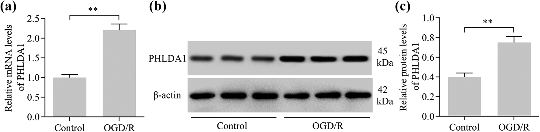
The effect of OGD/R injury on changes in PHLDA1 expression in HT22 hippocampal neurons. (a) Changes in the PHLDA1 mRNA in neurons suffering from OGD/R injury were detected via RT-qPCR (N = 3). (b and c) Changes in the PHLDA1 protein in neurons suffering from OGD/R injury were determined via the western blotting assay (N = 3). **p < 0.01.
Inhibition of PHLDA1 relieved OGD/R-evoked neuronal apoptosis and ROS production
To investigate the function of PHLDA1 in OGD/R injury, we implemented the loss-function-experiments of PHLDA1 using siRNA-mediated gene knockdown. The data showed that delivering siRNAs targeting PHLDA1 into HT22 neurons dramatically decreased the expression of PHLDA1 (Figure 2(a) to (c)). Exposure to OGD/R significantly impaired neuronal viability (Figure 2(d)). Intriguingly, the knockdown of PHLDA1 could effectively attenuate the deleterious effects of OGD/R on neuronal viability (Figure 2(d)). Moreover, the inhibition of PHLDA1 clearly reduced OGD/R-evoked neuronal apoptosis (Figure 2(e) and (f)), as detected by flow cytometry. Consistently, western blotting showed that inhibition of PHLDA1 also suppressed the increases in cleaved caspase-3 level caused by OGD/R (Figure 2(g)). OGD/R-induced increases in intracellular levels of ROS were also strikingly down-regulated via PHLDA1 knockdown (Figure 2(h)). To validate the role of PHLDA1 in OGD/R injury, we further detected the effects of PHLDA1 overexpression on OGD/R-induced neuronal apoptosis and ROS generation. Our results exhibited that the overexpression of PHLDA1 (Figure 3(a) and (b)) aggravated the deleterious effects of OGD/R on neuronal viability (Figure 3(c)). Moreover, OGD-evoked increases in neuronal apoptosis (Figure 3(d) and (e)) and ROS levels (Figure 3(f)) were also further exacerbated by PHLDA1 overexpression. In short, these data indicate that PHLDA1 inhibition protects against OGD/R-evoked neuronal apoptosis and oxidative stress.

The knockdown of PHLDA1 restrained OGD/R-evoked increases in neuronal apoptosis and oxidative stress. HT22 neurons were transfected with PHLDA1 and control siRNAs for 48 h and then subjected to OGD/R injury. The down-regulation of PHLDA1 in siRNA-transfected neurons with or without OGD/R injury was verified via (a) RT-qPCR and (b and c) western blotting assays (N = 3). (d) The effect of PHLDA1 inhibition on OGD/R-impaired neuronal viability was examined via the CCK-8 viability assay (N = 5). (e and f) The effect of PHLDA1 inhibition on OGD/R-evoked apoptosis was measured via the apoptosis assay (N = 3). (g) The effect of LHLDA1 inhibition on OGD/R-induced increases in cleaved capase-3 level was determined by western blotting (N = 3). (h) The effect of PHLODA1 inhibition on OGD/R-evoked oxidative stress was assessed via the ROS assay (N = 5). **p < 0.01; ***p < 0.001.

The up-regulation of PHLDA1 exacerbated OGD/R-induced neuronal apoptosis and oxidative stress. HT22 neurons were transfected with the PHLDA1 expression vector or control vector (CV) for 48 h prior to OGD/R injury. (a and b) The up-regulation of PHLDA1 was confirmed via western blotting (N = 3). (c) The effect of PHLDA1 overexpression on OGD/R-impaired neuronal viability was evaluated via the CCK-8 viability assay (N = 5). (d and e) The effect of PHLDA1 overexpression on OGD/R-induced apoptosis was assessed via the apoptosis assay (N = 3). (f) The effect of PHLDA1 overexpression on OGD/R-induced oxidative stress was monitored via the ROS assay (N = 5). **p < 0.01; ***p < 0.001.
Inhibition of PHLDA1 repressed OGD/R-induced release of proinflammatory cytokines
To further assess the role of PHLDA1 in OGD/R injury, we determined the effect of PHLDA1 inhibition on OGD/R-induced inflammatory response in HT22 neurons. The results showed that OGD/R exposure led to significant increases in the levels of IL-6, TNF-α and IL-1β (Figure 4(a) to (c)). Strikingly, the knockdown of PHLDA1 weakened OGD/R-induced increases in the levels of IL-6, TNF-α and IL-1β (Figure 4(a) to (c)). On the contrary, the overexpression of PHLDA1 reinforced OGD/R-induced increases in the levels of IL-6, TNF-α and IL-1β (Figure 4(d) to (f)). In short, these data indicate that the inhibition of PHLDA1 restrains OGD/R-induced inflammatory response in neurons.

PHLDA1 participated in the modulation of OGD/R-induced inflammatory response. The effect of PHLDA1 knockdown on the levels of (a) IL-6, (b) TNF-α and (c) IL-1β was determined via ELISA analysis (N = 5). The effect of PHLDA1 overexpression on the levels of (d) IL-6, (e) TNF-α and (f) IL-1β was quantified via ELISA analysis (N = 5). **p < 0.01; ***p < 0.001.
Inhibition of PHLDA1 enhanced Nrf2 activation in OGD/R-injured neurons
Nrf2 has been documented as a master regulator of neuronal apoptosis, oxidative stress and inflammatory response under OGD/R conditions. Herein, we determined the role of PHLDA1 in modulating Nrf2 signaling in OGD/R-injured neurons. Strikingly, our data showed that OGD/R-induced increases in Nrf2 nuclear translocation were enhanced by PHLDA1 knockdown (Figure 5(a) and (b)). Moreover, the knockdown of PHLDA1 dramatically up-regulated the transcriptional activity of Nrf2 (Figure 5(c)). In addition, the overexpression of PHLDA1 markedly blocked the activation of Nrf2 signaling in OGD/R-injured neurons (Figure 5(d) to (f)). Altogether, these findings imply that the inhibition of PHLDA1 potentiates Nrf2 signaling in OGD/R-injured neurons.

PHLDA1 regulated Nrf2 activation in OGD/R-injured neurons. (a and b) The effect of PHLDA1 knockdown on the levels of Nrf2 in nucleus was examined via the western blotting assay. Lamin B1 served as a loading control for nuclear protein (N = 3). (c) The effect of PHLDA1 knockdown on Nrf2 transcriptional activity was determined via the ARE luciferase reporter assay (N = 5). (d and e) The effect of PHLDA1 overexpression on the levels of Nrf2 in nucleus was assessed via the western blotting assay (N = 3). (f) The effect of PHLDA1 overexpression on Nrf2 transcriptional activity was monitored via the ARE luciferase reporter assay (N = 5). *p < 0.05; **p < 0.01.
PHLDA1 regulated Nrf2 signaling via regulation of GSK-3β
We next investigated the molecular basis of PHLDA1 in regulating Nrf2 signaling. Strikingly, we found that the knockdown of PHLDA1 significantly increased the phosphorylation of GSK-3β, a crucial regulator of Nrf2 activation, in OGD/R-injured neurons (Figure 6(a) and (b)). To confirm that PHLDA1 knockdown enhances Nrf2 signaling via regulating the phosphorylation of GSK-3β, we determined the effects of GSK-3β reactivation of PHLDA1-induced Nrf2 activation. We utilized the constitutively active vector of GSK-3β (GSK3β-S9A) to reactivate GSK-3β in OGD/R-injured neurons with PHLDA1 knockdown (Supplementary Figure 1). Our results demonstrated that the reactivation of GSK-3β clearly abolished the PHLDA1-knockdown-induced enhancing effects on Nrf2 activation (Figure 6(c)). Moreover, the reactivation of GSK-3β dramatically reversed the protective effects of PHLDA1 knockdown on the inhibition of OGD/R-induced neuronal apoptosis (Figure 6(d) and (e)), ROS production (Figure 6(f)) and proinflammatory cytokine release (Figure 6(g) to (i)). Moreover, we investigated the effect of GSK-3β on PHLDA1-overexpression-medaited inactivation of Nrf2 in OGD/R-injured neurons. Interestingly, we found that inhibition of GSK-3β by a chemical inhibitor abolished PHLDA1-overexpression-mediated suppressive effects on Nrf2 activation (Supplementary Figure 2). Collectively, these finding confirm that GSK3β contributes to PHLDA1-mediated Nrf2 signaling.

The knockdown of PHLDA1 enhanced Nrf2 signaling via regulation of GSK-3β. (a and b) The effect of PHLDA1 knockdown on the levels of total GSK-3β and phospho-GSK-3β was determined via the western blotting assay (N = 3). HT22 neurons were transfected with PHLDA1 siRNA and GSK-3β-S9A vector for 48 h prior to OGD/R injury. (c) The activation of Nrf2 signaling was assessed via the ARE luciferase reporter assay (N = 5). (d and e) Neuronal apoptosis was evaluated via the apoptosis assay (N = 3). (f) ROS levels were monitored via the ROS assay (N = 5). The levels of (g) IL-6, (h) TNF-α and (i) IL-1β were quantified via ELISA analysis (N = 5). *p < 0.05; **p < 0.01; ***p < 0.001.
Inhibition of PHLDA1 protected against OGD/R injury via enhancing Nrf2 signaling
To determine whether PHLDA1 inhibition protects against OGD/R injury via Nrf2, we explored the effect of Nrf2 inhibition on PHLDA1-knockdown-mediated effects in OGD/R-inured neurons. We showed that treatment with Nrf2 inhibitors clearly repressed the activation of Nrf2 signaling in OGD/R-injured neurons with PHLDA1 knockdown (Figure 7(a)). As expected, the inhibition of Nrf2 markedly reversed the suppressive effects of PHLDA1 on OGD/R-evoked neuronal apoptosis (Figure 7(b) and (c)), ROS production (Figure 7(d)) and proinflammatory cytokine release (Figure 7(e) to (g)). Altogether, these results confirm that PHLDA1 inhibition protects against OGD/R injury via Nrf2.

The inhibition of Nrf2 reversed PHLDA1-knockown-induced neuroprotective effects on OGD/R injury. HT22 neurons were transfected with PHLDA1 siRNA and pARE vector, followed by cultivation for 48 h with or without Nrf2 inhibitor (ML385) prior to OGD/R injury. (a) Nrf2 activation was monitored via the luciferase reporter assay (N = 5). (b and c) Neuronal apoptosis was assessed via the apoptosis assay (N = 3). (d) ROS levels were measured via the ROS assay (N = 5). The levels of (e) IL-6, (f) TNF-α and (g) IL-1β were evaluated via ELISA analysis (N = 5). **p < 0.01; ***p < 0.001.
Discussion
This work, for the first time, reports a vital role of PHLDA1 in mediating OGD/R-evoked neuronal injury. Our data showed that the inhibition of PHLDA1 protected neurons from OGD/R-induced apoptosis, oxidative stress and inflammatory response. Our data further illustrate that PHLDA1 exerts a protective effect in OGD/R-injured neurons via potentiating Nrf2 signaling through the regulation of GSK-3β (Figure 8). Our work underscores a potential role of PHLDA1 in mediating neuronal injury during cerebral ischemia-reperfusion injury.

A proposed schematic diagram for PHLDA1-mediated Nrf2 signaling in protecting against OGD/R injury.
PHLDA1 expression is elevated under harmful stimulus or pathological conditions. PHLDA1 expression is increased in response to doxorubicin-, 5-fluorouracil-, Ultraviolet-, or γ-ray irradiation-induced cellular damage. 25 PHLDA1 is induced by homocysteine, which contributes to the progression of atherosclerosis in hyper-homocysteinemia. 26 In Sprague-Dawley rats, PHLDA1 expression is up-regulated following fenvalerate-induced testicular damage. 27 Jiao et al. reported that the exposure of lipopolysaccharides resulted in the significant induction of PHLDA1 in macrophages. 28 Notably, myocardial ischemia-reperfusion injury also evokes increases in PHLDA1 expression in myocardial tissues. 12 In this study, we showed that PHLDA1 expression was also induced by OGD/R injury in neurons. Our data indicate that elevated expression in OGD/R-exposed neurons may contribute to the mediation of OGD/R injury.
PHLDA1 has been recognized as a critical pro-apoptotic protein. PHLDA1 is capable of enhancing the apoptosis of T cell hybridomas via the induction of Fas expression. 6 The overexpression of PHLDA1 promotes the apoptosis of metastatic melanoma induced by chemotherapeutic agents. 29 The knockdown of PHLDA1 inhibits cancer cell apoptosis and promotes drug resistance to receptor tyrosine kinase inhibitors. 30 The up-regulation of PHLDA1 increases detachment-mediated apoptosis of vascular endothelial cells. 26 PHLDA1 plays an essential role in mediating fenvalerate-induced cellular apoptosis in rat testes. 27 The up-regulation of PHLDA1 enhances the apoptosis of cardiomyocytes exposed to hydrogen peroxide. 12 Moreover, the inhibition of PHLDA1 alleviates ischemia-reperfusion-induced cardiac apoptosis in vivo. 12 In the present work, we showed that the inhibition of PHLDA1 attenuated the deleterious effects of OGD/R injury on neuronal viability, and decreased OGD/R-evoked neuronal apoptosis. On the contrary, the overexpression of LPHLDA1 enhanced the sensitivity of neurons to OGD/R-evoked apoptosis. Therefore, our work confirms a pro-apoptotic role of PHLDA1 in OGD/R-injured neurons and proposes it as an attractive target for inhibiting neuronal apoptosis.
PHLDA1 exerts an important role in mediating oxidative stress. The deficiency of PHLDA1 reduces the oxidative stress in atherosclerosis associated with up-regulation of the antioxidant enzyme peroxiredoxin-1. 11 The knockdown of PHLDA1 decreases hydrogen peroxide-induced ROS generation in cardiomyocytes. 12 In this work, our data showed that the knockdown of PHLDA1 alleviated OGD/R-evoked ROS production in neurons, while overexpression produced the opposite effect. Thus, our data confirm a vital role of PHLDA1 in mediating the oxidative stress induced by OGD/R injury.
PHLDA1 has been reported as a new regulator of inflammation. The up-regulation of PHLDA1 contributes to lipopolysaccharide-induced inflammation in macrophages via affecting cellular proliferation. 28 Notably, PHLDA1 exerts a critical role in mediating lipopolysaccharide-induced neuroinflammation in microglia. 23 The knockdown of PHLDA1 weakened the expression of proinflammatory cytokines in mice substantia nigra injected with lipopolysaccharide. 23 In agreement with these findings, our study showed that the knockdown of PHLDA1 markedly lessened the release of proinflammatory cytokines in neurons following OGD/R injury. Our work confirms that PHLDAL1 inhibition produces anti-inflammatory effects.
Nrf2 is a well-documented protective factor in cerebral ischemia-reperfusion injury. Intriguingly, our work identified PHLDA1 as a novel regulator of Nrf2 activation. Our data confirmed that the knockdown of PHLDA1 enhanced the levels of nuclear Nrf2 and increased the transcriptional activity of Nrf2 in OGD/R-injured neurons, indicating that PHLDA1 enhances the activation of Nrf2. Moreover, our results showed that PHLDA1 inhibition potentiated Nrf2 signaling via increasing the phosphorylation of GSK-3β, a critical regulator of Nrf2. The phosphorylation of GSK-3β decreases the activity of GSK-3β, which can be mediated by Akt. 31 Indeed, Akt/GSK-3β contributes to the mediation of Nrf2 activation. 32 Interestingly, PHLDA1 plays a role in mediating Akt activation. It is reported that PHLDA1 is capable of inhibiting Akt activation, via competitively binding to phosphatidylinositol.25,30,33 Therefore, PHLDA1 inhibition may induce GSK-3β inactivation via enhancing Akt activation. Furthermore, our data showed that the reactivation of GSK-3β markedly reversed the enhancing effect of PHLDA1 inhibition on Nrf2 activation. In addition, the inhibition of Nrf2 partially diminished PHLDA1-inhibition-mediated neuroprotective effects in OGD/R-injured neurons. To summarize, our work illustrates that PHLDA1 inhibition protects against OGD/R injury via modulation of the GSK-3β/Nrf2 pathway.
Keap1 has been proposed as the main and canonical regulator of Nrf2. 34 Recently, a new mechanism known as non-canonical has been described, where the activation of Nrf2 can be achieved by other factors, including GSK-3β. 15 In this work, our data have shown that PHLDA1 regulates Nrf2 activation via regulation of GSK-3β. Our results demonstrated that activation of GSK-3β reversed PHLDA1-inhibition-induced activation of Nrf2, while suppression of GSK-3β abolished PHLDA1-overexpression-induced suppression of Nrf2. However, whether Keap1 is involved in PHLDA1-mediated Nrf2 signaling is unclear. Nevertheless, our work confirms that GSK-3β contributes to modulation of PHLDA1-mediated Nrf2 signaling.
In conclusion, we have identified PHLDA1 inhibition as a possible approach for protecting against OGD/R injury. Our findings showed that PHLDA1 inhibition ameliorated OGD/R-induced neuronal apoptosis, oxidative stress and inflammatory response in cultured hippocampal neurons in vitro by enhancing Nrf2 activation through the regulation of GSK-3β. Our study supports PHLDA1 as a new modulator of the GSK-3β/Nrf2 pathway and highlights the PHLDA1/GSK-3β/Nrf2 pathway as a new mechanism for mediating OGD/R injury in neurons. However, the precise roles of PHLDA1/GSK-3β/Nrf2 pathway in mediating neuronal injury in cerebral ischemia-reperfusion injury need to be studied in vivo using animal models.
Supplemental material
Supplemental Material, sj-pdf-1-het-10.1177_09603271211014596 - Loss of PHLDA1 has a protective role in OGD/R-injured neurons via regulation of the GSK-3β/Nrf2 pathway
Supplemental Material, sj-pdf-1-het-10.1177_09603271211014596 for Loss of PHLDA1 has a protective role in OGD/R-injured neurons via regulation of the GSK-3β/Nrf2 pathway by F Yang and R Chen in Human & Experimental Toxicology
Footnotes
Author contributions
F Yang designed the work and drafted the article and R Chen performed the experiments and interpreted the data.
Declaration of conflicting interests
The author(s) declared no potential conflicts of interest with respect to the research, authorship, and/or publication of this article.
Funding
The author(s) received no financial support for the research, authorship, and/or publication of this article.
Supplemental material
Supplemental material for this article is available online.
References
Supplementary Material
Please find the following supplemental material available below.
For Open Access articles published under a Creative Commons License, all supplemental material carries the same license as the article it is associated with.
For non-Open Access articles published, all supplemental material carries a non-exclusive license, and permission requests for re-use of supplemental material or any part of supplemental material shall be sent directly to the copyright owner as specified in the copyright notice associated with the article.