
Abstract
Background
Three editions of International Classification of Headache Disorders (ICHD) diagnostic criteria for Tolosa-Hunt syndrome (THS) have been published in 1998, 2004 and 2013; in ICHD-3 beta, there have been considerable changes. The validity of these new diagnostic criteria remains to be established.
Methods
We retrospectively identified 77 patients with non-traumatic painful ophthalmoplegia (PO) admitted between 2003 and 2013. We reviewed patients’ age at onset and gender, time courses between onset of pain and development of cranial nerve palsy, the cranial nerves involved, imaging findings, therapeutic efficacy of steroid treatment and recurrence of attacks.
Results
THS was the most frequent type of PO (46/77). In THS patients, the third cranial nerve was most commonly involved (76.3%). The median time interval between pain and cranial nerve palsy was two days, although in five patients (10.9%) the interval ranged from 16 to 30 days. Definitely abnormal MRI findings were found in 24 patients (52.2%).
Conclusions
It is essential to rule out other causes of PO in diagnosing THS, with MRI playing a crucial role in differential diagnosis. It may be helpful to understand and master the entity of THS for researchers and clinicians to adjust the gradation and ranking of the diagnostic criteria.
Introduction
Painful ophthalmoplegia (PO) refers to orbital pain with any combination of ipsilateral third, fourth or sixth cranial nerve palsies: Occasionally the optic nerve and ophthalmic division of the fifth nerve are involved (1).
ICHD-3 beta diagnostic criteria for 13.7 Tolosa-Hunt syndrome.

ICHD-3 beta: International Classification of Headache Disorders, third edition (beta).
We reviewed the cases of non-traumatic PO admitted to our hospital during the last 10 years in order to reveal its miscellaneous causes, and evaluate the ICHD-3 beta diagnostic criteria for THS.
Methods
This project was approved by the research ethics committee of the Chinese PLA General Hospital, and all patients gave written informed consent.
We conducted a search of final diagnosis on the hospital information system of the Chinese PLA General Hospital, Beijing, between the dates 1 January 2003 and 31 January 2013, using the following keywords: ‘Tolosa Hunt’, ‘ophthalmoplegia’, ‘oculomotor muscle palsy, ‘cavernous sinus’ and ‘idiopathic orbital inflammation’. We included all inpatients who had directly entered or were referred from other local hospitals to the departments of neurology, ophthalmology and vascular surgery for orbital pain and acquired third, fourth and/or sixth cranial nerve palsies. Their clinical records were retrospectively analysed, the following being recorded for each case: patient’s age at onset and gender, the cranial nerves involved, the time course between onset of pain and appearance of palsy(ies), previous medical history, MRI (including contrast-enhanced cerebral MRI) and angiographic findings (including computed tomography angiography (CTA), magnetic resonance angiography (MRA) and digital subtraction angiography (DSA)), laboratory findings including specific serologic tests (e.g. levels of angiotensin-converting enzyme (ACE), antineutrophil cytoplasmic antibody (c-ANCA), antinuclear antibody (ANA), anti-double-stranded DNA (anti-dsDNA)) and cerebrospinal fluid (CSF) analyses, therapeutic efficacy of steroid treatment, and recurrence of attacks. All patients were followed up to confirm the final diagnosis, the minimum interval between onset and follow-up being 10 months.
The diagnostic criteria for THS and diabetic ophthalmoplegia were based on those of ICHD-II (3). We did not rule out patients from the diagnosis of THS whose interval between onset of pain and ophthalmoplegia was more than two weeks or the time of resolution after steroid treatment was more than 72 hours. We compared the performances of the criteria of ICHD-I, ICHD-II and ICHD-3 beta (2,3,6).
Statistical analysis was performed using SPSS 16.0 (SPSS Inc, Chicago, IL, USA). Continuous variables with normal distributions were summarised as means and standard deviations, continuous variables with non-normal distributions as medians and quartiles, and categorical variables as numbers and percentages. p values < 0.05 were considered significant.
Results
Painful ophthalmoplegia: Distribution by aetiology of age and gender.

In THS, the median time interval between onset of pain and cranial nerve palsy was two days (quartiles 0, 7.25; range 0–30 days); in five patients (10.9%), longer time intervals ranged from 16 to 30 days. Recurrent attacks occurred in 17 patients (37.0%), of whom five (10.9%) had experienced at least one attack of similar orbital pain without ophthalmoplegia.
The cranial nerves affected in patients with THS are shown in Table 3; most commonly affected was the third (n = 36, 78.3%), but multiple (≥2) nerves were involved in 25 patients (54.3%). The distribution of lesions demonstrated by MRI in THS patients is shown in Table 4. Definitely abnormal MRI findings occurred in 24 patients (52.2%). Typical images are shown in Figure 1. Nine (19.6%) of the 46 THS patients had associated rhinosinusitis and/or mastoiditis.
(a) The abnormal soft tissue lesion is nearly isointense with grey matter on the coronal spin-echo (SE) T1-weighted image. (b) The lesion is isointense to mildly hypointense with grey matter on the coronal turbo spin-echo (TSE) T2-weighted image. (c) The lesion shows marked enhancement after intravenous gadolinium. The cavernous sinus shows enlargement and the lateral wall bulges. (d) The lesion extends towards the ipsilateral orbital apex on the post-gadolinium SE axial T1-weighted image. Tolosa-Hunt syndrome (N = 46): distribution of cranial nerve palsy(ies). Tolosa-Hunt syndrome (N = 46): Distribution and extent of granulomatous lesion on magnetic resonance imaging.


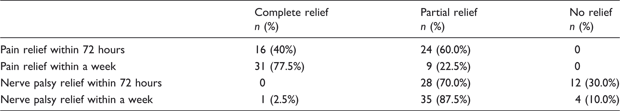
Tolosa-Hunt syndrome (N = 46): Response to steroid treatment.
ICHD diagnostic criteria for Tolosa-Hunt syndrome.

ICHD: International Classification of Headache Disorders; ICHD-II: International Classification of Headache Disorders, second edition; ICHD-3 beta: International Classification of Headache Disorders, third edition (beta); MRI: magnetic resonance imaging.
Relative performance of ICHD diagnostic criteria for THS in painful ophthalmoplegia.

Other diseases shown in Table 2.
ICHD: International Classification of Headache Disorders; THS: Tolosa-Hunt syndrome; ICHD-II: International Classification of Headache Disorders, second edition; ICHD-3 beta: International Classification of Headache Disorders, third edition (beta).
Discussion
The clinical features of THS and the efficacy of steroid treatment were described by Tolosa in 1954 (7) and Hunt in 1961 (8). THS is seen at almost any age (9) and without gender preference. The involvement of cranial nerves is variable (10). Our findings were similar to those of Tessitore and Tessitore (10), that the third cranial nerve was most commonly involved (85% of cases; our data 78%), then the sixth (70%; our data 41%), the ophthalmic branch of the fifth (30%; our data 28.0%) and the fourth cranial nerve (29%; our data 30%). Previous studies reported bilateral cranial nerve involvement in 4.1%–5.0% of cases (10); we found this in 4.3%. The recurrence rate of 37% in our study was higher than previously reported (21%) (11).
Diagnostic criteria for THS have been published by the International Headache Society in each of the three editions of ICHD (2,3,6) (Table 6), changing each time. In all editions they represented expert consensus based on case series. In the light of our data, we express our understanding about the changes and our doubts about the criteria.
First of all, the aetiology of THS is still unknown, which undoubtedly remains a barrier to formulation of specific and reliable criteria. While pathological examination has revealed a nonspecific inflammatory process, there is no specific biomarker to confirm the diagnosis of THS. Other causes of PO, all of which must be excluded, may be complicated: They include a range of vascular, neoplastic, nonspecific inflammatory and infective causes (1,12–15). According to our data, THS is the most likely cause, but intracranial aneurysm and diabetic ophthalmoplegia are not uncommon aetiologies of PO. Our study suggests that presentation with sudden-onset or pulsating headache should raise suspicion of an aneurysm of the PCA or ICA. There is a rare situation in THS that an ICA aneurysm may be directly induced by inflammatory infiltration into its intracavernous segment (16,17). Diabetic ophthalmoplegia is an important cause of PO (18,19) which, according to ICHD-II diagnostic criteria (Table 6), was difficult to differentiate from THS with a history of diabetes or abnormality of glucose tolerance, especially in patients with negative MRI findings (19). Third cranial nerve palsy without sparing of pupillary function and pain preceding the signs of neuropathy hint heavily at the diagnosis of THS but, while it is a difficult option for diabetic patients, a short experimental course of steroid therapy may be necessary for those whose diagnosis is uncertain. Beyond these, the differential diagnosis of THS includes other vascular diseases (such as cavernous sinus thrombosis and dural arteriovenous fistula (20)), neoplastic diseases (such as meningiomas (21), neurilemmoma, nasopharyngeal carcinoma, pituitary adenoma, lymphomas (22), and miscellaneous metastatic tumours (23,24)), infectious diseases (such as fungal infection, tuberculosis, syphilis, and actinomycosis), and specific granulomatous diseases (such as Wegener’s granulomatosis, sarcoidosis, giant cell arteritis and systemic lupus erythematosus). ‘Ophthalmoplegic migraine’ is a recurrent demyelinating or inflammatory cranial neuropathy, or a variant of THS with inflammation extending along the cranial nerve, and still controversial (12,25).
Second, with technological advances in imaging, the role of MRI in the criteria has been strengthened, from no special mention in ICHD-I through ‘and/or demonstration by MRI or biopsy’ in ICHD-II to ‘demonstrated by MRI or biopsy’ in ICHD-3 beta (2,3,6). Because the lesions of THS are located in the skull base, inaccessible, close to vital structures and small, it is not easy to obtain pathological samples. MRI therefore plays a crucial role in the differential diagnosis of THS (11,15,26,27). La Mantia et al. reviewed previously reported cases from 1998 to 2002 (4): in 85 patients who fulfilled ICHD-II diagnostic criteria for THS, 44 (51.8%) had apparent MRI or biopsy evidence of a cavernous sinus granuloma and 41 (48.2%) had no MRI abnormality. In our data similarly, 24 of 46 patients (52.2%) had MRI abnormalities. But in the series of Hung et al. there were strong similarities between patients with normal and abnormal MRI findings in terms of clinical manifestations, laboratory findings, responses to corticosteroid treatment and outcomes (28).
Dynamic contrast-enhanced and high-resolution MRI has undeniable superiority in exhibiting lesions that may be too subtle for conventional MRI sequences (29,30). Still we question the requirement in ICHD-3 beta for definite evidence in THS of focal granuloma. When lesions are seen on MRI, their location and extent are variable. Whatever the extent of spread of the lesions through the cavernous sinus/parasellar region or orbital apex/superior orbital fissure, or even intraorbitally, the disease entity may be uniform. Even orbital pseudotumour may be the same entity in a different location, though this remains controversial (31). ICHD-3 may need to relax its limitations on lesion location. Meanwhile, patients with negative MRI findings cannot be diagnosed as THS without biopsy evidence despite the fact that they might meet the criterion ‘Not better accounted for by another ICHD-3 diagnosis’ and despite our and previous findings that this applies to almost 50% of THS patients. ICHD is a worldwide standard for diagnosis for clinicians and researchers: Is it appropriate to set such a strict criterion at the present technological stage?
Third, ICHD-3 beta retains the temporal relation between pain and ophthalmoplegia: that headache precedes cranial nerve palsies by ≤2 weeks or develops with it. This, like other criteria, was an expert consensus based on case series. If the severe orbital pain of THS hints at involvement of the dura mater of the skull base, then it may be the case that the lesion extends to the adjacent ocular motor nerve. But in our study, five patients (10.9%) showed time intervals between onset of pain and nerve palsy of >15 days, and five THS patients (10.9%) experienced recurrent attacks of identical orbital pain without ophthalmoplegia. Do we need to study this temporal relation further?
Fourth, ICHD-3 downgraded the diagnostic role of therapeutic response to steroid treatment. At least in part, this was due to a general policy of not including response to treatment among ICHD-3 diagnostic criteria because diagnosis could not then precede treatment. It is true that there were many controversies about ‘Pain and paresis resolve within 72 hours when treated adequately with corticosteroids’ (criterion D in ICHD-II) (11). According to our data, 60% and 70% of patients respectively obtained partial pain and nerve palsy relief within 72 hours after steroid treatment. It was not appropriate to define the time course of relief strictly as in ICHD-I and ICHD-II, but should ICHD-3 beta have taken it away altogether? Treatment response to corticosteroids is still a very characteristic feature of THS, and resolution after steroid treatment is required to confirm the final diagnosis of THS (32).
Fifth, Iemma et al. reported a CT-scan based study of 1345 neurological and neurosurgical patients, of whom 75 (5.6%), with no previous history of paranasal sinusitis, exhibited inflammatory changes in the paranasal sinuses (33). In our study, nine of 46 THS patients (19.6%) had associated rhinosinusitis and/or mastoiditis. This is a significantly higher rate in THS than in Iemma et al.’s report of a broad group of patients. Do intracranial inflammations correlate with these extracranial inflammations? Have they the same aetiopathogenesis? Further studies of these questions are needed.
According to the diagnostic rates in our study among those receiving steroid treatment (ICHD-I 87.5%, ICHD-II 60.0% and ICHD-3 beta 47.5% (Table 7)), stricter limitations were applied for THS in the newer editions of ICHD. ICHD-3 has been published in beta version expressly to allow field-testing and revision, as appropriate, before ICHD-3 is finally published in 2015 or 2016. What can we do for the diagnostic criteria of THS? Grading and ranking diagnostic criteria while relaxing the time limitation between onset of pain and ophthalmoplegia, reintroducing relief after steroid treatment, with an appropriate time course (despite the accepted theoretical objections to including therapeutic response in diagnostic criteria) and adding details of MRI plus contrast enhancement to the diagnostic criteria all may be helpful to clinicians and researchers whose task is to understand and master the essence of this strange disease, THS.
In our opinion, graded diagnostic criteria may include the following:
Essential characteristics: painful ophthalmoplegia and recurrence of attacks; Primary characteristics: granulomatous inflammation (demonstrated by MRI or pathology) and good response to corticosteroid therapy; Secondary characteristics: localisation and/or extent of the inflammatory lesion and the temporal relation between onset of pain and ophthalmoplegia.
Ranking the diagnostic criteria may give rise to categories of definite, probable and possible TSH. Currently this categorisation is foreign to ICHD, but if patients with normal MRI findings or time interval between onset of pain and ophthalmoplegia of >2 weeks or whose diagnosis depends on pain and paresis resolving within 72 hours after steroid treatment cannot be diagnosed as definite TSH, and are not better accounted for by another ICHD-3 diagnosis, then probable or possible THS are, at least, working alternatives. Further studies may be required to operationalise such criteria, and certainly to field-test them.
Conclusions
According to our study, THS, intracranial aneurysm and diabetic ophthalmoplegia are the most common aetiologies of PO. It is essential to rule out all other causes of PO to confirm the diagnosis of THS. MRI plays a crucial role in the differential diagnosis of THS. But, since almost half of patients both in our and a previous series had normal MRI, it is very questionable whether ICHD-3 beta diagnostic criteria should rely on it (or biopsy) so completely. In the absence of other specific and reliable criteria, response to steroids is likely to remain important in supporting diagnosis in clinical practice, despite that, ideally, diagnosis should always precede treatment. Graded and ranked diagnostic criteria of THS, introducing categories of definite, probable and possible, may lead to better understanding and mastery of this strange entity.
Our study was a retrospective analysis. We look forward to a prospective multicentre study of THS.
Clinical implications
Some reported cases of 13.7 Tolosa-Hunt syndrome (THS) had additional involvement of the fifth nerve (commonly the first division) or optic, seventh or eighth nerves. Sympathetic innervation of the pupil is occasionally affected. The syndrome has been caused by granulomatous material in the cavernous sinus, superior orbital fissure or orbit in some biopsied cases. Careful follow-up is required to exclude other causes of painful ophthalmoplegia such as tumours, vasculitis, basal meningitis, sarcoid or diabetes mellitus. Pain and paresis of 13.7 THS resolve when it is treated adequately with corticosteroids.
Footnotes
Funding
This research received no specific grant from any funding agency in the public, commercial, or not-for-profit sectors.
Conflict of interest
None declared.
Acknowledgements
The authors would like to thank all of the referring clinicians.