
Abstract
To understand diseases of wild urban jackrabbits (Lepus townsendii), we autopsied 130 individuals that died near roadways in Calgary, Alberta, Canada. Renal hamartomas were present in 8 of 130 hares (6.2%; 95% confidence interval: 3.2%–11.7%). Most were unilateral (7/8); one case had bilateral lesions. Hamartomas are benign, tumor-like lesions comprised of normal tissue elements in abnormal amounts and arrangements. Macroscopically, hamartomas were white, tan, or pink-red, well-circumscribed, singular or multilobular, expansile nodules in the cortex or corticomedullary junction. Histologically, renal hamartomas consisted of well-demarcated mature stromal tissue with fibrous tissue and occasionally, adipocyte differentiation. These results represent a unique temporal and geographical cluster of a renal anomaly in an urban wildlife population. Renal hamartomas were not identified in other large studies of diseases in free-ranging leporids including hares. Contributing factors to this cluster remain unknown.
Growing interest in diseases of urban wildlife is concurrent with the expansion of urban areas and their human populations. Despite apparent population declines in their historical range of western North America, white-tailed jackrabbits (Family: Leporidae; synonym, white-tailed prairie hares; Lepus townsendii, hereafter referred to as hares) frequently inhabit cities. 19 Urban densities of hares can be significantly higher than nearby rural populations. 19 Hares exhibit crepuscular activity, high fecundity rates, and biannual pelage color change and consume a diet consisting of grass, forbs, and shrubs. 17 They are prey for small to medium urban carnivores and may be considered pests due to their propensity to damage plants. Previous studies have investigated the presence of specific pathogens or the general causes of death in free-ranging leporid populations.5,7,9,10 However, despite the ubiquity of wild urban leporids worldwide, and their apparent recent population decline, little research has been published on their diseases and health status, particularly in urban settings. 19
Urban wildlife disease research often focuses on zoonotic pathogens or notable epizootic disease events (eg, rabbit hemorrhagic disease). 10 However, incidental pathogens and lesions can alter behavior, increase predation risk, or reduce reproductive success and body condition in leporids.1,8,20 Furthermore, poor health in individual hosts has been correlated with an increased odds of carrying zoonotic pathogens. 15 Understanding the common pathogens and lesions of urban leporids will aid in developing baseline health data in the face of ongoing population declines and determine the risk they may pose to domestic animals and humans. As part of a larger study on the diseases of urban leporids in a major Canadian city, the objective of this study was to characterize hamartomas identified in the kidney of hares.
From October 2019 to July 2020, the City of Calgary submitted a convenience sample of hare carcasses (with location data) found near roads within Calgary, Alberta, Canada. We included only carcasses in good postmortem condition with minimal decomposition and with the majority of organs present (some organs were missing due to trauma/scavenging). Carcasses were stored frozen at (−20°C) until sampling.
As part of the larger project, we conducted a systematic gross examination on each carcass and collected samples for histological analysis. We collected data on demographic characteristics (body mass, sex, body condition, maturity, and pregnancy status). We classified sexual maturity in females based on pregnancy or uterine striation, and in males by location and color of testes. 11 We developed and applied a 3-point body condition scoring system (1: poor, 2: moderate, 3: good) that was further refined with observations of carcasses during initial autopsies (Supplemental Table S1).
Following identification of macroscopic kidney lesions, we fixed representative samples in 10% neutral-buffered formalin, then processed these tissues by routine methods. A veterinary pathologist (JLR) certified by the American College of Veterinary Pathologists examined 4-μm-thick tissue sections stained with hematoxylin and eosin and Masson’s trichrome using light microscopy. We used Epitools (Wilson method) to calculate the prevalence and confidence interval (https://epitools.ausvet.com.au/ciproportion) and Open Street Maps (https://openstreetmap.org), QGIS (QGIS Development Team), Canada Landcover 2020 dataset (Government of Canada), and Impervious Surface Map 2021 (City of Calgary) to map cases. We used R (R Development Core Team, Vienna, Austria) to calculate descriptive statistics of demographic characteristics (sex, body condition, body mass). We fit univariable logistic regression models with presence of hamartoma as the outcome variable and body condition (dichotomized to poor and moderate/good), sex, and body mass as the explanatory variables. We used a significance level of α ≤ 0.05 for all statistical analyses.
The University of Calgary’s Animal Care Committee approved this study (AC19-0107).
Between October 2019 and July 2020, we autopsied 130 hares. There were 57 females and 70 males (sex could not be determined for 3); 101 individuals were sexually mature and 24 were immature (maturity could not be determined for 5). Forty-one individuals were in poor body condition, 71 were in moderate body condition, and 15 were in good body condition (body condition could not be determined for 3). The mean body mass for males was 2.97 kg (interquartile range [IQR]: 2.70–3.20 kg) and 3.28 kg for females (IQR: 3.15–3.70 kg). Missing data were the result of autolysis or carcass condition (eg, organs lost due to trauma or scavenging).
The apparent prevalence of renal hamartoma was 6.2% (8/130; 95% confidence interval: 3.2%–11.7%; Fig. 1; Supplemental Table S2). Masses were unilateral in 7 cases; one case had bilateral masses. The presence of renal hamartoma was not significantly associated with sex, body condition, or body mass (P ≥ .1).
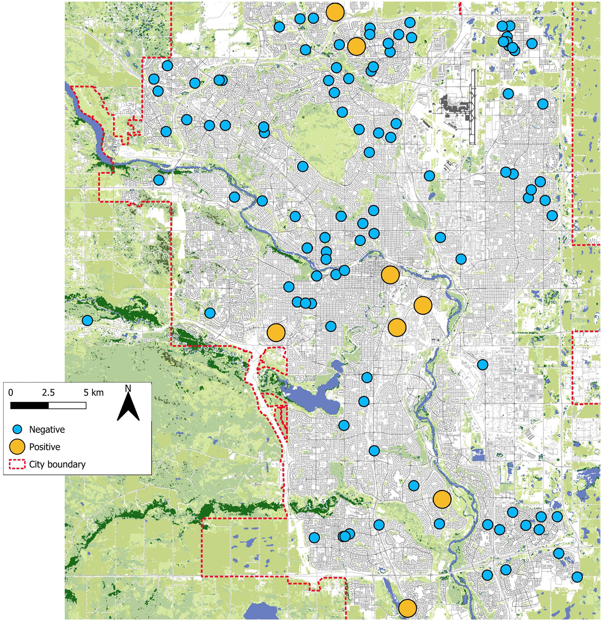
Map of the city of Calgary indicating the location of cases of white-tailed jackrabbits (Lepus townsendii) with renal hamartoma (yellow) and those without (teal). Cases are distributed across the city with no apparent small-scale geographical clustering.
Grossly, the renal cortex or corticomedullary junction contained well-circumscribed, singular or multilobular, expansile, firm, white, tan, or pink-red nodules that frequently elevated the renal capsule and ranged in size from 1 to 15 mm in diameter (Fig. 2). Histologically, hamartomas were well demarcated with mild compression of adjacent tissue. They consisted of well-differentiated fibroblasts and abundant mature collage that stained blue with Masson’s trichrome stain and surrounded and entrapped renal tubules. In 3 masses, there were mature adipocytes (3/9 masses, 33%). Tubules were dilated, forming cystic structures that contained proteinaceous fluid and/or cellular debris in 8 masses (8/9, 89%). Foci of lymphoplasmacytic inflammation were present in 6 masses (6/9, 67%). A spectrum of cellularity existed; four were paucicellular (4/9, 33%), four were moderately cellular (4/9, 56%), and one was highly cellular (1/9, 11%) (Supplemental Fig. S1). All tumor-like lesions were benign with no observed malignant transformation or metastasis.
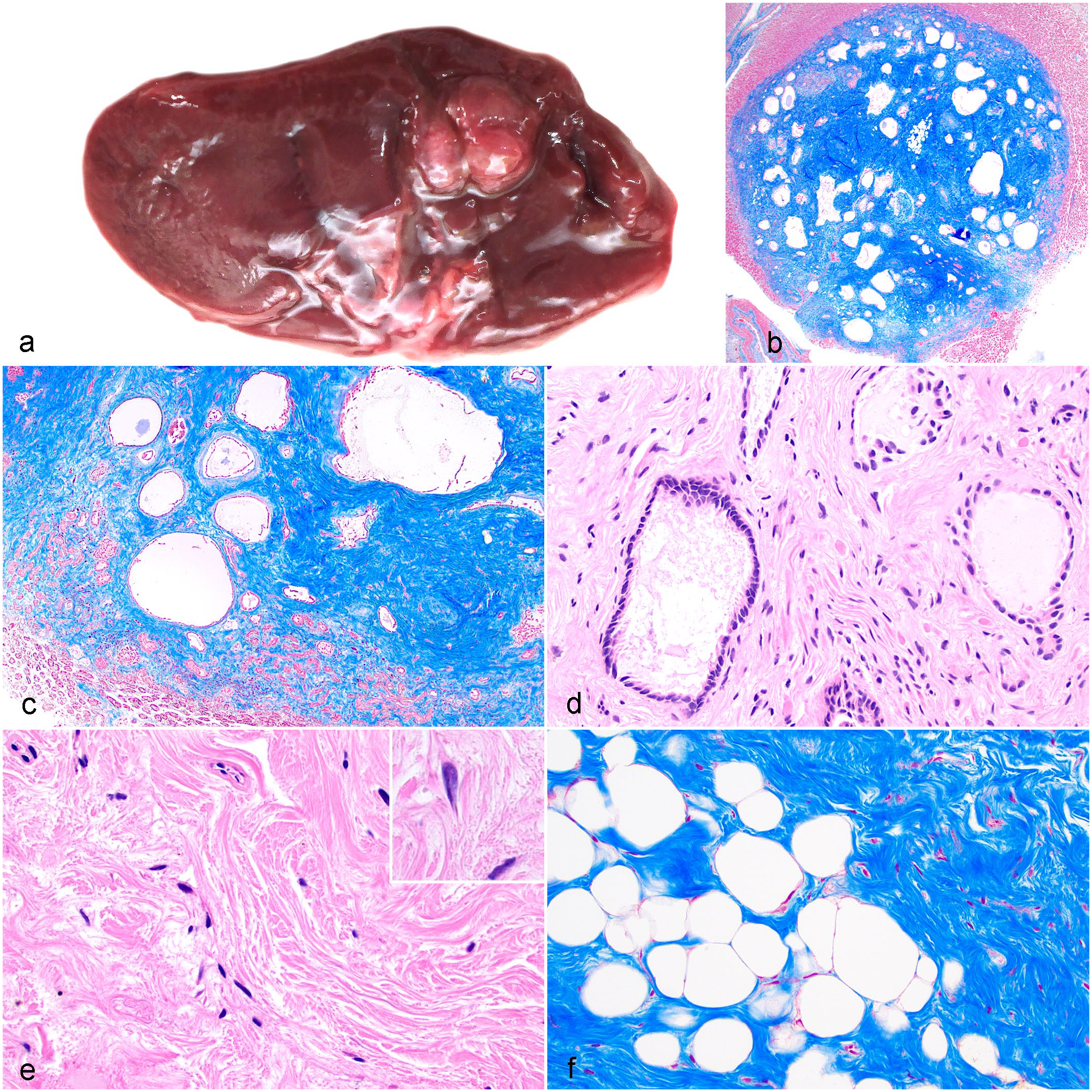
Renal hamartoma, kidney, wild, urban white-tailed jackrabbit (Lepus townsendii). (a) A pink-red, expansile hamartoma is present in the corticomedullary junction of the right kidney. Case 6. (b) The mass is well-demarcated, expansile, and compresses adjacent renal tissue. The mass has low cellularity and is mainly comprised of abundant mature collagen that surrounds many large cysts. Masson’s trichrome. (c) Junction of the hamartoma with normal tubules. Renal tubules (bottom) and large cysts are surrounded by abundant collagen. Masson’s trichrome. (d) Cysts are lined by low cuboidal epithelium and surrounded by collagen and few fibroblasts. Hematoxylin and eosin (HE). (e) Fibroblasts are spindloid with elongated nuclei and are surrounded by abundant bundles of mature collagen. Inset: fibroblasts are bland and uniform with no features of malignancy. HE. (f) Some areas of the mass contain bland, mature adipocytes with no features of malignancy. Masson’s trichrome.
Acute trauma was the cause of death in 7 cases; we could not determine the cause of death in 1 case. Among the cases, there were 6 females and 2 males; all were sexually mature and none of the females were pregnant. The body condition was poor in 4 cases (little to no visible fat stores) and moderate in 4 (moderate visceral and little subcutaneous fat stores).
This study identified a temporal and geographical cluster of wild urban hares affected by renal hamartomas. Hamartoma is defined as a “tumor-like malformation composed of an abnormal mixture of normal tissue elements or an abnormal proportion of a single element.” 13 Classification of the masses in our cases created a diagnostic challenge, similar to that described for a case of hamartoma in a wild cottontail rabbit. 4 Differential diagnoses for macroscopic renal masses in lagomorphs include hamartoma, nephroblastoma, angiomyolipoma, fibroma, renal adenocarcinoma, teratoma, metastatic neoplasia including lymphoma, and granuloma.
We considered the diagnosis of nephroblastoma since these are embryonic tumors that can include mesenchymal tissues such as cartilage, skeletal muscle, adipose tissue, and bone. 13 However, our cases did not have the characteristic epithelial and blastemal components of this tumor type. 18 Nephroblastomas are common in domestic rabbits (Oryctolagus spp.), 18 and there is a case described in a wild cottontail rabbit (Sylvilagus sp.). 4 Since the masses lacked an endodermic component, we excluded teratoma as a diagnosis. We dismissed angiomyolipoma based on the Masson’s trichrome staining characteristics; the majority of the lesions were comprised of collagen with typical blue staining versus pink that would be expected for smooth muscle cells. 3 Hamartoma, with predominance of mature fibroblasts, collagen, and, in 3 cases, adipocytes is the diagnosis of best fit for these lesions.
Neither renal hamartomas nor renal neoplasias were identified in large disease studies of several hundred wild leporids including hares.5–7,10 Similarly, no renal hamartomas were identified in a large sample of domestic rabbits. 2 This lack of previous identification in related wild and domestic leporids supports the uniqueness of our observations.
Since we did not identify malignant transformation, invasion, or metastases in our cases, and all but one case died of trauma, we concluded these tumor-like nodules were benign, incidental lesions. However, the small size of hare carcasses, environmental attrition, and scavenging could limit the detection of more severely affected cases. 14
Hamartomas are considered congenital lesions, likely occurring from improper tissue-development. 13 All our cases were in sexually mature hares; likely these lesions developed early in life and, due to their benign nature, persisted as the hare reached sexual maturity. Our study included only 24 immature individuals, so the absence of this lesion in that category may be a reflection of the low numbers examined.
As expected, based on our sample collection method, most individuals in our study died of acute trauma, presumably from encounters with motor vehicles. This convenience sample may have been biased toward individuals with demographic or disease characteristics that increased their vulnerability to vehicle trauma and inadvertently targeted sampling to geographical areas with road infrastructure within the urban environment. 16 The use of roadkill eliminated the need for active culling and tapped into an abundant supply of carcasses. This approach was ethically preferable and allowed us to focus resources on examining a large sample size versus the resources that would have gone into a live animal collection program. Although roadkill may be a biased convenience sample of the population of interest, it is an abundant and easily obtainable source of wildlife carcasses and can be an important resource for disease surveillance programs. Other large studies of passively collected lagomorphs, including those in Canada, did not identify any cases of renal hamartomas in hares.5,7,10 Studies of neoplasia in other species have benefited from focused and systematic sample collection methods like ours, rather than traditional passive surveillance methods. 14
No demographic risk factors were associated with the presence of hamartoma; however, this could be the result of the low number of animals with the lesion. The reason for this cluster of renal hamartomas remains unknown. One possibility is a lack of genetic diversity or inbreeding within this urban population, potentially due to a founder effect. 14 However, in this same sample, the co-evolved commensal lung organism, Pneumocystis sp., exhibited unexpectedly high genetic diversity, suggesting a population genetic bottleneck is unlikely. 12 In addition to genetic factors, general causes of neoplasia in wildlife include anthropogenic toxins and immunosuppression. 14 Future research could explore potential genetic homogeneity and anthropogenic environmental toxin exposure among these individuals to understand the factors contributing to hamartoma formation in this population.
Understanding urban wildlife and their diseases of concern aligns with a One Health approach that explores the intersections between animal, human, and environmental health. By investigating the health of an understudied, yet common urban mammalian species, we discovered an unusual cluster of individuals affect by tumor-like lesions. Given that wildlife neoplasia is likely under-reported, 14 our estimated prevalence is probably an underestimate. Diseases in general, including those with potential subclinical effects, should be considered as an important factor contributing to the apparent hare population decline across North America. 19
Supplemental Material
sj-pdf-1-vet-10.1177_03009858251367402 – Supplemental material for Geographical cluster of renal hamartomas in wild urban white-tailed jackrabbits (Lepus townsendii)
Supplemental material, sj-pdf-1-vet-10.1177_03009858251367402 for Geographical cluster of renal hamartomas in wild urban white-tailed jackrabbits (Lepus townsendii) by Summer T. Hunter, Marie-Anne Brundler, Sylvia L. Checkley, Susan C. Cork, Carolyn Legge, J. Scott Weese and Jamie L. Rothenburger in Veterinary Pathology
Footnotes
Acknowledgements
We wish to thank the City of Calgary Road Maintenance team for their collection and delivery of hare caresses, the team at the University of Calgary’s Diagnostic Services Unit, Megan Jones for her helpful feedback and encouragement to pursue this manuscript and Nigel Caulkett, whose ideas and enthusiasm were critical to the initiation of this research project. Fabien Mavrot kindly created the map of cases. The City of Calgary, where this work was completed, is located on the traditional territories of the people of the Treaty 7 region in southern Alberta and is also home to the Metis Nation of Alberta.
Supplemental material for this article is available online.
Author Contributions
STH and JLR designed and performed the autopsies and sample collection; STH, JLR, SLC, SCC, and JSW contributed to the study design; JLR, CL, and M-AB performed histologic evaluations; JLR performed statistical analysis; the manuscript was written by STH and JLR with contribution from the other authors.
Declaration of Conflicting Interests
The authors declared no potential conflicts of interest with respect to the research, authorship, and/or publication of this article.
Funding
The authors disclosed receipt of the following financial support for the research, authorship, and/or publication of this article: This research was supported by the University of Calgary and the City of Calgary.
References
Supplementary Material
Please find the following supplemental material available below.
For Open Access articles published under a Creative Commons License, all supplemental material carries the same license as the article it is associated with.
For non-Open Access articles published, all supplemental material carries a non-exclusive license, and permission requests for re-use of supplemental material or any part of supplemental material shall be sent directly to the copyright owner as specified in the copyright notice associated with the article.