
Abstract
Thirty-eight cases of renal tubular cell neoplasms were diagnosed in 184 captive, adult (> 1-year-old), black-footed ferrets (Mustela nigripes) examined from 1985 to 1996. This prevalence (20.7%) is one of the highest reported for this neoplasm in a population of animals. These tumors rarely metastasized (1/38), and usually were incidental postmortem findings, associated clinical disease being present in only 3 (8%) of the 38 cases. The prevalence of renal tubular cell neoplasms found at postmortem examination increased linearly with age, up to 67% in ferrets >8 years old. Both males (prevalence = 19%) and females (prevalence = 24%) were affected. Multiple renal tumors were common, and seven ferrets (18.4% of affected animals) had bilateral tumors. The cause of this neoplastic syndrome could not be determined. Since most of the animals affected by this condition were in their postreproductive years of life, the impact of this neoplastic syndrome on the captive propagation of this species is negligible.
In 1987, the last free-ranging, black-footed ferrets (Mustela nigripes) were captured near Meeteetse, Wyoming. With this capture began a propagation program that would not only succeed in saving this highly endangered species, but would also greatly contribute to increase knowledge of the biology of the black-footed ferret. One of the first concerns raised during the beginning of this propagation program was the potential low level of genetic diversity associated with the small number of founders, and the prolonged isolation of the original population. 16 To detect the expression of potential detrimental phenotypes, causes of death and other pathologic findings have been carefully monitored over the years. One of the most unusual findings from this surveillance was high prevalence of renal neoplasms. 11 Despite the unusually high prevalence of this type of tumor in this group of black-footed ferrets, to the authors' knowledge, this syndrome has not been described previously in this species. In addition, the potential impact of these neoplasms on the captive management of black-footed ferrets has not been evaluated. We describe the clinical presentation, pathology, behavior, and epidemiology of renal neoplasia in this species, and evaluate the potential detrimental effect of this condition on conservation of the black-footed ferret.
Materials and Methods
This study was conducted in 1997 on the entire captive population of black-footed ferrets. This population was established after the capture of the last remaining 18 ferrets from the Meeteetse population in the mid 1980s. The study population on which the observations reported here were made has been described fully elsewhere, as has the retrieval and methods of evaluation of case records and tissue blocks for tumors. 11 Because immature animals (<1 year old) were not affected by renal neoplasms, only animals that survived for at least 1 year and died in captivity were included in this study (from 1985 to 1996). Forty-three (19%) of the 227 ferrets meeting these criteria were not included in this study either because of insufficient information or to inability to retrieve the full necropsy report. Consequently, the results of the postmortem examination of 184 ferrets were used in this study.
Postmortem reports were retrieved from seven diagnostic laboratories, and were reviewed for descriptions of macroscopic or microscopic lesions, or both, that were consistent with renal neoplasia. All retrievable paraffin-embedded blocks with renal tissue (114), including 36 of the 38 cases in which renal neoplasia was reported, were recovered and exported from the USA into Canada under the Convention for International Trade in Endangered Species (CITES) permit number: CA-CW-IM-0016-98. Six-micron-thick sections were cut from each block and were stained with HE. 14 Each section was screened microscopically for neoplastic changes by the same pathologist (S. Lair).
Results
Thirty-eight of the 184 ferrets examined in this study were affected with renal tubular cell neoplasia, for prevalence at death of 20.7%. Prevalence in adult males and adult females was 19 and 24%, respectively. The prevalence of renal tubular cell neoplasms increased with age linearly, from 0% for ferrets <4 years old to 67% in ferrets >8 years old (Fig. 1).
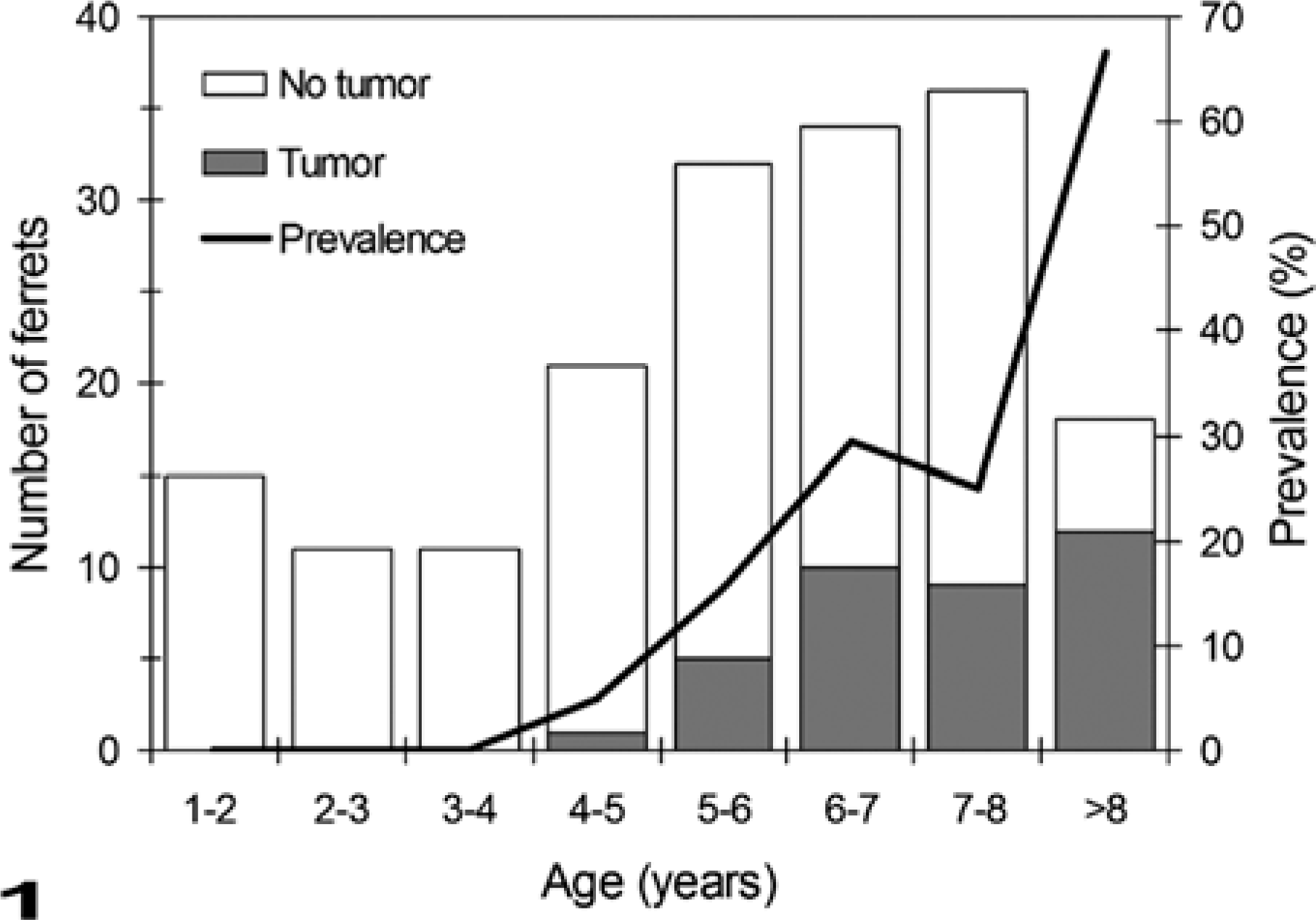
Age distribution of ferrets in the captive population with renal cell neoplasms (n = 171; age was available for 98 males and 73 females).
These neoplasms were seldom associated with clinical signs of disease. All but three of the 38 cases were incidentally found at postmortem examination. Emaciation was observed in two ferrets with large renal neoplasms, whereas the third ferret with clinical signs of disease died after 6 days of decreased food intake. In one of these animals, the renal tumor was diagnosed by palpation and radiographic examination 8 months before the appearance of the first clinical signs of disease.
The renal tubular cell neoplasms were well demarcated, spherical masses that usually protruded from the renal surface (Figs. 2, 3). The size of these neoplastic growths varied from pinpoint foci <1 mm in diameter to large tumors replacing most of the kidney (up to 22 × 16 mm). Up to five neoplastic masses were detected per animal (mean ± SE = 1.8 ± 0.2 tumors/affected ferret), and tumors were observed in both kidneys in seven of the 38 ferrets. On cut section, the neoplasms were composed of a homogenous, firm, white to yellow tissue. Central areas of necrosis and hemorrhage were present in larger tumors.
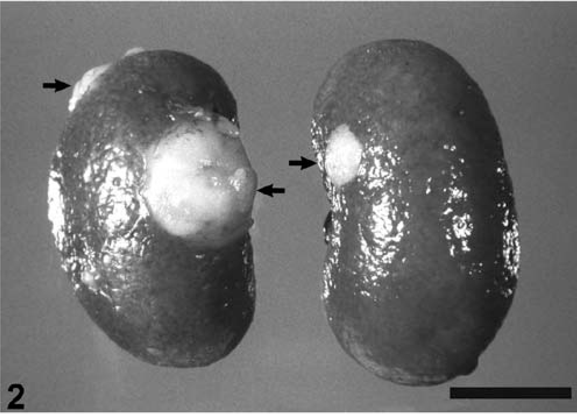
Kidney; 8-year-old male, black-footed ferret. Notice multiple, well demarcated, renal tubular-cell neoplasms (arrows). Bar = 1 cm.
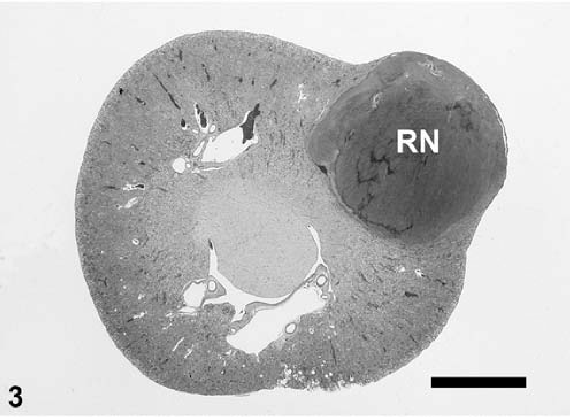
Kidney; 5-year-old female, black-footed ferret. Notice well-demarcated renal tubular cell neoplasm (RN) protruding from the renal cortex. HE. Bar = 2.7 mm.
Microscopically, renal tubular cell neoplasms were composed of densely packed, highly cellular nests of rudimentary tubular structures separated by sparse to moderately abundant fibrous stroma (Figs. 3, 4). Of the 36 cases examined microscopically, 21 had a predominantly tubular pattern, 10 had a solid organization, and five were formed by similar proportions of tubular units and solid sheets of cells. Neoplastic cells usually were infiltrating into adjacent renal parenchyma. Mature glomeruli, reflecting the expansive and infiltrative nature of these tumors, were frequently encountered in the neoplastic tissue. Larger tumors often were partially surrounded by compacted regional interstitial connective tissue. Neoplastic cells shared some morphologic similarities with renal tubular cells. Two types of neoplastic cells were observed. The most common type (present in 29 of the 36 cases) was small and polygonal, with scant, usually slightly basophilic cytoplasm and poorly defined borders (Fig. 4). The second type of neoplastic cells (present in 10 cases) had more-abundant, finely granular, acidophilic cytoplasm with poorly defined borders (Fig. 4). The level of cellular anaplasia was, in most of the cases, low to moderate. The nuclei of both types of neoplastic cells were round to ovoid and finely vesicular, with up to fivefold anisokaryosis. The chromatin was usually coarsely granular, and the nucleoli were small and pale. Except for the case in which metastases were observed, the mitotic index was low (0–2 mitotic figures/high-power [400X] field).
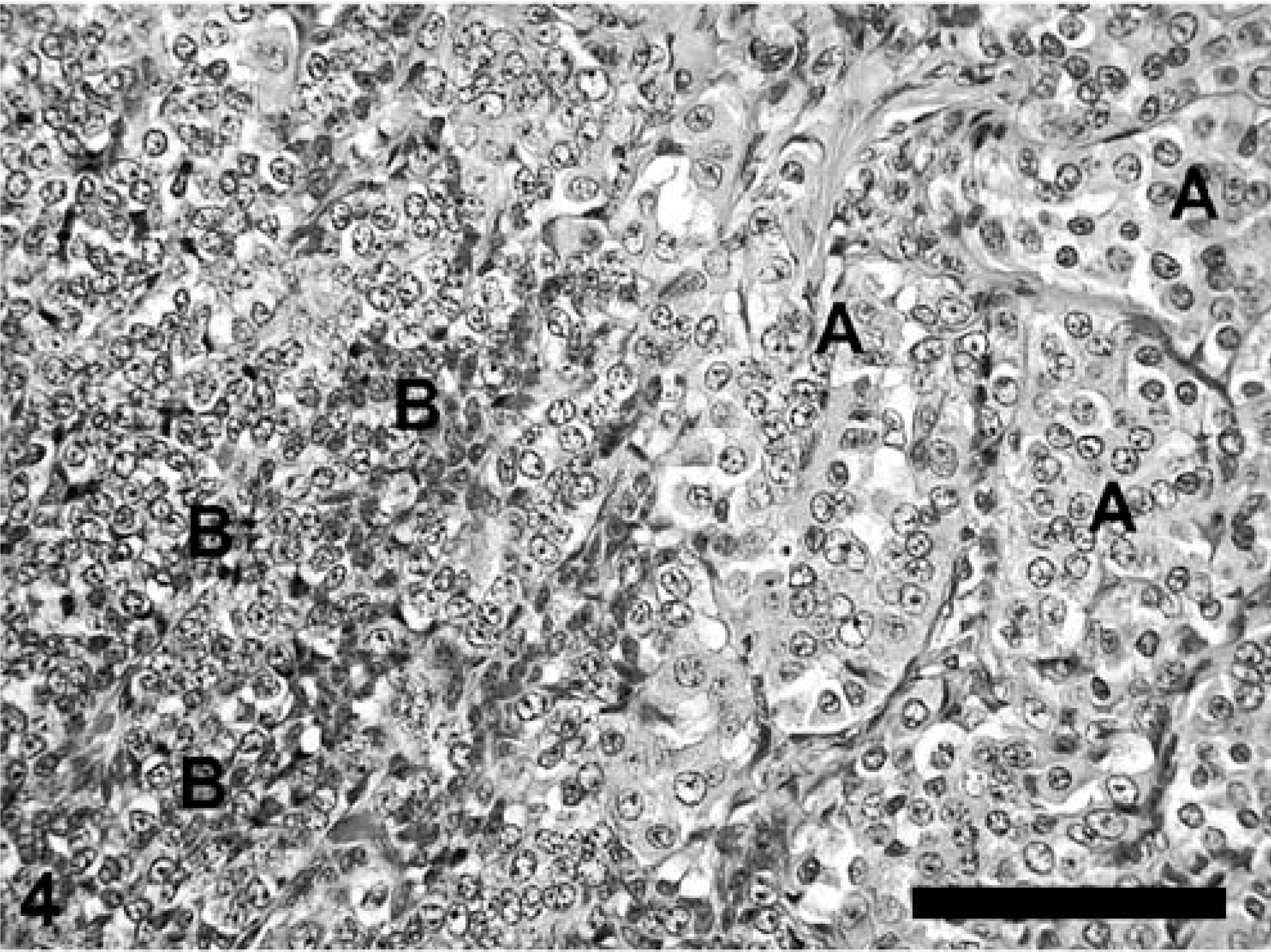
Kidney; 7-year-old male, black-footed ferret; renal tubular cell neoplasm. Two cellular phenotypes are present: A, neoplastic cells with abundant finely granular acidophilic cytoplasm forming rudimentary tubular structures; and B, small densely packed rhomboid cells with scant slightly basophilic cytoplasm and poorly defined borders. HE. Bar = 125 µm.
A common feature (in 29 of 36 cases) was the presence of osseous metaplasia in the center of the neoplastic masses that was characterized by the presence of networks of osseous trabeculae. These trabeculae were formed of partially mineralized osteoid matrix surrounded by well-differentiated osteoblasts and osteoclasts. A few hematopoietic cells were present in this osseous tissue. The tumors were frequently associated with a scirrhous reaction (in 28 of the 36 cases), and central necrosis was present in nine cases.
Metastases were observed in only one case. This ferret had multiple nodules in the kidneys, on the parietal peritoneum, and in the liver. The well-delimited hepatic and serosal nodules markedly compressed the adjacent parenchyma, and were composed of densely packed neoplastic cells that were similar to neoplastic renal tubular cells of the primary renal tumors.
Discussion
All neoplasms encountered in the kidneys of black-footed ferrets examined during this study were histologically similar; distinction between adenomas and carcinomas could not be made histologically. Consequently, these neoplasms are considered to be part of the same process. Multiple renal tumors were common, and were bilateral in at least 18% of the cases. This is a higher value than that in human beings, where bilateral renal tumors have a prevalence of 0.5–1.5%. 3 Multiple renal tubular cell neoplasms rarely have been described in domestic species, with the exception of cattle. 9 Metastases were detected in only 1 of 38 ferrets. Metastatic rates for this neoplasm also are low in mice 20 and rats, 2 though they are high in dogs 13 and human beings. 19 The generally low mitotic index is consistent with the low metastatic rates of these tumors.
The prevalence of renal tubular cell neoplasms observed in this group of ferrets was very high, compared with that of other species of mammals. 5, 17, 20 Renal tubular-cell neoplasms also are rare in domestic ferrets (Mustela putorius furo), where they have been reported only occasionally. 12, 21 This contrasts with the high prevalence reported here, and suggests that the black-footed ferret is predisposed to develop such tumors. An epidemiologic study looking at potential risk factors for renal tubular cell neoplasm in black-footed ferrets indicated a statistically significant association between the presence of this neoplasm and aging, 11 which is not an uncommon relationship for neoplastic diseases.
Predisposing factors for renal tumors in human beings include cigarette smoking, obesity, and exposure to petroleum products. 22 High rates of renal carcinomas have been described in a natural setting in free-ranging rats exposed to lead. 10 Since lead was not detected in the kidneys of any of the four black-footed ferrets tested, this heavy metal is unlikely to be causally associated with renal neoplasia in this species (data not presented). Numerous other xenobiotic compounds have been documented experimentally to induce renal epithelial tumors in laboratory animals. 20 The absence of geographic clustering among breeding facilities of cases of renal tubular cell neoplasms in black-footed ferrets does not favor the implication of local environmental factors in the development of this condition. 11
Epidemiologic studies have suggested a possible hormonal association with the cause of renal neoplasms in dogs, 13 mice, 20 and human beings. 1 The absence of sex predisposition and the lack of a relationship between renal tubular cell neoplasms and reproductive parameters do not support a potential hormonal influence in the development of this condition in black-footed ferrets. 11
Our study was not designed to explore potential infectious causes in the etiopathogenesis of this neoplasm. However, the absence of temporal or geographic case clustering and the absence of cases in young and middle-aged animals would be unusual for a disease associated with an infective agent. 11
Hereditary renal tubular cell neoplasms have been proposed in rhesus macaques (Macaca mulatta), 18 German Shepherd Dogs, 15 two strains of laboratory rodents, 6, 7 and human beings with von Hippel-Lindau disease. 8 Neither association between renal neoplasms and inbreeding nor evidence of familial clustering were documented in this group of ferrets. 11 These results and the advanced age at onset observed in our population of ferrets suggest that this condition is not a hereditary syndrome attributable to a single-gene disorder in this species. However, the high level of genetic homogeneity in the studied population might account for the failure to detect familial clustering. Interestingly, this type of tumor was not observed in the five black-footed ferrets from the South Dakota population that died at an old age. 4 Even if the number of animals examined from this other population of ferrets is too small to draw conclusions, this finding suggests that any predisposition for renal neoplasia is specific to the Meeteetse population rather than a characteristic of black-footed ferrets in general.
Since the cause of this tumor remains unclear, methods of prevention are unknown. Renal masses detected either by radiography or palpation in an adult black-footed ferret should strongly suggest a renal tubular cell neoplasm. Nephrectomy, the recommended treatment for this tumor in human beings 3 and dogs 13 is not recommended in black-footed ferrets owing to the common bilateral distribution, the low metastatic rate, and the advanced age at onset of this neoplasm in this species. Ferrets with renal tumors have remained free of clinical signs of disease for several months after the diagnosis, and most die of another cause.
Since the few animals in which this neoplasm was clinically relevant were in their postreproductive years of life, the impact of the high prevalence of this neoplastic condition on the captive propagation of this species is negligible.