
Abstract
Over a 16-year period, 190 tumors and tumorlike lesions from 179 pet rabbits were submitted for histopathologic examination. A total of 23 different tumor types and 1 tumorlike lesion were diagnosed. The most common diagnoses were trichoblastoma, collagenous hamartoma, and Shope fibroma. Viral-induced tumors were Shope fibroma (19) and Shope papilloma (2). Common nonviral epithelial tumors included trichoblastoma (59), squamous cell carcinoma (5), squamous papilloma (4), trichoepithelioma (3), and apocrine carcinoma (3). Common mesenchymal tumors were lipoma (10), liposarcoma (3), myxosarcoma (9), malignant peripheral nerve sheath tumor (8), fibrosarcoma (7), and leiomyosarcoma (4). Malignant melanoma was diagnosed in 8 rabbits. Collagenous hamartomas were diagnosed in 26 rabbits. Mesenchymal proliferations occurred significantly more often in male rabbits than in females. Collagenous hamartomas and myxosarcomas occurred exclusively in male animals, and 3 rabbits had multiple collagenous hamartomas. Immunohistochemistry was applied in cases in which a definite diagnosis could not be reached on hematoxylin and eosin slides. Follow-up information was received in 19 cases. Carcinomas recurred (2 of 3) or metastasized (1 of 3), whereas sarcomas frequently recurred (7 of 12). One malignant melanoma (1 of 3) and one poorly differentiated round cell neoplasm recurred (1 of 1). This is the first comprehensive retrospective analysis on skin neoplasia in pet rabbits.
Rabbits have increased in popularity as companion animals; 12 however, the majority of publications on neoplasia in this species have been derived from spontaneous or induced tumors in animals kept under laboratory conditions. 10, 14, 31, 48 In the laboratory setting, the rabbit population is composed of relatively few breeds. Furthermore, the rabbits are housed under controlled conditions and often are sacrificed at an early age (2 years or less), whereas the normal life span of a rabbit is 5 to 10 years. These properties sharply contrast with those of pet rabbits that present to local veterinarians for treatment and routine care. It is unclear whether data derived from such studies are applicable to pet rabbits.
Cutaneous neoplasms in rabbits can be divided into virus-induced and non–virus-induced tumors. Virus-induced tumors include rabbit Shope fibroma and papillomas. Shope fibroma, caused by Rabbit fibroma virus, a Leporipoxvirus, was first described in 1932, 40 and it is transmitted by fleas and mosquitoes. 16, 34, 45 These tumors most commonly occur during autumn. 34, 45 Approximately 7 to 12 days after the infection, animals develop one or multiple firm nodules that spontaneously regress over the course of a few weeks. 33, 34 In newborn, young juvenile, or immunosuppressed rabbits, the tumors may persist and metastasize, leading to the death of the animal. 16, 33, 34
Shope papilloma is also transmitted by biting arthropods, yet it is caused by a papovavirus. 16, 22 The virus is distinct from the rabbit oral papilloma virus, 16, 47 and it only infects the haired skin. 22 While some papillomas spontaneously regress, others progress to squamous cell carcinoma, with subsequent metastasis to lymph nodes and to the lung. 22 Regression or progression of the papilloma is determined by host factors, such as breed, immunosuppression, and previous infection. 22
Epithelial nonviral skin neoplasms have been cited in sporadic case reports or few case series and include basal cell tumors 11, 24, 36, 37, 48 and squamous cell carcinomas. 14, 48 Single case reports describe trichoepithelioma, 1 tricholemmoma, 28 and sebaceous gland carcinoma 32, 42 in rabbits.
Equally few mesenchymal tumors have been described, and these include lipomas, 25 neurofibrosarcoma, 3 hemangiosarcoma, 30 myxosarcoma, 35 malignant fibrous histiocytoma, 51 and extraskeletal osteosarcoma. 19, 35 Lymphosarcoma is considered the most common tumor of young rabbits, and numerous cases of multicentric lymphosarcoma with or without skin involvement have been published over the years. 5, 9, 17, 27, 39, 46, 49 Other tumors described include a few cases of malignant melanoma 2, 18, 20, 43 and a case report on eosinophil granulocytic sarcoma. 29
Three cases of collagenous hamartomas have been published to date. 13, 26, 50
The goal of this retrospective study is to describe the skin tumors in pet rabbits diagnosed at the surgical biopsy service at the Laboratory of Pathology and Toxicology, School of Veterinary Medicine, University of Pennsylvania (Philadelphia, PA), over a 16-year period. In accordance with the current literature on tumors in domestic animals, 6– 8, 15 both neoplasms and tumorlike lesions, such as hamartomas, are included here.
Materials and Methods
All records that included surgical specimens from pet rabbits submitted to the Laboratory of Pathology and Toxicology, School of Veterinary Medicine, University of Pennsylvania between 1 January 1990 and 31 March 2006 were reviewed. Only records that documented neoplasms and hamartomas that originated in the haired skin or mucocutaneous junction were included. A single case with multiple collagenous hamartomas submitted to the necropsy service of the University of Pennsylvania's School of Veterinary Medicine also was included. Mammary gland tumors were excluded. Two cases submitted by pharmaceutical companies and one case from a university research facility were excluded, as the animals were housed in a laboratory setting and therefore did not fulfill the criteria of a pet rabbit.
Questionnaires were mailed to the referring veterinarians of all animals diagnosed with a malignant neoplasm. The questionnaire included the verification of the animal's signalment, mode of treatment, behavior of the neoplasm (recurrence, metastasis), and the date and cause of the death.
Specimens were routinely processed for histopathologic evaluation and stained with haematoxylin and eosin (HE). To clarify morphology in sections obscured by melanin, additional sections were bleached prior to staining by immersion in 0.25% potassium permanganate (Fisher Scientific, Pittsburgh, PA). The glass slides were reviewed by two board-certified pathologists (W.v.B. and E.A.M.). Diagnoses were made in accordance with the Histological Classification of Tumors of Domestic Animals. 6, 15 Shope fibromas were diagnosed according to criteria described previously. 33
In cases in which a definitive histologic diagnosis could not be determined based on the HE slide, immunohistochemistry was performed. Immunohistochemical staining for vimentin (monoclonal anti-mouse; no pretreatment; 1 : 40; Dako, Carpinteria, CA), desmin (monoclonal anti-mouse; no pretreatment; 1 : 25; Dako), smooth muscle actin (monoclonal anti-mouse; no pretreatment; 1 : 30; Dako), cytokeratins AE1/AE3 (monoclonal anti-mouse; proteinase K pretreatment; 1 : 500; Chemicon, Ternecula, CA;), BLA36 (monoclonal anti-mouse; microwave pretreatment; 1 : 20; Dako), CD3 (monoclonal anti-mouse; microwave pretreatment; 1 : 50; Dako), CD79a (monoclonal anti-mouse; microwave pretreatment; 1 : 1000; Dako), and S100a (monoclonal anti-mouse; microwave pretreatment; 1 : 25; Lab Vision Neomarkers, Fremont, CA) were performed on 8 tumors. Vimentin, desmin, and smooth muscle actin were applied to 4 spindle cell sarcomas; cytokeratins AE1/AE3, vimentin, and S100a were applied to 3 tumors with a preliminary diagnosis of anaplastic carcinoma or malignant melanoma and BLA36, CD3, and CD79a, and cytokeratins AE1/AE3 were applied to 1 round cell neoplasm.
Sex distribution among mesenchymal tumors, epithelial tumors, and the overall submissions to the Laboratory of Pathology and Toxicology were compared using Fisher's exact test.
Results
Between January 1990 and March 2006, 507 lagomorph specimens were received by the surgical biopsy service. A total of 217 (43%) were from male animals, 255 (50%) were from female animals, and in 35 cases (7%) the sex of the rabbit was unknown. A total of 190 masses were diagnosed in 179 pet rabbits. A total of 24 different types of proliferations were diagnosed. A total of 9 animals had more than one mass. A total of 4 rabbits had two identical tumors: 1 rabbit had two trichoblastomas, and 3 rabbits had two malignant melanomas. Three rabbits had two different masses (squamous cell carcinoma/collagenous hamartoma; myxosarcoma/lipoma; collagenous hamartoma/lipoma). Two rabbits had three masses (collagenous hamartoma/collagenous hamartoma/leiomyosarcoma; trichoepithelioma/trichoepithelioma/collagenous hamartoma). Tumors and tumorlike lesions occurred in 21 different rabbit breeds. Anatomical locations of the proliferations diagnosed three times or more are given inTable 1, and sex and age distribution are given inTable 2. Mesenchymal proliferations were diagnosed significantly more often in male rabbits (n = 56; 79%) compared with the overall submissions of lagomorph specimens to the pathology service (males: 40%; P < .0001) or compared with the epithelial tumors in rabbits (males: 55%; P < .0038).
Localization of skin tumors and harmartomas in pet rabbits.
∗ n = number of tumors.
Tumor type, sex, and age distribution(in years) of rabbits with cutaneous masses.
∗ n = number of rabbits.
A total of 59 questionnaires were mailed to referring veterinarians. Follow-up information was received in 19 cases. In the other cases, veterinarians did not reply to the questionnaire, had discarded medical records, or the animal was never again presented to the practice.
Viral-induced tumors
Shope fibroma
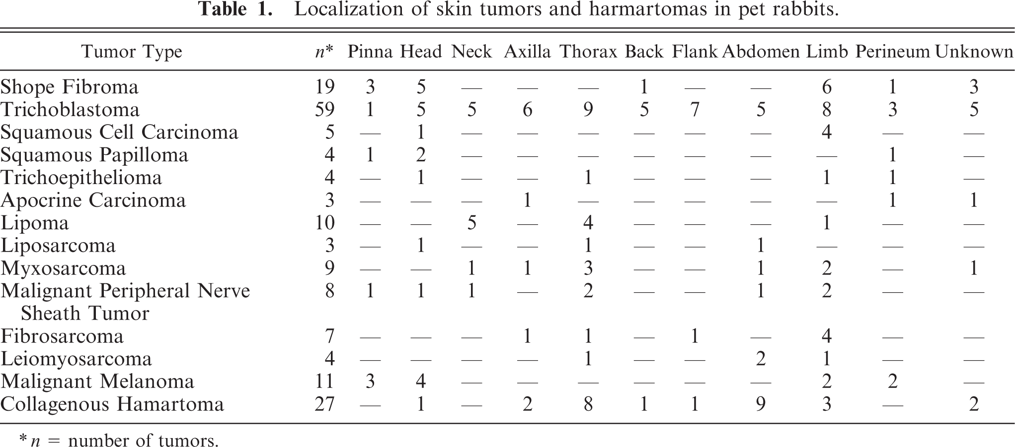
Shope fibroma was diagnosed in 19 rabbits. The most common locations were the limbs (6), head (5), and pinna (3). Cases were predominately submitted between July and November (16 of 19). A total of 12 cases were submitted from veterinarians in New Jersey, 4 were from Pennsylvania, and 1 each from Maryland, New Hampshire, and Virginia. Shope fibromas (Fig. 1) consisted of well-demarcated proliferations of highly pleomorphic ovoid to stellate-shaped cells with abundant pale eosinophilic cytoplasm. In a few cases, intracytoplasmatic eosinophilic inclusion bodies could be identified in these cells. Mitoses were frequent. Infiltration by heterophils, macrophages, lymphocytes, and plasma cells was a consistent finding. The overlying epidermis was hyperplastic and frequently ulcerated, and eosinophilic intracytoplasmic inclusion bodies were present in most cases. Even in cases with complete ulceration of the epidermis, the cell morphology of the atypical mesenchymal cells and the degree of inflammation were characteristic enough to separate Shope fibromas from sarcomas.
Skin; rabbit. Shope fibroma. Note hyperplastic and spongiotic epidermis with intracytoplasmic eosinophilic inclusion bodies (inlet). HE.
Shope papilloma
Shope papilloma was diagnosed on the pinna of 2 rabbits (one 6-year-old female and one 4-year-old male). These tumors had an exophytic papillated proliferation of squamous epithelium. Viral cytopathic effect consisted of nuclear chromatin margination, cytoplasmic clearing, and rare intranuclear inclusion bodies.
Nonviral epithelial tumors
Trichoblastoma
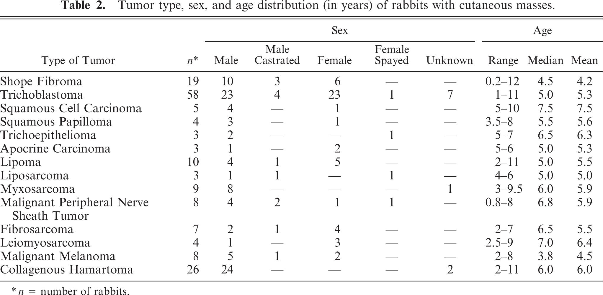
Trichoblastoma was the most common tumor in this study, and it was diagnosed in 58 rabbits. Trichoblastomas occurred as solitary neoplasms, apart from an instance of a 3-year-old male rabbit that had one tumor removed from the ear and one from the tail. On histologic examination (Fig. 2), there was a well-demarcated proliferation of epithelial cells arranged in anastomosing cords, ribbons, and lobules supported by a fine fibrovascular stroma. The cells were cuboidal to columnar with little eosinophilic cytoplasm, ovoid nuclei, and frequently were arranged in palisades. The epidermis was elevated and frequently ulcerated.
Skin; rabbit. Trichoblastoma with lobules of basaloid cells supported by a fine fibrovascular stroma. HE.
Squamous cell carcinoma
Squamous cell carcinoma was diagnosed in 5 animals. Two tumors originated from the subungual region and invaded P3. Three squamous cell carcinomas originated from the haired skin (metatarsus, hind limb, and medial canthus of the eyelid). The tumor on the metatarsus invaded the subjacent bone. All cases had a marked desmoplastic response. No viral cytopathic effect was noted in any of the tumors. One tumor recurred within a few weeks following surgery. The animal survived for 18 months after surgery and died for unknown reasons. Another rabbit with squamous cell carcinoma survived for 5 months after the surgery and died for unknown reasons.
Nonviral squamous papilloma
Nonviral squamous papilloma was diagnosed in 4 rabbits. Tumors occurred on the third eyelid (2), the pinna, and on the scrotum. The histologic changes consisted of exophytic papillated projections of mildly pleomorphic squamous epithelium that lacked features of viral cytopathic effect.
Trichoepithelioma
Trichoepithelioma was diagnosed in 3 rabbits. One 7-year-old male rabbit had 2 tumors (tail and lip). Both tumors were submitted for histopathologic exaulation. These tumors consisted of a well-demarcated, encapsulated, multilobular proliferation of cells with differentiation into the three segments of the hair follicle.
Apocrine carcinoma
Apocrine carcinoma was diagnosed in 3 animals. Histopathologic changes (Fig. 3) were characterized by a well-demarcated proliferation of cuboidal to columnar cells with basilar-oriented, moderately pleomorphic nuclei, which formed tubules, acini, or cysts. Mitoses averaged 2 to 6 per ten 40× fields. There was a moderate amount of fibrovascular stroma, and the neoplasms contained large areas of necrosis. In one rabbit, the apocrine carcinoma recurred 10 months after surgery and metastasized to another skin site and to the liver. This led to the euthanasia of the animal 13 months after the surgery.
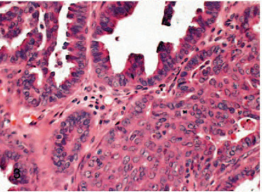
Skin; rabbit. Apocrine carcinoma with moderately pleomorphic tubules of cuboidal to columnar cells with few mitotic figures. HE.
Meibomian adenoma
Meibomian adenoma was diagnosed in one 4-year-old male rabbit and one female rabbit of unknown age. Histologically, there was a proliferation of mature sebocytes and few reserve cells.
Sebaceous carcinoma
Sebaceous carcinoma was diagnosed in a female rabbit of unknown age. The subcutis of the pinna was effaced by a multilobular proliferation of pleomorphic cuboidal to elongated cells with indistinct cell borders and variably vacuolated amphophilic cytoplasm. Mature sebocytes were scattered throughout the proliferation. Mitoses averaged 6 per ten 40× fields.
Nonviral mesenchymal tumors
Lipoma
Lipoma. was diagnosed in 10 animals. Locations were neck (5), thorax (4), and hind limb (1).
Liposarcoma
Liposarcoma was diagnosed in 3 rabbits. Microscopic examination (Fig. 4) revealed a well-demarcated, partly encapsulated proliferation composed of round to ovoid cells, with one variably sized cytoplasmic vacuole and eccentric mildly pleomorphic round to oval nuclei. In areas of high cellularity, the cells were more spindle shaped and pleomorphic and lacked cytoplasmic vacuoles. Mitoses were rare. One neoplasm had a prominent background of myxoid extracellular ground substance (myxoid variant). A subcutaneous thoracic liposarcoma in a 4-year-old female spayed rabbit was considered nonresectable, and the animal was treated palliatively with adriamycin. Despite treatment, the neoplasm increased in size, and the rabbit died 5 months after the initial diagnosis. On postmortem examination, the neoplasm had invaded the thoracic cavity, leading to pleural effusion and atelectasis.
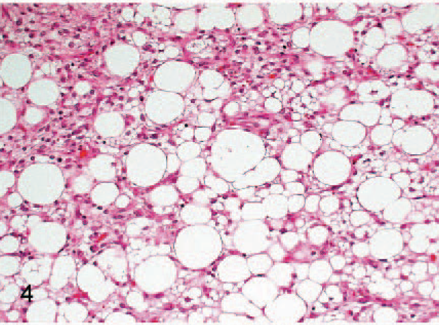
Skin; rabbit. Liposarcoma. Note mildly pleomorphic spindle cells with eccentric nuclei and variably sized lipid vacuoles. HE.
Myxosarcoma
Myxosarcoma was diagnosed in 9 cases (8 male rabbits and 1 rabbit with sex unknown). None of these tumors occurred in females. On histologic examination, the tissue was effaced by poorly demarcated whorls and bundles of stellate-shaped cells in abundant myxoid background. Pleomorphism was mild, and mitoses were rare. In areas of increased cellularity, cells were more spindle shaped to elongated and had fibrillar eosinophilic cytoplasm, and pleomorphism was more pronounced.
Malignant peripheral nerve sheath tumor
Malignant peripheral nerve sheath tumor was diagnosed in 8 animals. On histologic examination (Fig. 5), a well-demarcated, nonencapsulated dermal proliferation composed of short interwoven fascicles and perivascular whorls apposed and elevated the epidermis. Pleomorphism was mild; mitoses were infrequent. Minor portions of the neoplasm had a myxoid background. In 1 rabbit the malignant peripheral nerve sheath tumor recurred 6 weeks after the surgery. The recurrence was resected. The tumor again recurred 18 weeks after the second surgery. The rabbit was then lost to follow-up.
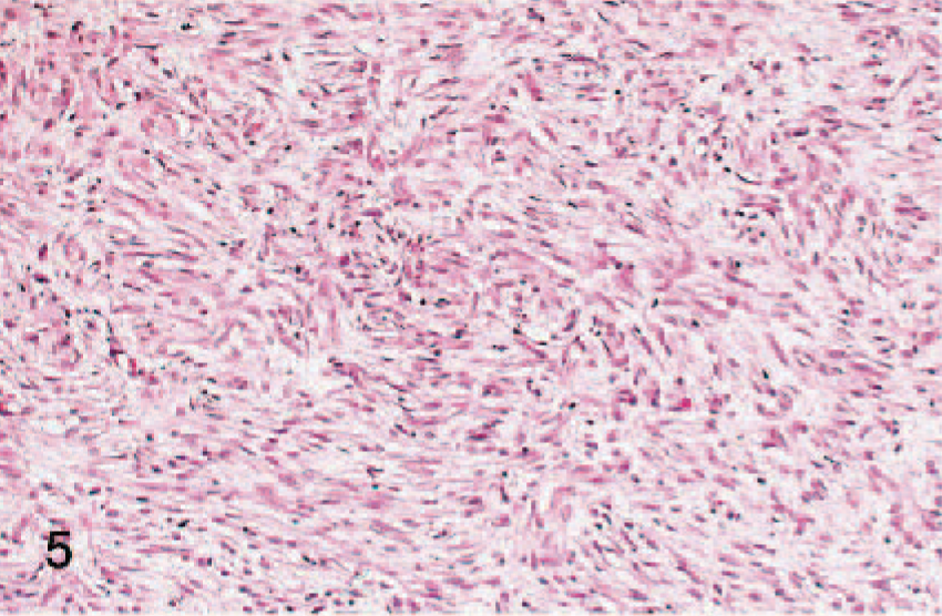
Skin; rabbit. Malignant peripheral nerve sheath tumor. Note short, interlacing fascicles of moderately pleomorphic spindle cells. HE.
Fibrosarcoma
Fibrosarcoma was diagnosed in 7 rabbits and most commonly occurred on the limbs (4). The dermis, panniculus, and subcutis were effaced by a highly cellular proliferation of long interwoven streams of spindle-shaped to elongated, moderately pleomorphic cells. Mitoses averaged 2 per 40× field. Collagen fibers were embedded between neoplastic cells. Frequently, small portions of the neoplasm contained a myxoid background. The neoplasm extended and elevated the mildly hyperplastic epidermis. Follow-up information was received on 4 fibrosarcomas. Tumors recurred 4 months, 5 weeks, and 5.5 months after the surgery, respectively. The first rabbit was euthanatized immediately after the recurrence. The other two animals survived up to 7 months and 9 months after surgery, respectively, and both died for unknown reasons. A fourth rabbit with fibrosarcoma died 2 weeks after the surgery for unknown reasons.
Leiomyosarcoma
Leiomyosarcoma was diagnosed in 4 rabbits. On microscopic examination there was a poorly demarcated neoplastic dermal proliferation composed of streams and bundles that extended to the mildly hyperplastic epidermis. Arrector pili muscles were embedded in the neoplasm. The cells were elongated and had indistinct cell borders, finely fibrillar cytoplasm, and elongated angular nuclei with coarsely stippled chromatin. Anisocytosis and anisokaryosis were moderate. Mitoses averaged 2 per ten 40× fields. Due to the small sample size and tissue artifact, one of the leimyosarcomas could not be diagnosed on HE with certainty. On immunohistochemistry, neoplastic cells stained positive for vimentin and smooth muscle actin but were negative for desmin. One of the rabbits with leiomyosarcoma died 2 weeks after the surgery for unknown reasons. Another rabbit, which had a tumor on its carpus, had the affected leg amputated. The animal died 8 weeks after amputation for unknown reasons.
Leiomyoma
Leiomyoma was diagnosed in a 12-year-old male rabbit at the lip. In contrast to the leiomyosarcomas, the proliferation was well demarcated and partially encapsulated. Cellular pleomorphism was mild, and the mitotic index was low (1 per ten 40× fields).
Anaplastic sarcoma
Anaplastic sarcoma was diagnosed in a 12.5-year-old male rabbit at the ventral abdominal skin and in a 6-year-old male rabbit in the axilla. Both anaplastic sarcomas had similar morphologic changes. These tumors were characterized by a proliferation of stellate-shaped to spindle-shaped cells forming whorls and bundles. The cells had indistinct cell borders, fibrillar eosinophilic to glassy gray, sometimes vacuolated cytoplasm, and round to ovoid nuclei with prominent nucleoli. Anisocytosis and anisokaryosis were high; mitoses averaged 1 to 4 per 40× field. Karyomegalic and multinucleated cells were frequent. Large areas of the neoplasm were necrotic, and minor portions had a myxoid background. On immunohistochemical examination, neoplastic cells in both cases stained positively for vimentin and negatively for smooth muscle actin and desmin. Both animals with anaplastic sarcomas developed recurrences within 4 and 12 weeks after surgery, respectively. In the former case, the recurrence was resected, recurred again, and the rabbit was euthanatized 7 months after the initial surgery for humane reasons related to the tumor. In the latter case, the animal died 5 months after the surgery for unknown reasons.
Osteosarcoma
Osteosarcoma was diagnosed on the flank of a 7-year7old male rabbit. The tumor was not associated with any underlying bone and was composed of a well-demarcated, nonencapsulated, highly cellular proliferation of spindle-shaped cells forming sheets and whorls. The cells were highly variable in size and shape; multinucleated cells and mitoses were frequent. Osteoid, bone, and cartilage were embedded in the neoplasm. The osteosarcoma recurred 7 months after the initial surgery. The recurrent tumor was resected and recurred again after 1 month. The animal died 16 months after the initial surgery for unknown reasons.
Hemangiosarcoma
Hemangiosarcoma was diagnosed in a 9-year-old male castrated rabbit and a 6-month-old male castrated rabbit. The neoplasms were localized to the elbow and to the neck, respectively. The dermis and subcutis contained a proliferation of highly pleomorphic endothelial cells forming irregular blood-filled cavities and clefts.
Rhabdomyosarcoma
A rhabdomyosarcoma occurred in the abdominal skin of a 6-year-old male rabbit. On histopathology (Fig. 6), the neoplasm was composed of interwoven bundles and whorls of spindle-shaped to elongated cells. The cells had indistinct cell borders, fibrillar eosinophilic cytoplasm, and centrally located ovoid to elongated nuclei with coarsely stippled chromatin. Anisocytosis and anisokaryosis were marked, multinucleated cells and karyomegalic cells were frequent, and mitoses averaged 2 per ten 40× fields. Large areas of the neoplasm were necrotic and partially mineralized; individual neoplastic cells stained strongly positive for desmin (Fig. 7). Focally neoplastic cells also stained positive for smooth muscle actin. The rabbit had metastasis to another skin site within 1 month after the surgery and was then lost to follow-up. The metastatic tumor was not submitted for histopathologic examination.
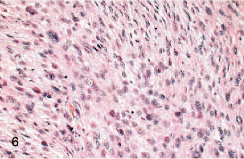
Skin; rabbit. Rhabdomyosarcoma with highly pleomorphic spindle cells with abundant fibrillar eosinophilic cytoplasm. Note the multinucleated cells. HE.
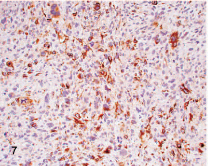
Skin; rabbit. Rhabdomyosarcoma. Note the strong cytoplasmic staining of neoplastic cells for desmin. Immunohistochemical staining for desmin.
Other nonviral tumors
Malignant melanoma
Malignant melanoma was diagnosed in 8 rabbits. In 3 cases, rabbits had two malignant melanomas, and both tumors were simultaneously submitted for histology. The locations of the melanomas were ear pinna (3), eyelid (3), head (1), perivulvar haired skin (1), scrotum (1), thigh (1), and stifle (1). Sheets or packets of epitheloid to spindle-shaped cells with distinct cell borders, amphophilic to gray cytoplasm, and round to ovoid nuclei with prominent nucleoli effaced the dermis. Anisocytosis and anisokaryosis were marked, and the mitotic index was high (4 to 20 per ten 40× fields). Intraepithelial nests were rare. Multiple small cytoplasmic vacuoles (balloon cell morphology) were noted in 2 cases. There was lymphatic invasion in 4 tumors. All tumors contained abundant melanin. One rabbit developed local recurrence 2 months after the surgery and died 3.5 months after the surgery. Another rabbit died 3 months after the surgery for unknown reasons without developing a grossly evident recurrence or metastasis. A third rabbit was euthanatized 7 weeks after the surgery for its poor clinical condition. There was no grossly visible recurrence or metastasis.
Three neoplasms that were presumed to be amelanotic malignant melanomas on HE were submitted for immunohistochemical examination. Pleomorphic epitheloid cells formed packets and sheets and resembled malignant melanomas, yet the cells lacked melanin pigment. Neoplastic cells stained negative for cytokeratins AE1/AE3 and positive for vimentin. S100a staining was inconclusive. The preliminary diagnosis could therefore neither be verified nor be excluded by the use of immunohistochemistry, and a final diagnosis of malignant neoplasm was given.
Lymphoma
Lymphoma occurred in the flank of a 4-year-old female rabbit. There was a well-demarcated dermal proliferation of neoplastic lymphoblasts forming sheets of cells. Pleomorphism and mitotic rate were high; epitheliotropism was not noted.
Poorly differentiated round cell neoplasm
A poorly differentiated round cell neoplasm was diagnosed on the head of a 10-year-old male rabbit. On histology, the tissue was effaced by sheets of neoplastic cells. The cells were round to ovoid, had indistinct cell borders, pale eosinophilic cytoplasm with round to ovoid, sometimes cleaved nuclei, and small nucleoli. Anisocytosis and anisokaryosis were moderate. Mitoses were frequent (2 per 40× field). Immunohistochemistry for CD3, CD79a, BLA36 and cytokeratins AE1/AE3 was negative. The rabbit died 4 weeks after the diagnosis due to local tumor growth.
Tumorlike lesions
Collagenous hamartoma
Collagenous hamartoma was diagnosed in 26 rabbits, making it the second most common proliferation in this survey. Collagenous hamartomas were diagnosed throughout the 16-year period of this survey and affected 11 different breeds of rabbits. A total of 24 rabbits were male, and the sex of the animal was unknown in 2 instances. No cases were recorded in female rabbits. Three of the rabbits had multiple grossly similar nodules. One of these rabbits was euthanatized at the age of 9 years due to severe Pasteurella pneumonia, and it was submitted to postmortem examination. Numerous (approximately 50) firm spherical (0.5–1 cm in diameter) nodules were randomly scattered throughout the dermis over the thorax and abdomen. In 4 cases, rabbits were described as having two grossly similar masses. In 1 case, both lesions were submitted. The most common locations of the hamartomas were in the dermis of the abdomen (9) and thorax (8). Histopathologic examination (Fig. 8) revealed a dermal nodule of excessive collagen blending into the surrounding dermis. Collagen fibers were slightly thicker or of equal thickness as in the adjacent tissue. Fusiform fibrocytes were embedded between collagen fibers. Hair follicles were frequently compressed or atrophied, and the epidermis was mildly elevated.
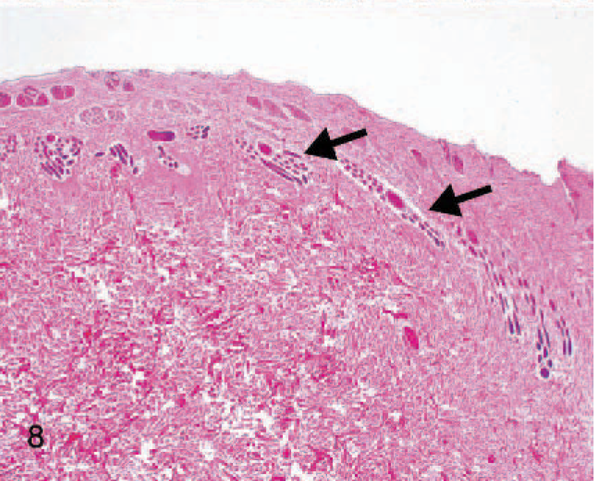
Skin; rabbit. Collagenous hamartoma. Note excessive dermal collagen, leading to atrophy and compression of hair follicles (arrows) and elevation of the epidermis. HE.
Discussion
This is the first comprehensive documentation of cutaneous tumors and tumorlike lesions in pet rabbits. To date, large studies on rabbit neoplasia were derived from laboratory animals and predominately contained data on young and juvenile animals. 10, 31, 48 The studies focused on uterine and mammary tumors and only described a few individual skin tumors. 10, 13, 14, 48
A total of 24 different types of skin tumors and tumorlike lesions were diagnosed in this survey. The three types most commonly diagnosed were trichoblastoma (59), collagenous hamartoma (27), and Shope fibroma (19), and these comprised more than half of the tumors. Except for Shope fibroma, this stands in vast contrast to the current literature. Lymphoma, 14 squamous cell carcinoma, 48 and Shope fibroma 25 are cited as the most common skin tumor among rabbits. Few individual case reports of trichoblastomas or basal cell tumors have been published to date. 11, 24, 36, 37, 48 To the authors' knowledge, only 3 cases of collagenous hamartomas have been cited. 13, 26, 50
Shope fibromas occurred on the limbs, head, and pinna, and were predominately diagnosed in autumn. This correlates well with previous studies. 16, 34
Epithelial tumors consisted largely of adenomas and only few carcinomas. To the authors' knowledge, apocrine carcinomas and meibomian adenomas have not previously been described. Papillomas, regardless of whether they are viral induced or not, commonly occurred on the ear or the eyelid. These are also common locations for canine papillomas. 8
Eleven different types of mesenchymal tumors were diagnosed. To the authors' knowledge, liposarcomas, fibrosarcomas, leiomyosarcomas, leiomyomas, and rhabdomyosarcomas have not been described previously. The sarcomas commonly apposed and interdigitated with the hyperplastic epidermis, and they frequently contained minor portions with a myxoid ground substance. At low magnification, these tumors thus shared features with Shope fibromas. The unique cellular features of Shope fibroma 33, 40 and the lack of inclusion bodies, however, clearly differentiates these two groups. The myxoid change frequently observed in the sarcomas seems to be a feature inherent to mesenchymal proliferations in rabbits, regardless of whether they are viral in origin or not, and therefore cannot be used as a helpful diagnostic feature.
Malignant melanomas were diagnosed in 8 animals. No melanocytomas were seen in this study and, to date, melanocytomas have not been described in lagomorphs. This contrasts with dogs, which develop more melanocytomas than malignant melanomas on the haired skin. 41 One rabbit developed a local recurrence. In 3 cases, the animals presented with 2 malignant melanomas at different sites, and in 4 tumors, lymphatic invasion was determined on histologic examination. This underscores the aggressive behavior observed in previous case descriptions, 18, 20, 43 and suggested by the high cellular pleomorphism and mitotic rate on histologic examination.
Interestingly, sarcomas, benign mesenchymal tumors, and hamartomas occurred significantly more often in male rabbits. This sex imbalance was not seen in the overall submissions of lagomorph specimens to the University of Pennsylvania or the nonviral epithelial skin neoplasms. Most drastic is this sex bias in myxosarcomas and collagenous hamartomas, which were seen exclusively in male animals.
For the myxosarcomas and other sarcomas, hormonal promotion might explain the male predominance. Sex hormones are a well-known tumor promoter. 23 However, it is unclear why this effect should be limited to sarcomas and not equally lead to the development of epithelial tumors.
Collagenous hamartomas are nonneoplastic nodules of exuberant collagen. 7 Therefore, a potential mode of pathogenesis is a hormonal induction in intact male rabbits, leading to increased activation of fibroblasts. This concept is supported by the fact that some of the animals had two or multiple collagenous hamartomas occurring simultaneously at different sites. Sex hormone–induced uterine leiomyomas have been described in humans, 4 dogs, and Guinea pigs, 21 and it is tempting to speculate that collagenous hamartomas in rabbits could possibly be reversed through castration. The occurrence of multiple simultaneous collagenous hamartomas is also reminiscent of nodular dermatofibrosis in German Shepherd dogs. 44 Multiple different breeds of rabbits were affected in our study, and we do not know of any concurrent disorders. Additional studies should be able to clarify this interesting phenomenon.
Most lesions could be classified according to the current World Health Organization Classification of Tumors in domestic animals and the present literature on neoplasia in rabbits. 33, 40 The neoplasms closely resembled common canine and feline tumors. Due to anaplasia or small sample size, 8 neoplasms could not be readily classified on HE. Only limited numbers of antibodies were available for immunohistochemical examination. Polyclonal anti-rabbit antibodies, frequently used in daily diagnostics, could not be applied to rabbit tissue. Thus, immunohistochemistry proved to be of limited value and helped to establish a diagnosis only in 2 of 8 cases.
Finally, the data must be interpreted in light of their retrospective nature. Medical records were frequently incomplete or discarded as a result of the long interval between the submission of the cases and the initiation of this study. The neoplasms with follow-up information behaved as expected from current knowledge on the behavior of canine and feline skin neoplasms. 7 Carcinomas recurred (2 of 3) or metastasized (1 of 3), whereas sarcomas frequently recurred at the surgery site (7 of 12). Only in 1 case did a sarcoma (rhabdomyosarcoma) metastasize to a distant site. The presumed metastasis was not reconfirmed by histologic examination. This suggests that the neoplasms in rabbits do behave comparably to the corresponding neoplasms in dogs and cats. It therefore seems feasible to apply the same criteria of malignancy.
Footnotes
Acknowledgement
We are grateful to Jackie Ferracone for the preparation of the immunohistochemical stains.