
Abstract
There is limited published data regarding cardiovascular disease in nondomestic felid populations. To address this knowledge gap, necropsy cases of tigers and lions with representative myocardial samples submitted to a diagnostic laboratory were histologically assessed with hematoxylin and eosin and Sirius red stains. A total of 32 submissions (15 tigers, 17 lions) were identified in a 4-year period. All tigers and lions had some degree of coronary artery lesions in the left ventricle and/or interventricular septum. Major findings included moderate to marked arteriosclerosis in 8 tigers (53%) and 4 lions (24%) and moderate to marked perivascular fibrosis in 10 tigers (67%) and 9 lions (53%). Moreover, 10 tigers (67%) and 8 lions (47%) had coronary artery lesions with variable degrees of perivascular cardiomyocyte degeneration and/or loss. To our knowledge, this is the first report describing coronary artery pathology in captive tigers and lions.
Cardiovascular disease is considered the leading cause of human mortality worldwide, with coronary artery disease (CAD) being the most common. CAD itself encompasses many presentations, but the most common form in humans is arteriosclerosis and is defined by the chronic thickening of arterial walls. This progressive thickening can occur from deposition of amorphous materials or fibromuscular proliferations. When there is distinct deposition of lipoproteins within the arterial walls, the more specific term of atherosclerosis is used. These changes can cause occlusive disease, leading to acute ischemic myocardial infarction.6,13 The underlying pathophysiology of arteriosclerosis is thought to be multifactorial and complex, with both genetic and lifestyle components implicated. 19 Another relatively common subtype of CAD in humans is microvascular coronary disease. This is a form of nonobstructive CAD that is also considered multifactorial, with alterations in both vascular architecture (such as perivascular fibrosis) and endothelial dysfunction implicated in its pathogenesis. 4 Although atherosclerosis is relatively uncommon in domestic species, arteriosclerosis has been reported in several veterinary species.1,18
The African lion (Panthera leo) and tiger (Panthera tigris) are common nondomestic felids in zoological and conservation settings. Free-ranging populations of both species have been markedly affected in the wild, leading to increased importance in captive conservation efforts and disease investigation. Common causes of captive morbidity and mortality in nondomestic felids include infectious disease, renal disease, dental disease, and neoplasia.9,10,16 Currently, there is limited published data regarding cardiovascular disease, especially coronary artery pathology and regional myocardial lesions, in nondomestic felid populations. Thus, the aims of our retrospective study are to determine the relative prevalence of intramural coronary artery pathology and identify the most common myocardial lesions in a cohort of captive nondomestic felids.
Necropsy cases submitted from 2019 to 2022 to the Veterinary Diagnostic Laboratory at Colorado State University were considered for inclusion in this study. Inclusion criteria consisted of tigers and lions with at least 1 representative sample of the ventricular walls and/or interventricular septum obtained through routine postmortem sampling. Tissue sections were stained with hematoxylin and eosin, and Sirius red, for evaluation. Intramural coronary arteries and myocardium were evaluated for arteriosclerosis, perivascular fibrosis, interstitial and replacement myocardial fibrosis, and cardiomyocyte degeneration and loss. Perivascular fibrosis was further subdivided based on increased perivascular connective tissue thickness, presence of perivascular dissecting or replacement fibrosis, and perivascular cardiomyocyte degeneration and/or loss. Severity of lesions was assessed semiquantitatively (Table 1). Arteriosclerosis within tissue sections was further graded based on the proposed scheme by Ryser-Degiorgis et al 18 as follows: grade 1, ≤5 vessels mildly to moderately affected; grade 2, ≤5 vessels moderately to severely affected or >5 vessels but <50% of the vessels mildly to moderately affected; and grade 3, >50% of the vessels mildly to severely affected. Prevalence was calculated and determined based on the most severe lesion in any of the heart sections. Location of lesions (right ventricle vs interventricular septum and left ventricle), age (≤10 years, >10 years), and sex (male, female) were evaluated for an association with lesions of at least mild severity or greater using separate Fisher’s exact tests, with P ≤ .05 considered significant.
Semiquantitative scoring scheme for intramural coronary artery lesions and myocardial pathology.
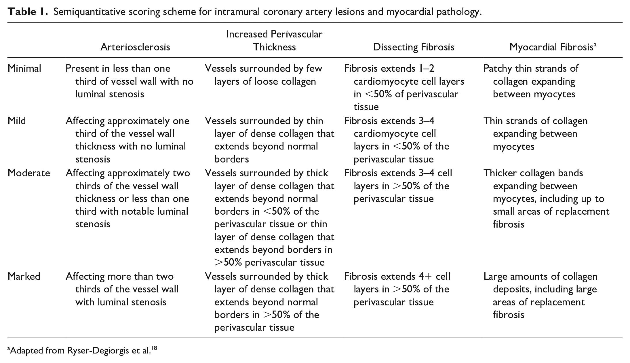
Adapted from Ryser-Degiorgis et al. 18
A total of 32 submissions (15 tigers, 17 lions) were identified with representative sections of right ventricle, interventricular septum, and left ventricle (tigers: n = 10, 12, and 14, respectively; lions: n = 17, 15, and 16, respectively). Three out of 32 cases had 1 heart section available, 6/32 had 2 heart sections available, and 23/32 samples had all 3 sections available for evaluation. Of the tiger cases, 5 were male, 10 were female, and the average age was 12.3 years (range 2–20 years, age of 1 tiger was not reported). Of the lion cases, 5 were male, 10 were female, and the sex was not reported for 1 lion. The average age of the lions was 14.3 years (range 9–21 years, ages of 2 lions were not reported). All lions and tigers were captive in a wildlife rehabilitation facility. In both tiger and lion cases, 30 animals were euthanized, 1 died due to other clinical concerns, and 1 had incomplete information on death. The most common reported cause of morbidity in tigers was chronic kidney disease (CKD, n = 8). In lions, the most commonly reported morbidity was infection by canine distemper virus (n = 6). Canine distemper virus (n = 5) and neoplasia (n = 3) were the second most common causes of disease in tigers and lions, respectively. By Fisher’s exact test, there was no significant association between age or sex and presence of cardiac lesions in either lions or tigers.
A summary of the prevalence of all lesions is reported in Table 2 (individual case data are provided in Supplemental Table S1). All tigers and lions had some degree of coronary artery lesions in the left ventricle and/or interventricular septum (Figs. 1, 2; representative normal coronary arteries are provided in Supplemental Fig. S1). Only one lion had lesions in the right ventricle (moderate myocardial fibrosis). Arteriosclerosis, perivascular fibrosis, and myocardial fibrosis were significantly associated with the left ventricle and interventricular septum in tigers (Fisher’s exact test, P = .0005, < .0001, and .0020, respectively) and in lions (Fisher’s exact test, P = .0003, < .0001, and .0066, respectively).
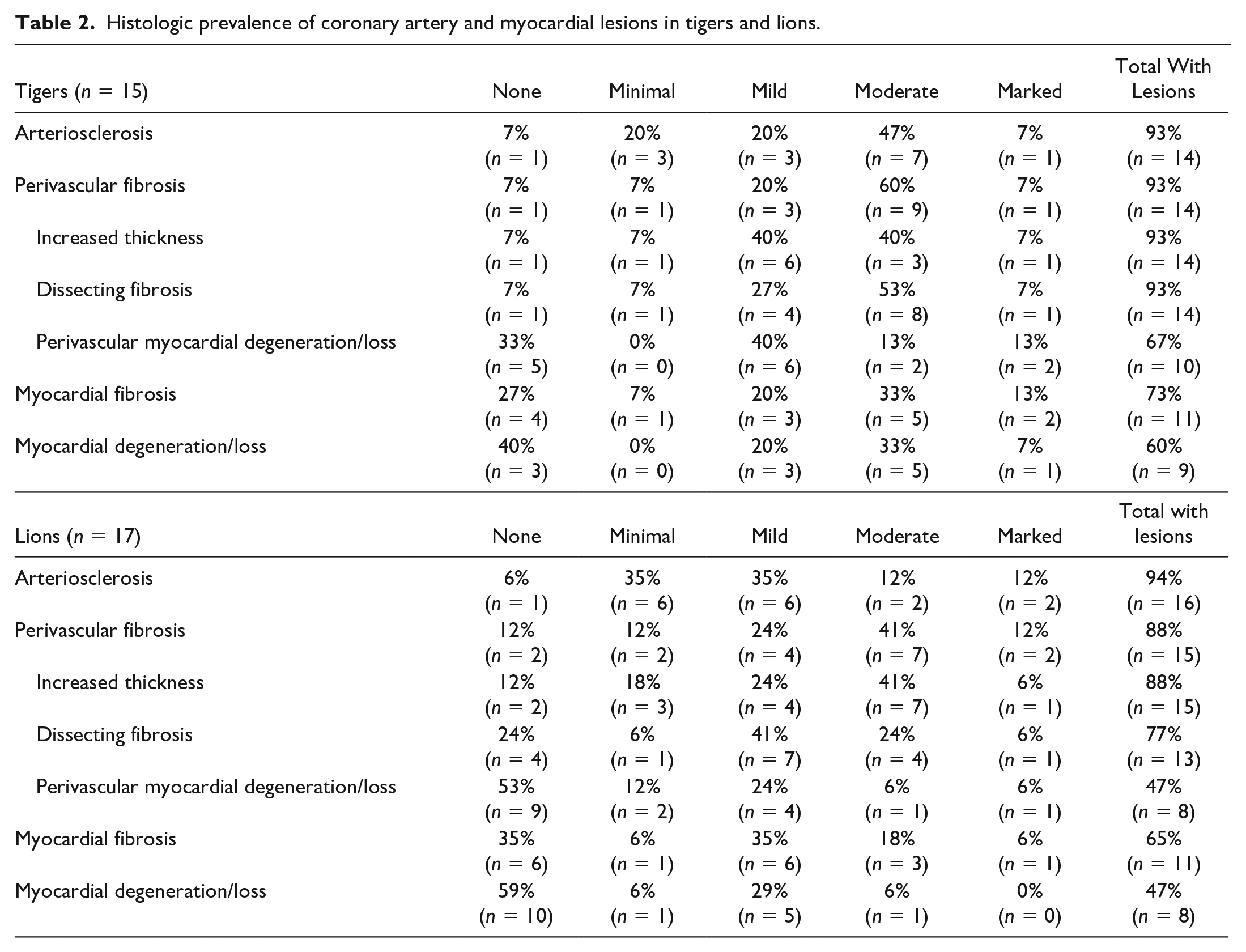
Histologic prevalence of coronary artery and myocardial lesions in tigers and lions.
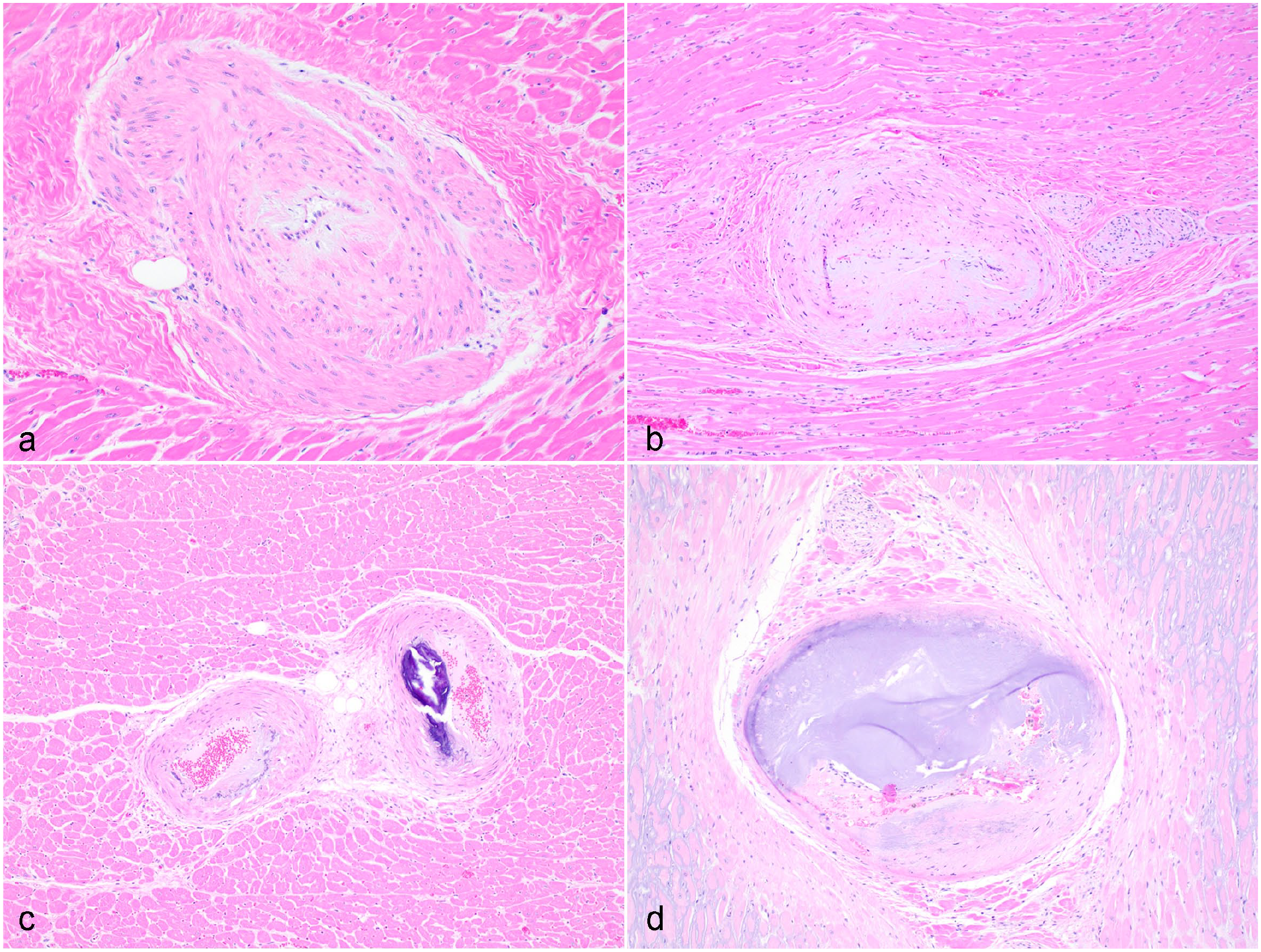
Heart, coronary arteriosclerosis in lions and tigers. Intramural coronary vessels from (a, b) lions and (c, d) tigers, demonstrating (a, c) moderate and (b, d) marked arteriosclerotic changes. Hematoxylin and eosin.
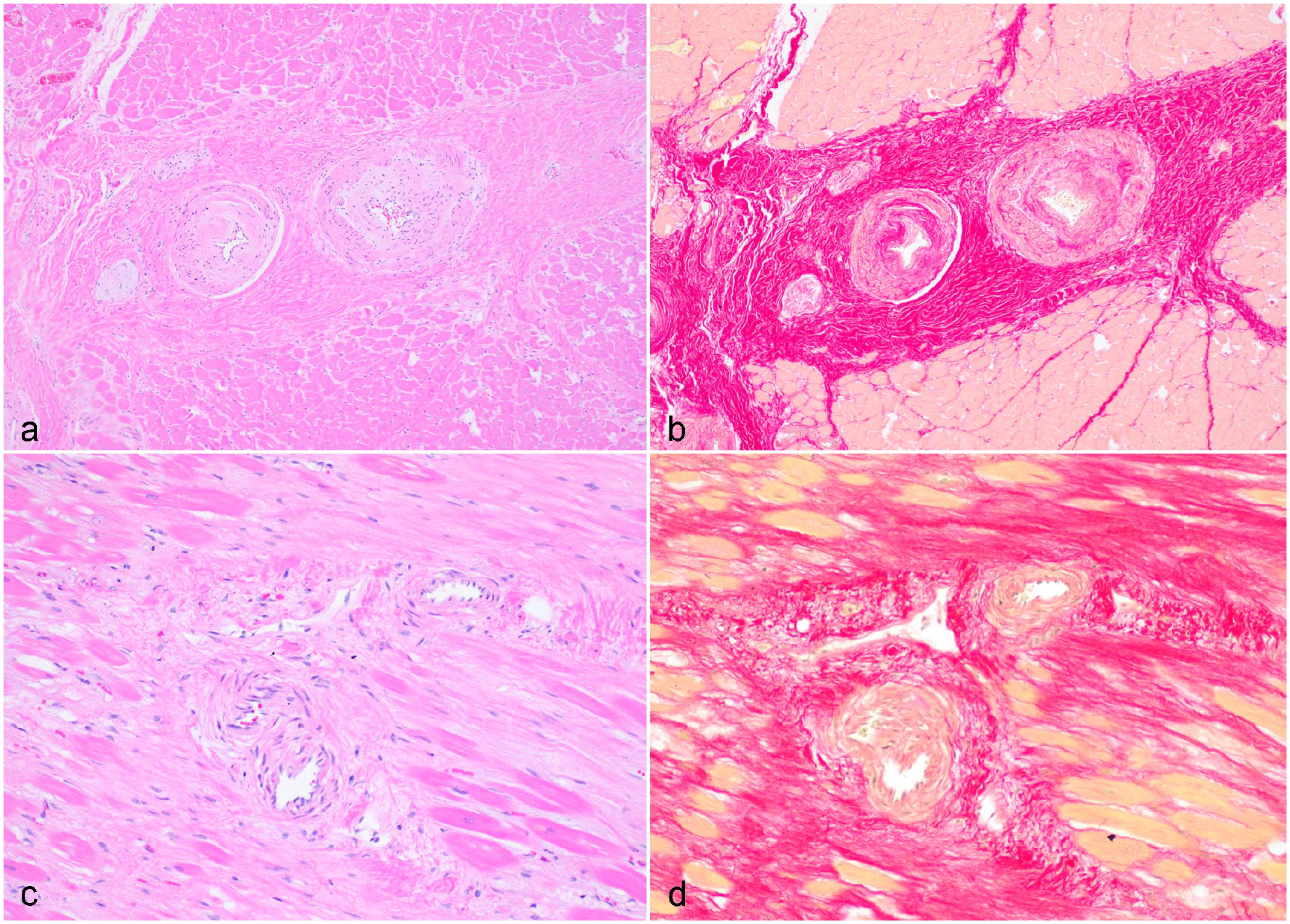
Perivascular fibrosis in lions and tigers. (a) Heart, lion. Intramural coronary vessels with markedly increased perivascular connective tissue thickness and moderate dissecting fibrosis. Hematoxylin and eosin (HE). (b) Step section of the heart in (a) stained with Sirius red to highlight the fibrosis. (c) Heart, tiger. Markedly increased perivascular connective tissue thickness and marked dissecting fibrosis. HE. (d) Step section of the heart in (c) stained with Sirius red to highlight the fibrosis.
The most common lesions included moderate to marked arteriosclerosis (8 tigers [53%] and 4 lions [24%]) and moderate to marked perivascular fibrosis (10 tigers [67%] and 9 lions [53%]). Ten tigers (67%) and 8 lions (47%) had coronary artery lesions with some degree of perivascular cardiomyocyte degeneration and/or loss. Based on the Ryser-Degiorgis grading schematic for arteriosclerosis, 50% of tigers and 31% of lions had grades 2 to 3 arteriosclerotic lesions. 18 Interestingly, Anitschkow cells, which were also included in the Ryser-Degiorgis grading scheme, were not identified in any of the evaluated samples.
To our knowledge, this is the first report describing intramural coronary artery pathology in captive tigers and lions. Fourteen of 15 tigers had changes consistent with arteriosclerosis, which has not been reported in tigers before. In humans, arteriosclerosis is commonly associated with age.17,20 In our study, tissues were examined from an age-skewed population. No statistically significant relationship was identified, but this result may be limited by our population distribution. Additional risk factors in humans for CAD include obesity. 12 There is limited clinical information available regarding our population, but necropsy notes indicate obesity in some of the animals examined, which may play a role in presentation. All felids were from the same facility, which may suggest an additional but undetermined environmental component. All animals died or were euthanized due to other clinical concerns, so the clinical significance of these lesions remains to be determined.
Interestingly, 8/15 tigers and 2/17 lions had clinical and/or histologic evidence of CKD, which is a common cause of morbidity and mortality in captive large felids. In humans, CKD is associated with an increased risk of coronary heart disease development. 2 Moreover, lower kidney function has been demonstrated to be associated with severity of CAD in humans, with decreased estimated glomerular filtration rate being strongly associated with severity of CAD. 15 Proposed mechanisms of this relationship include high burden of cardiovascular risk factors in populations with CKD and novel risk factors, including systemic inflammation, oxidative stress, anemia, and abnormal calcium-phosphate metabolism. 14 Only 3/8 tigers and 0/2 lions with CKD changes had moderate to marked arteriosclerotic changes, further suggesting the likely multifactorial and complicated nature of the pathophysiology of arteriosclerosis in these species. In this study, 10/15 tigers and 9/17 lions had moderate to marked perivascular fibrosis as defined by increased perivascular connective tissue deposition and/or dissection into surrounding cardiomyocytes, with or without cardiomyocyte degeneration and loss. In humans, perivascular fibrosis has been linked to hypertension. 17 Unfortunately, due to the limited clinical information available for these cases and overall challenges with obtaining blood pressure in captive wild animals, the relationship between coronary artery lesions and hypertension was unable to be assessed in this population. Interestingly, in humans with hypertrophic cardiomyopathy, there is increased evidence of perivascular fibrosis when compared with other cardiomyopathies, which can lead to impaired coronary blood flow.5,14 Hypertrophic cardiomyopathy and hyperthyroidism in domestic felids also has been linked with abnormal small intramural coronary arteries.1,8 Due to lack of known cardiovascular disease and functional endocrine parameters in these cases, neither hypertrophic cardiomyopathy nor hyperthyroidism were considered primary differentials. Mild to marked myocardial fibrosis (interstitial and replacement) was identified in 10/15 tigers and 10/17 lions. Interstitial and replacement fibrosis are common histologic features in domestic feline and human cardiomyopathies. In addition, studies have correlated myocardial collagen content with left ventricular stiffness, which may compromise pumping function and predispose to conduction disturbances.3,7,11 All changes were significantly associated with the interventricular septum and/or left ventricle compared with the right ventricle. This suggests that ideal postmortem cardiac evaluation should at least encompass interventricular septum and/or left ventricle samples in both lions and tigers.
Information on the overall incidence and descriptions of cardiac disease in nondomestic felids is limited. The most well-characterized feline cardiomyopathy, hypertrophic cardiomyopathy, was first reported in 3 lions in a retrospective study and accounted for nearly 40% of the cardiovascular diseases in this study. 16 Despite this, the overall incidence of cardiac disease was relatively rare (8 out of 111 cases). A recent study has described arteriosclerosis, perivascular fibrosis, and replacement fibrosis in free-ranging Eurasian lynx. 18 Both studies suggest that an underlying genetic component should be considered. Unfortunately, there is limited genetic information regarding the population in our study, so the role that genetics may play in our population is unknown.
In conclusion, we describe and report intramural coronary artery and myocardial pathology in captive tigers and lions. Changes were most frequently identified in the left ventricle and interventricular septum, which emphasizes the importance of evaluating multiple heart sections in postmortem examination. Unfortunately, a definitive cause of coronary artery pathology in this population was not determined and all animals were euthanized or died due to other clinical concerns. Furthermore, our study is limited due to its retrospective nature, small sample size, limited cardiac tissue samples available for evaluation, and lack of morphometric parameters. However, the severity of the lesions in some cases may suggest an emerging, clinically silent cardiomyopathy. Possible contributing or inciting factors include obesity, hypertension, genetics, environmental, and aging, but the pathogenesis is likely multifactorial.
Supplemental Material
sj-pdf-1-vet-10.1177_03009858241246984 – Supplemental material for Intramural coronary artery and myocardial pathology in captive tigers (Panthera tigris) and African lions (Panthera leo)
Supplemental material, sj-pdf-1-vet-10.1177_03009858241246984 for Intramural coronary artery and myocardial pathology in captive tigers (Panthera tigris) and African lions (Panthera leo) by Rebecca L. Makii and Juan Muñoz Gutiérrez in Veterinary Pathology
Footnotes
Acknowledgements
We thank the tissue trimming and histology laboratory staff at Colorado State University for their technical assistance with this project.
Author Contributions
RLM carried out data curation and analysis. The manuscript was written by RLM. JMG conceived the study, assisted with study design, and edited the manuscript. All authors read and approved the final manuscript.
Declaration of Conflicting Interests
The author(s) declared no potential conflicts of interest with respect to the research, authorship, and/or publication of this article.
Funding
The author(s) disclosed receipt of the following financial support for the research, authorship, and/or publication of this article: Funding for this study was provided by the Colorado State University Institutional Start-Up Support.
Supplemental material for this article is available online.
References
Supplementary Material
Please find the following supplemental material available below.
For Open Access articles published under a Creative Commons License, all supplemental material carries the same license as the article it is associated with.
For non-Open Access articles published, all supplemental material carries a non-exclusive license, and permission requests for re-use of supplemental material or any part of supplemental material shall be sent directly to the copyright owner as specified in the copyright notice associated with the article.