
Abstract
To comprehensively evaluate the occurrence of renal lesions in a variety of nondomestic felids, necropsy cases from 1978 to 2008 were reviewed from a municipal zoo and a large cat sanctuary for those in which the kidneys were examined histologically. Seventy exotic felids were identified (25 tigers, 18 lions, 6 cougars, 5 leopards, 3 snow leopards, 3 clouded leopards, 3 Canadian lynx, 2 ocelots, 2 bobcats, 2 cheetahs, 1 jaguar), and their histologic renal lesions were evaluated and compared. The most common lesion was tubulointerstitial nephritis (TIN); 36 of 70 (51%) cats were affected to some degree. Lymphocytic interstitial nephritis was the most common lesion in the tigers (9 of 25, 36%) and was rarely seen in other species. Although the renal pelvis was not available for all cats, 28 of 47 (60%) had some degree of lymphocytic pyelitis. There was no significant association between the presence of pyelitis and that of TIN. Only 1 cat had pyelonephritis. Renal papillary necrosis was present in 13 of 70 (19%) cats and was significantly associated with historical nonsteroidal anti-inflammatory drug treatment (odds ratio, 7.1; 95% confidence interval, 1.9 to 26.8). Only 1 cat (lion) had amyloid accumulation, and it was restricted to the corticomedullary junction. Primary glomerular lesions were absent in all cats. Intraepithelial pigment was identified in many of the cats but was not correlated with severity of TIN. Despite several previous reports describing primary glomerular disease or renal amyloidosis in exotic felids, these lesions were rare to absent in this population.
Chronic kidney disease is the most common disease affecting older domestic cats. 15 The reported prevalence of kidney disease in domestic cats ranges from 1.6 to 31%. 2,15 Despite numerous reports of renal disease in exotic felids, 1,3,8,14,16,19 we could find no reports that describe and compare the occurrence of renal lesions across multiple species of exotic felids.
Previous reports of renal lesions in exotic felids have focused mainly on renal AA amyloid deposition in association with systemic AA amyloidosis. 14,16,19 Large studies have reported glomerulosclerosis in captive cheetahs 1 and glomerulonephritis in the Iberian lynx (Lynx pardinus). 8 Renal papillary necrosis (RPN) has been reported in 3 lions and a tiger, 3 and there is a single case report of pyelonephritis in a Siberian tiger (Panthera tigris altaica). 7
This retrospective report evaluated kidney lesions in exotic felids that were necropsied at the University of Tennessee Veterinary Teaching Hospital before 2009.
Materials and Methods
Necropsy records from the University of Tennessee’s pathology service were searched for submissions of exotic felids from a municipal zoo and a large cat sanctuary from January 1978 through December 2008. All the cats in this study were housed at a municipal zoo or a large cat sanctuary at the time of their death. Most of the animals were similarly housed, in earth-floored enclosures, and fed all-meat diets. The large cat sanctuary serves as a rescue organization, so the prior history of many of these cats is unknown. Cases were selected for further study if the kidneys were available for histologic examination. Neonatal animals (< 1 month old) were excluded.
The reason for death or euthanasia was obtained from the medical record, necropsy request form, or necropsy results. The medical records for each case were evaluated to determine if nonsteroidal anti-inflammatory drugs (NSAIDs) had been administered in the 2 months preceding the animal’s death. Medical records were also screened for clinical pathology and/or serology data or other factors that may have contributed to the development of renal lesions.
Histologic Evaluation
The kidneys were evaluated histologically by 2 board-certified pathologists (K.M.N., S.J.N.) based on based on sections stained with hematoxylin and eosin, periodic acid–Schiff (PAS), and Masson’s trichrome. Histologic renal lesions were described using established criteria. 10 The major renal findings were characterized as tubulointerstitial nephritis (TIN), lymphocytic interstitial nephritis (LIN), or other. Lesions that primarily involved the interstitium and tubules were designated as TIN. Specific features of TIN included lymphoplasmacytic interstitial inflammation and fibrosis with tubular degeneration, necrosis, atrophy, and basement membrane thickening. 10 Commonly, sclerotic or shrunken glomeruli with a thickened Bowman capsule were embedded in these areas of fibrosis and atrophy. If a primarily lymphocytic interstitial infiltrate was present without tubular changes, the term LIN was used. For both TIN and LIN, the degree of change was categorized as mild, moderate, or marked, based on the extent of renal involvement. Additional histologic findings (RPN, eosinophilic granulomas, etc) were assessed when present. When possible, kidneys were evaluated on a scale of 0 to 3 (no lesion to severe lesions) for inflammation in the renal pelvis. Active cases of pyelonephritis were defined as pyelitis with a primarily neutrophilic inflammatory infiltrate in the renal papilla and concurrent medullary tubular necrosis with variable degrees of extension into the cortex. 10 Histochemical stains (Congo red with and without potassium permanganate digestion, Prussian blue, Hall’s bile) were used as needed to further characterize the renal lesions. Intraepithelial pigment was classified as lipofuscin or iron, using PAS- and Prussian blue–stained slides, respectively.
Data Analysis
The association of lesions, as measured on the nominal or interval scales with all cats, individual species, and subspecies, was evaluated with either chi-square or Fisher exact test, depending on whether the expected cell value was < 5. Logit estimators with a correction factor of 0.5 were used to calculate the odds ratio (OR) when one or more cells in a 2 × 2 table contained 0. Results are presented as ORs with 95% confidence intervals (CIs). The association of age with species and specific lesions was evaluated with a one-way analysis of variance or a nonparametric equivalent, depending on the fit of data to a normal distribution. Continuous data were tested for an approximation to the normal distribution by the method of Shapiro–Wilk and are expressed as mean ± 1 standard deviation or median and range, depending on the fit to a normal distribution. A P value of < .05 was used to determine statistical significance in all comparisons.
Results
Kidney sections were available from 70 nondomestic felids (see Tables 1–3). Known ages ranged from 6 months to 25 years, with a median age of 14 years.
Summary of Renal Findings in Tigers (Panthera tigris) a
a n = 25. NSAID, nonsteroidal anti-inflammatory drug; RPN, renal papillary necrosis; NA, not available; Y, yes; N, no; LIN, lymphocytic interstitial nephritis; TIN, tubulointerstitial nephritis; D, died naturally; E, euthanatized; IVDD, intervertebral disc disease; DJD, degenerative joint disease.
b White tiger.
c Embolic pattern.
Summary of Renal Findings in African Lions (Panthera leo krugeri) and Asian Lions (Panthera leo persica) a
a n = 18. NSAID, nonsteroidal anti-inflammatory drug; RPN, renal papillary necrosis; As, Asian; Afr, African; NA, not available; Y, yes; N, no; LIN, lymphocytic interstitial nephritis; ACA, adenocarcinoma; TIN, tubulointerstitial nephritis; CMJ, corticomedullary junction; D, died naturally; E, euthanatized; DJD, degenerative joint disease.
Summary of Renal Findings in Other Exotic Felids a
a n = 27. NSAID, nonsteroidal anti-inflammatory drug; RPN, renal papillary necrosis; NA, not available; Y, yes; N, no; TIN, tubulointerstitial nephritis; D, died naturally; E, euthanatized; SCC, squamous cell carcinoma; IVDD, intervertebral disc disease; DJD, degenerative joint disease.
b Black phase leopard.
c Neutrophilic.
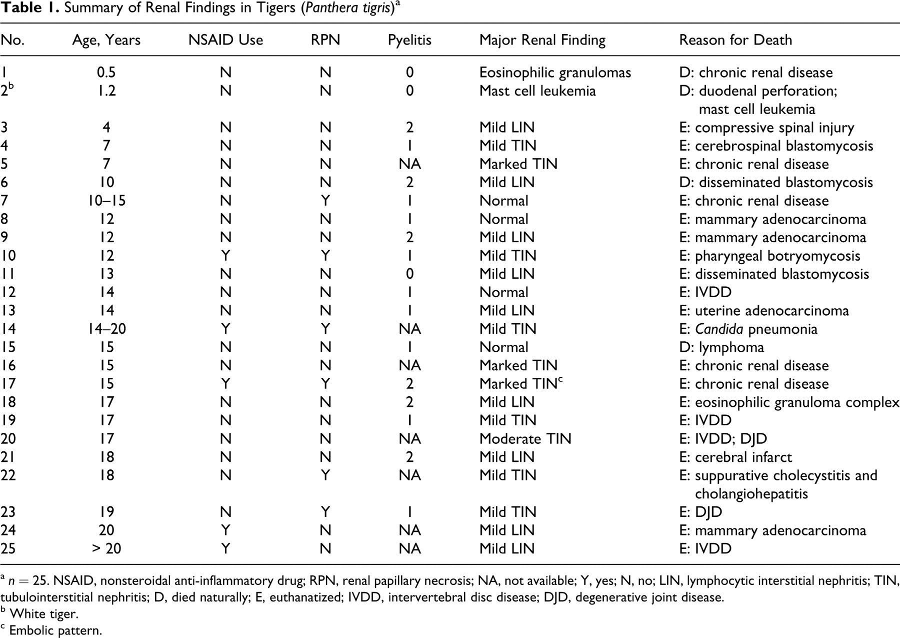
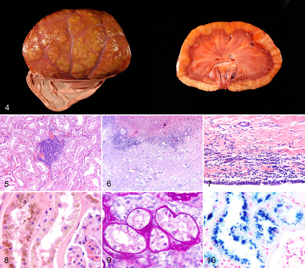
Renal lesions were present in 52 cats (74%). TIN was the most common lesion, with 36 of 70 cats (51%) affected. Mild TIN was seen in 17 cats (24%) (Fig. 1 ). Moderate TIN was seen in 7 cats (10%) (Fig. 2 ). Marked TIN was seen in 12 cats (17%) (Figs. 3, 4).
There was no significant difference between the prevalence of TIN in tigers and lions. However, the presence of TIN was significantly associated with increased age of the cat (OR, 1.3; 95% CI, 1.1–1.4). Similarly, with regard to age, cats with mild (15.6 ± 3.4 years) or moderate (17.3 ± 1.8 years) TIN were significantly older than the cats with normal kidneys (11.1 ± 5.6 years) (P = .019 and P = .012, respectively). The odds of developing TIN increased by 1.238 with every year of age.
Mild LIN was seen in 11 cats (16%) (Fig. 5 ). The cats with mild LIN included 9 of 25 tigers (36%) and 2 of 18 lions (11%); LIN was not seen in the other species. A single cougar had moderate lymphoplasmacytic interstitial nephritis, which was limited to the corticomedullary junction; there was no concurrent tubular disease.
RPN was seen in 13 cats (19%) (Fig. 6 ), including 6 of 25 tigers (24%), 2 of 6 cougars (33%), 4 of 5 leopards (80%), and 1 of 3 clouded leopards (33%). None of the lions, snow leopards, Canadian lynx, bobcats, ocelots, cheetahs, or the jaguar had RPN. One tiger (case No. 7) had RPN without concurrent nephritis. The presence of RPN was significantly associated with a history of NSAID treatment (OR, 5.4; 95% CI, 1.3–22.0).
In 47 cats, the renal pelvis was available for examination. In 28 of these cats (60%), there was some degree of pyelitis. Mild lymphocytic pyelitis (grade 1) was present in 13 cats (28%), and moderate lymphocytic pyelitis (grade 2) was noted in another 13 cats (28%) (Fig. 7 ). Two cats (4%), both leopards, had marked pyelitis (grade 3). In 1 leopard (case No. 50), the inflammation was lymphocytic; in the other (case No. 52), it was neutrophilic. Both leopards also had concurrent RPN. Of the 5 leopards, 4 (80%) had some degree of pyelitis, which was not observed in any of the lions, bobcats, or the jaguar. There was no significant association between the presence or severity of pyelitis and the presence or severity of TIN.
Pigment was commonly seen in the renal tubular epithelial cells (Fig. 8 ). According to PAS-stained slides, 46 of 70 cats (66%) had some degree of lipofuscin accumulation (Fig. 9 ). According to Prussian blue staining, 19 of 63 cats (30%) had some degree of iron accumulation in the renal tubular epithelium (Fig. 10 ). The presence or degree of pigment (lipofuscin or iron) accumulation was not correlated with age or the presence of TIN.
Miscellaneous kidney lesions included cats affected by each of the following: eosinophilic granuloma, mast cell tumor, metastatic mammary adenocarcinoma, and renal cortical adenoma. In 1 lion (case No. 38), amyloid was present and restricted to a band at the corticomedullary junction. The lack of digestion following potassium permanganate treatment suggested that it was not AA amyloid; there was no concurrent inflammation or tubular disease. Varying degrees of tubular lipid, mineralization, and small renal cysts were seen sporadically; they were not associated with other renal lesions.
Of the 11 cats for which death or euthanasia was attributed to chronic renal disease, 9 had marked TIN, 1 had extensive renal eosinophilic granulomas, and 1 had RPN. All these cats had azotemia, with serum or plasma blood urea nitrogen values ranging from 65 to 345 mg/dl and creatinine values ranging from 3.6 to 14.2. Serum or plasma calcium values were generally within the reference intervals for domestic cats, but inorganic phosphorus values were typically increased (7 cats had values > 8.0 mg/dl). Clinical pathology data from other cats that died or were euthanatized for other reasons were generally incomplete and not further interpreted.
Discussion
The initiating cause (or causes) of chronic renal disease in domestic cats is often unknown. TIN is traditionally attributed to hematogenous infectious agents but can be secondary to ascending pyelonephritis, primary glomerular disease, or a previous episode of acute renal failure. 10,13 The lesions of TIN and chronic pyelonephritis can overlap and be difficult to differentiate. Because only 1 cat in this study had active pyelonephritis, it is unlikely that the majority of TIN cases occurred as a sequela to pyelonephritis. It has also been proposed that chronic TIN may be the result of immune-mediated damage to the renal tubules and interstitium. 13 Similarly, some have speculated about the role of routine vaccination in the development of chronic renal disease. 15 Most of these cats were vaccinated, but the type and frequency of vaccination were not available for most cats. Leptospirosis is known to cause TIN. A single study reported 7 of 22 wild Iberian lynx in Spain were seropositive for leptospirosis, but none of those animals had gross or histologic evidence of nephritis. 12 Three felids with marked TIN were screened for antibodies to several Leptospira serovars, and all were negative. Leptospirosis has not been diagnosed in any species from either of the facilities where these cats were housed (E. C. Ramsay, personal communication). Regardless of the inciting cause, the lesions are progressive, and the ongoing inflammation, fibrosis, and tubular degeneration further impair renal function. 15
In this study, the most common renal lesion was TIN, which was seen to varying degrees in 51% of cats. Mild TIN was most common, being present in 24% of cats. In none of the cases was there an obvious inciting cause. No species predisposition was identified in this study, but as in domestic felids, 15 TIN was significantly more likely to occur in older felids. Of the 11 cats in this study that died or were euthanatized owing to chronic renal disease, 9 had marked TIN, 1 had extensive renal eosinophilic granulomas, and 1 had RPN. The clinical pathology changes seen in these cats are similar to those seen in domestic cats.
LIN was seen in 11% of cats. It was the most common lesion in the tigers, affecting 38% of cats, but was less common or absent in other felids. The apparent predisposition of tigers to LIN was not statistically significant.
Some degree of pyelitis was present in 60% of cats in which the renal pelvis was available for examination. The cause of the pyelitis was not apparent in any of the cases. There was no significant association between TIN and pyelitis. Chronic or previous episodes of pyelonephritis could have resulted in the pyelitis and/or TIN. In typical cases of chronic/resolved pyelonephritis, there is a band of fibrosis that extends from the pelvis to the renal capsule. In many cases, however, this band of scar tissue was not apparent, which complicated the distinction between a previous episode of pyelonephritis and TIN. 10 Only 1 cat in our study (leopard, case No. 54) had lesions consistent with active pyelonephritis. This same leopard had concurrent RPN and marked TIN. In the single report of Staphylococcus intermedius–associated pyelonephritis in a Siberian tiger, 7 RPN was not specifically mentioned, but the authors did indicate significant necrosis in the deep medulla.
The pathogenesis of RPN in cases of pyelonephritis or urinary obstruction is believed to occur via mechanical compression of the thin-walled medullary blood vessels. 10 Renal medullary amyloidosis may also compress medullary blood vessels, resulting in RPN. 10 In one study of captive cheetahs with systemic AA amyloidosis, 38% had involvement of the renal medulla and 25% of those had concurrent RPN. Some cheetahs with RPN also had pyelonephritis. Glomerular amyloid was present in 21% of cheetahs. 14 Neither of the 2 cheetahs in the current study had RPN or amyloidosis. Similarly, in a study of black-footed cats (Felis negripes) with systemic AA amyloidosis, 91% had medullary amyloid and 21% had concurrent RPN. 19 Glomerular amyloid was present in 63% of black-footed cats in that study. 19 In another report, 4 closely related Siberian tigers had renal medullary AA amyloidosis; 2 of those tigers had concurrent RPN and chronic TIN. 16
The only felid (Asian lion, case No. 38) in this study with amyloid accumulation did not have RPN, nor did it have amyloid in any of the other tissues examined (adrenal, lymph node, skin, testes, lung, spleen, liver, heart, gastrointestinal tract, urinary bladder, diaphragm, gall bladder, brain). In this lion, the amyloid was restricted to a band at the corticomedullary junction. The amyloid did not digest following treatment with potassium permanganate; therefore, it was not AA amyloid. Further characterization of the amyloid was not performed. The underlying reason for the renal amyloid in this lion is unclear. The lion had concurrent mild TIN.
Dehydration alone or in combination with other drugs (NSAIDs, phenothiazine, monensin, roxarsone) has been associated with RPN in racing Greyhounds, horses, lambs, and calves. 10 The pathogenesis of NSAID-associated RPN is attributed to decreased production of PGE2, which has vasodilatory effects on arterioles of the juxtamedullary nephrons. The result is decreased perfusion of the renal crest. Additionally, NSAIDs may be directly toxic to cells in the interstitium of the renal medulla. 10 RPN has been reported in 3 lions and 1 tiger; in that report, the inciting cause was attributed to chronic dehydration and poor water quality. 3 Another publication described bilateral RPN attributed to dehydration in 2 tigers. 11 Bilateral RPN was also reported in a white tiger with persistent metanephric ducts and unilateral hydronephrosis. 4 In addition, RPN has been seen in 2 domestic cats and 1 tiger; 1 cat and the tiger were treated with NSAIDs. 20 In the current study, RPN was seen in leopards, clouded leopards, cougars, and tigers but not in any of the lions, bobcats, Canadian lynx, snow leopards, cheetahs, ocelots, or the jaguar. All 5 cats with RPN that had received NSAIDs in the past 2 months were treated with meloxicam, which was also used in 5 of the 6 NSAID-treated cats that did not develop RPN; the sixth cat (case No. 38) was treated with aspirin. Meloxicam doses and treatment regimens were identical in those animals that did develop RPN and those that did not, suggesting that other factors (ie, hydration status) may have contributed to the development of RPN in some animals. This is the first study to report a statistically significant association between RPN and NSAID use in exotic felids.
Despite several studies describing the occurrence of glomerular lesions in exotic felids, none of the cats in the present study had primary glomerular lesions. In one study, glomerulosclerosis was reported in 71 of 87 captive cheetahs (82%), with 30% having moderate to severe lesions. 1 Only 2 cheetahs were included in the current study, and neither had histologic evidence of renal disease. A membranous glomerulopathy was described in a tiger with chronic renal disease, 6 and glomerulonephritis was reported in a single Canadian lynx but not described further. 5 Progressive membranous glomerulonephritis was reported in 26 of 27 Iberian lynx (96%) from one study and was believed to have a familial and immune-mediated component. 8 No Iberian lynx were included in the current study.
Many of the lions in this study had some intraepithelial renal tubular pigment. In most animals, the pigment was PAS positive, which is consistent with lipofuscin, 17 which is often called the wear-and-tear pigment; that is, it represents a group of intralysosomal pigments formed from the peroxidation and polymerization of unsaturated fatty acids derived from the breakdown of membranes and organelles. 9 The accumulation of lipofuscin is most commonly seen in postmitotic cells in the brain, heart, or skeletal muscle or in tissues undergoing cachectic or senile atrophy. 9 Because lipofuscin is the result of degradation of unsaturated fatty acids, it is possible that its presence in this study could be related to the renal tubular lipidosis seen in some of the cats. To our knowledge, lipofuscin has not been reported in the chronic renal lesions of domestic felids. Subjectively, increased amounts of lipofuscin were present in areas with more severe TIN; however, this association was not statistically significant.
In some animals, the pigment stained strongly with Prussian blue, which is consistent with iron. Iron was most commonly seen in the lions, but this apparent predisposition was not statistically significant. The presence of iron-containing hemosiderin in renal tubules is due to resorption of hemoglobin from the glomerular filtrate by the proximal tubular epithelium; hence, iron accumulation would be more likely in animals with a history of hemolysis. 10,13 Hemosiderin may, however, be incidentally found in the renal tubules of dogs that do not have a history of hemolysis. 13 None of the lions in this study had a known history of a hemolytic event. Only one of the lions was known to be anemic (case No. 42; packed cell volume, 17); a packed cell volume value was available for only 8 of the 18 lions. It is likely that some degree of tubular hemosiderosis can be an incidental finding in lions. Studies in humans and in animal models have demonstrated that urinary and tissue iron increase with renal disease. Additionally, reducing the excretion of iron decreases the rate of progression of renal disease. 18 There was no statistically significant association between the presence of iron and the presence of TIN.
The presence of mineral and tubular lipid in the kidneys was common, even in otherwise normal kidneys. Additionally, tubular cysts were occasionally seen in leopards, cougars, lions, and tigers. Renal cysts may form secondary to obstructive tubular lesions, basement membrane anomalies, or focal hyperplastic tubular lesions. 10 In no instances was the presence of cysts severe enough to warrant a diagnosis of polycystic kidney disease.
Given the paucity of reports concerning causes of death or euthanasia in exotic felids, the following were also assessed and included: intervertebral disc disease, degenerative joint disease, chronic renal failure, blastomycosis, and neoplasia. Neoplasms included mammary adenocarcinomas, uterine leiomyoma or adenocarcinomas, squamous cell carcinoma, pancreatic carcinoma, lymphoma, and an oligodendroglioma. There was no apparent association between the cause of death or euthanasia and the presence or severity of TIN or LIN.
The most common renal lesion in exotic felids was TIN, and the development of TIN was significantly associated with increasing age. Mild LIN was the most common lesion in the tigers but was seen only rarely in other species. For the first time in exotic felids, a significant association between historical NSAID use and RPN was identified. Primary glomerular disease and renal amyloidosis were rare to absent in this population.
Footnotes
Acknowledgements
We would like to thank Misty Bailey for a critical review of the manuscript, Anik Vasington for assistance with the digital images, and the staff and curators of the Knoxville Zoo and Tiger Haven for their help with the animals as well as with organizing the data.
The authors declared no potential conflicts of interests with respect to the authorship and/or publication of this article.
The authors received no financial support for the research and/or authorship of this article.