
Abstract
This retrospective study describes 8 cases of intestinal hemangioma diagnosed in horses during postmortem examination or surgical biopsy at the University of Tennessee College of Veterinary Medicine. In all cases, the intestine was the sole organ affected, and lesions were focal (3/8) or multifocal (5/8). Nodules were most commonly within the small intestine (7/8), particularly the jejunum (5/7). One case was in the left dorsal colon, which is the first report of hemangioma in the large colon of a horse. Lesions were discrete, raised, smooth, black to red, and ranged from 2 to 15 mm in diameter. Microscopically, all lesions were cavernous type and mural, most frequently within the muscularis (6/8). A majority of cases occurred in middle aged to older horses (average age of 19.3 years), and no breed or sex predilections were identified. The hemangiomas were considered incidental findings.
Hemangiomas, benign tumors of endothelial cells, can potentially develop at any location due to the ubiquitous nature of blood vessels. 10 In horses, these tumors are infrequent and most commonly occur in the skin, often in younger animals, and sometimes as congenital lesions.4–6,14,15 A single case report in 1960 described a “venous angioma” in the cervical spinal cord of a 9-year-old mare. 12 A case series from 1987 included 10 cases of “vascular malformations and angiomatous lesions.” 15 Only 1 case in that series was in the intestine, and it was a solitary cavernous hemangioma in the colon of a 4-month-old thoroughbred colt. Two other reports in young horses differ in that the proliferations included arterioles and venules 11 or muscular arteries. 18 The locations in those 2 reported cases were the meninges 11 and the subcutis, 18 and the diagnoses were hemangiomatosis and hemangioma, respectively. In retrospect, those are both likely to be vascular malformations and not neoplasms.
Two previous reports of multiple hemangiomas in the intestines of aged horses were both incidental findings; 1 during necropsy following euthanasia for severe lameness 8 and 1 during surgical biopsy following colic due to a pedunculated lipoma. 20 The lesions in both of these cases consisted of discrete intramural nodules of thin-walled blood-filled spaces lined by flat endothelial cells, consistent with hemangiomas. One of these cases also involved the ovaries; both reports used the term “angiomatosis” for the multifocal discrete lesions resembling hemangiomas. 8 The intent of the current retrospective review is to describe eight additional cases of equine intestinal hemangioma as well as provide insight into their clinical significance.
All instances of intestinal hemangioma in horses diagnosed through the University of Tennessee College of Veterinary Medicine biopsy or necropsy services from 2005 to 2022 were identified via a pathology database search. The search was made using the terms “hemangioma” and “angioma” and specifying equine as the species. A total of 1092 equine necropsies and 359 equine surgical pathology cases were submitted during the time period in question. Thirty-two of the 359 equine biopsies submitted were intestinal. The gross and histologic findings, patient history, and signalment including breed, sex, and age were recorded. Microscopic findings of hemangioma were reviewed and confirmed by 1 board-certified anatomic pathologist (L.C.).
A summary of the 8 cases is shown in Table 1. Horses examined postmortem (7/8) or via surgical biopsy (1/8) were found to have intestinal hemangioma, all of which were deemed incidental findings. Cases included 5 geldings and 3 mares with an average age of 19.3 years (range of 10-30 years). Affected breeds were Tennessee Walking Horse (3/8), Quarter Horse (1/8), Thoroughbred (1/8), and mixed breed (3/8). Lesions were identified in the small intestine (7/8) and the large colon (1/8). Small intestinal hemangioma occurred most often in the jejunum (5/7), and the colonic hemangioma was in the left dorsal colon; lesions in the remaining 2 cases were in unspecified small intestinal locations. Grossly, the lesions were focal (3/8) or multifocal (5/8) (Fig. 1a), black (6/7) or red (1/7), round to oval (7/8) or irregularly shaped (1/8), raised, smooth, discrete nodules measuring 2 to 15 mm in diameter. The surgical biopsy submission did not specify the color of the lesion. One multifocal case was described as being “throughout the intestines” (case 2) while others did not specify exact locations relative to other lesions. Additionally, the total number of lesions was not provided for the multifocal lesions, except for one (case 7), which had “half a dozen.” Microscopically, all lesions were cavernous-type hemangiomas and were within the muscularis layer only (3/8) (Fig. 1b), submucosa only (2/8), muscularis and serosa (2/8), or submucosa and muscularis (1/8) (Table 1). No cases were transmural as the lesions only spanned either 1 or 2 layers (Table 1). A few multifocal cases included hemangiomas that varied in location and number of layers spanned, but because not all nodules identified grossly were reviewed histologically, the frequency of hemangiomas in these locations is not known. Of those that were multifocal, more than one nodule was examined histologically in 2/5 cases and had similar features, aside from location within different intestinal layers in case 2. More specifically, 2 nodules were examined in case 6, and at least 4 nodules were examined in case 2. The masses were composed of variably sized, blood-filled spaces lined by flat endothelial cells. Occasionally, the vascular spaces contained fibrin thrombi (3/8) and were separated and surrounded by pre-existing intestinal wall smooth muscle (2/8), collagenous stroma (2/8), or both (3/8) (Fig. 1c). A trichrome stain was used to demonstrate the presence of smooth muscle within the collagenous stroma (Fig. 1d). Some horses (5/8) were reported to have signs referable to the gastrointestinal system, but in all cases, signs were attributed to additional findings including enteritis (4/8), colitis (2/8), ulcerative typhlitis (1/8), and dental attrition (1/8).
Age, distribution, and location of intestinal hemangioma in 8 horses, as well as reported gastrointestinal clinical signs and relevant final diagnoses.
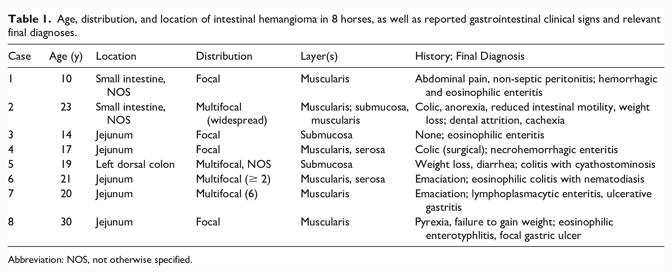
Abbreviation: NOS, not otherwise specified.
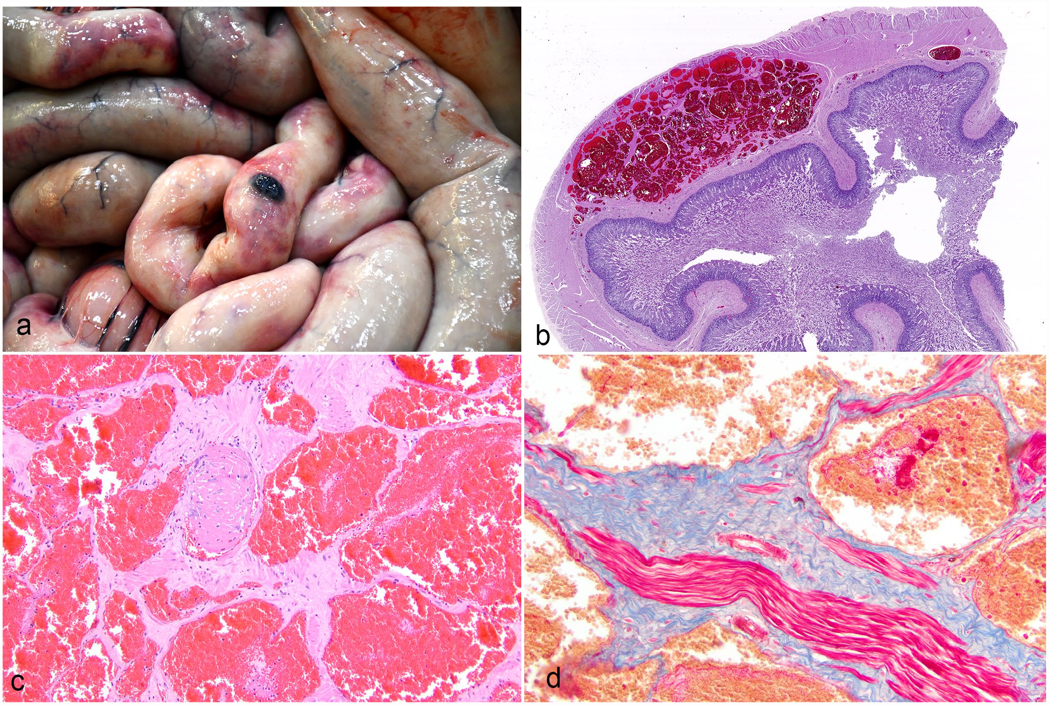
Hemangioma, jejunum, horse. (a) Raised, ovoid, black, mural nodule. Case 8. (b) Well-demarcated, nodule within the muscularis consisting of variably sized, cavernous, blood-filled channels. Case 8. Hematoxylin and eosin (HE). (c) Vascular channels are discrete, lined by a single layer of flat endothelium, and separated by trabeculae of collagen and smooth muscle. One contains a fibrin thrombus. Case 8. HE. (d) The collagen and smooth muscle stain blue and red, respectively, with Masson’s trichrome. Case 8.
Intestinal hemangioma is a rare finding in horses, and there does not appear to be a sex or breed predilection. The majority of cases, including all those described in the current study, are in middle-aged to geriatric horses. There is 1 previously published case in a 4-month-old, 14 in which the clinical signs may have led to an earlier diagnosis. It is possible that the lesions in the older horses developed at an earlier age; however, it is difficult to support this possibility as no cases of intestinal hemangioma in young horses appeared in our retrospective search, which included all ages of horses. In other species, hemangioma of the intestine is a rare finding associated with more complications.1-31-3,13 One 11-year-old male castrated dog with an ileal hemangioma was presented with an acute onset of abdominal pain and melena, which led to severe anemia. 1 In that case, the lesion consisted of a distinct, encapsulated, irregularly shaped, 5 cm diameter solitary mass that was sponge-like with multiple fluid-filled cavities on cut section.
While still considered relatively rare, more information exists regarding intestinal hemangioma in humans. In people, intestinal hemangiomas can occur at any age as either a solitary lesion or as part of various syndromes, such as blue rubber-bleb nevus syndrome and Osler-Weber-Rendu syndrome.3,9,13 Patients with intestinal hemangioma may not have associated signs 7 but typically present with gastrointestinal hemorrhage, often resulting in melena or hematochezia, and sometimes, iron deficiency anemia.3,13 More rarely, complications such as torsion, intussusception, or perforation occur.2,3,9 Unlike the intestinal hemangiomas in horses, those in people can take on many gross appearances, including but not limited to nodular, circumferentially infiltrative, serpentine, and polypoid, with diameters ranging from millimeters to 15 cm.7,9 Similar to horses, the jejunum is the most frequently affected intestinal segment reported.2,9,13
It should be noted that in the human medical literature, there is some confusion regarding classification of vascular lesions, and some papers have drawn attention to the frequent misuse of the terminology for lesions that are actually vascular malformations rather than true hemangiomas, which is important since distinguishing the different pathological processes provides valuable information regarding prognosis and treatment.3,19 A similar confusion exists in veterinary medicine, and the terms “hemangioma” and “vascular hamartoma” are often used interchangeably. Scott and Miller’s Equine Dermatology asserts that vascular hamartoma likely should be classified as hemangioma, 16 and Jubb, Kennedy, and Palmer's Pathology of Domestic Animals suggests that distinguishing these 2 terms and other benign vascular lesions is of minimal clinical importance in animals. 10 For these reasons, 1 case in the current study that was originally diagnosed as a vascular hamartoma was designated as a hemangioma. Similarly, we chose not to give the horses with multiple hemangiomas the diagnosis of angiomatosis, because the lesions were identical to the solitary hemangiomas. Some of the previously reported cases we referenced used the term angiomatosis,8,11,20 but we included them in our literature review because their description closely resembled our cases of solitary hemangioma. Angiomatosis is a controversial term used for a variety of vascular lesions in veterinary medicine, some focal and infiltrative and others multifocal and discrete. Inflammation is a component of bovine cutaneous angiomatosis while juvenile bovine angiomatosis has been reclassified as disseminated cavernous hemangiomas. 10 Just as we do not diagnose a horse with multiple lipomas with lipomatosis, we suggest using the term intestinal hemangioma for these proliferations, even if they are multiple.
Primary intestinal hemangiosarcoma has not been reported in horses. Hemangiosarcoma has been diagnosed in the intestine of horses as part of a disseminated or multicentric manifestation, and patients typically present with anemia along with clinical signs related to the affected organs and other non-specific systemic signs. Grossly, these disseminated tumors are red to black, friable masses that most commonly involve 2 or more of the following locations: lung and pleura, skeletal muscle, spleen, heart, kidney, and brain. 17 Based on these findings, the presence of dark red nodular lesions solely affecting the intestines without evidence of hemorrhage or related clinical signs is unlikely to represent hemangiosarcoma.
Hemangioma of the intestine should be considered as a diagnosis when one or more small, dark red, raised, smooth, well-demarcated lesions are confined to the intestines. These lesions can be diagnosed via surgical biopsy, but due to their incidental nature, it is likely that most remain undiagnosed until postmortem examination.
Footnotes
Declaration of Conflicting Interests
The author(s) declared no potential conflicts of interest with respect to the research, authorship, and/or publication of this article.
Funding
The author(s) received no financial support for the research, authorship, and/or publication of this article.