
Abstract
In horses, osteosarcoma is a rare tumor, with the majority of reported cases occurring in the head, and, more specifically, in the mandible of young horses. The following report documents 8 cases of equine osteosarcoma, the majority occurring in male horses aged 7 years or older with a lack of metastasis identified in any horse. Six arose in the maxilla or mandible and one in the proximal tibia. The predominant subtype was fibroblastic osteosarcoma with fewer osteoblastic type tumors. All had osteoid and most had a chondromucinous matrix. Surgical excision was attempted in the majority of cases. An inability to completely excise the tumor and progressive disease typically resulted in euthanasia. To the authors' knowledge, this case series also documents the first report of an equine extraosseous osteosarcoma within the subcutaneous tissue caudal to the shoulder. Surgical excision appears successful with no recurrence of disease 14 months later. Further investigations of equine osteosarcoma and various chemotherapeutic agents are warranted to present additional treatment options.
Osteosarcomas are the most common bone tumor in humans, dogs, and cats, comprising 80% of malignant bone tumors in dogs and 70% in cats.4 In horses, osteosarcoma is a rare tumor, with the majority of cases occurring in the head, and, more specifically, in the mandible of young horses, with few case reports documenting osteosarcoma within the appendicular skeleton and vertebral column.2–5 Osteosarcomas within the maxilla and mandible cause pronounced disruption of the face and dental arcade and interference with normal mastication.1 The etiology and pathogenesis of osteosarcomas is unknown, although trauma, viral infection, exposure to radiation, and genetic factors have been hypothesized as predisposing factors.2,3 Trauma as a predisposing factor is supported by the fact that the majority of osteosarcomas in humans and small animals occur within weight-bearing bones such as the proximal humerus, distal radius, distal femur, and proximal tibia.2,3 It is postulated that osteosarcomas occur in the mandible of large animal species because of the trauma associated with persistent chewing.3 This report documents a series of 8 cases of equine osteosarcoma (Table 1).
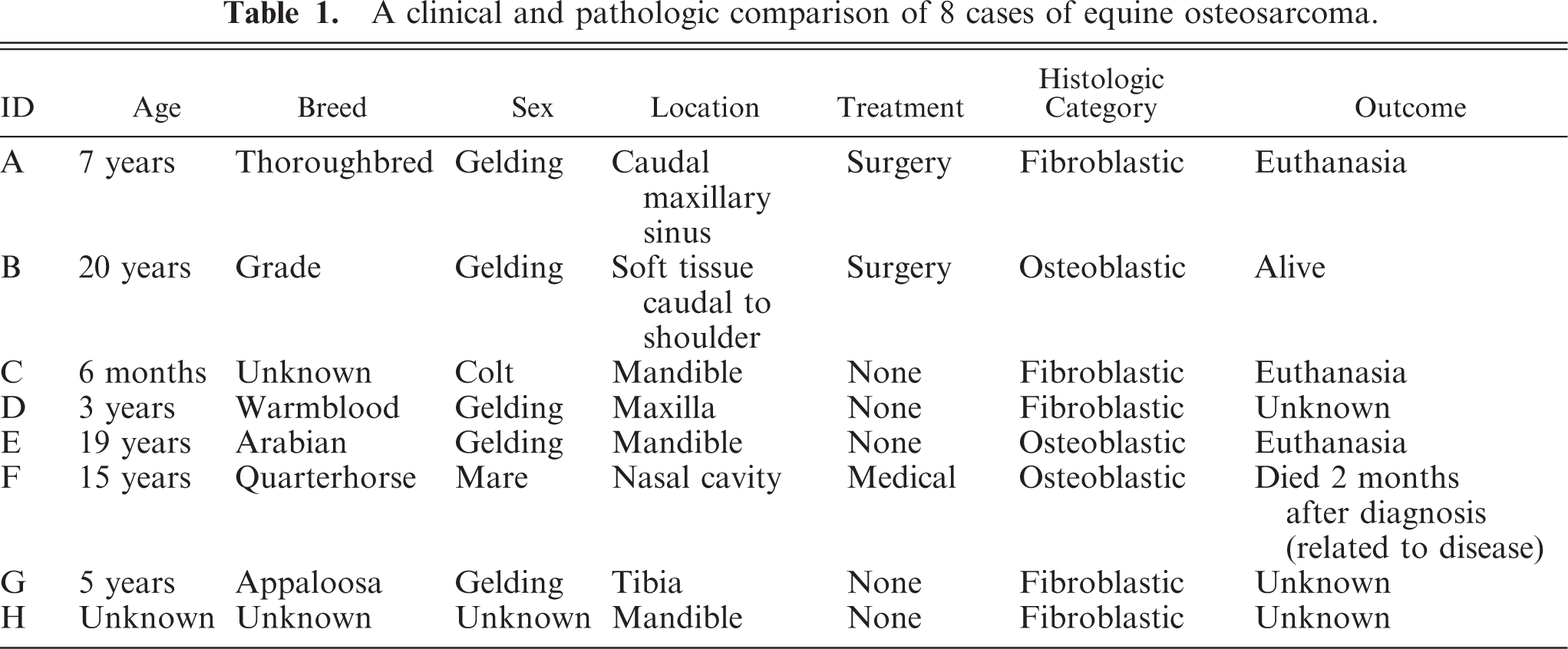
A clinical and pathologic comparison of 8 cases of equine osteosarcoma.
The majority of these equine osteosarcoma cases (6/8) occurred on the head, with 50% (3/6) occurring on the maxilla and 50% (3/6) occurring on the mandible. Overall, the gross appearance consisted of firm, expansive, and often invasive mineralized masses that distorted the normal tissue architecture with pronounced adjacent periosteal bone formation and central areas of necrosis (Fig. 1). Histologically, 66% (6/8) of the cases demonstrated fibroblastic cell features (Fig. 2). The cells were elongate and spindloid with indistinct cell borders blending with abundant fibrillar eosinophilic matrix. The remaining 2 cases had osteoblastic differentiation characterized by polygonal neoplastic cells with indistinct cell borders and abundant eosinophilic cytoplasm. All cases contained a variable amount of osteoid and chondromucinous matrix separating cells.
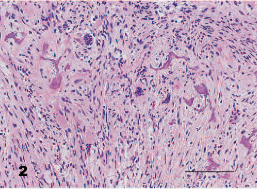
Caudal maxillary sinus; 7-year-old Thoroughbred gelding. The mass is composed of sheets of streaming neoplastic spindloid cells within a minimal fibrovascular stroma. Associated with the mass are numerous multinucleated cells (osteoclasts) and variably sized amorphous pools of eosinophilic material (osteoid). HE. Bar = 110 μm.
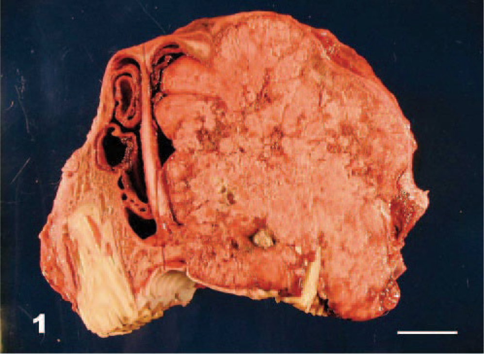
Maxilla; 4-year-old Warmblood gelding. There is complete effacement of the left maxilla, frontal sinuses, and nasal cavity by a multilobulated firm white to tan mass measuring 12 cm × 20 cm × 10 cm.
Clinically, the horses ranged in age from 6 months to 20 years, with a median age of 7 years. No breed predilection was noted, and 86% (6/7) of the cases were neutered males. Four of the horses were euthanized because of progressive disease after incomplete excision, 1 died from presumed respiratory obstruction, 1 horse survived, and the remaining 2 horses were lost to follow-up. Surgery as the sole treatment does not appear effective. The most common reasons for euthanasia were impaired respiratory and masticatory function.
A single case of an extraosseous osteosarcoma located within the soft tissue caudal to the right shoulder occurred in a mixed-breed 20-year-old gelding. There was no known history of trauma or vaccine injection at the site of the neoplasm. The neoplasm was a firm mass measuring approximately 22 cm × 14 cm × 14 cm that had multifocal areas of mineralization and necrosis on cut surface. Surgical excision was performed, and there has been no recurrence 14 months later. Histologically, neoplastic cells were osteoblastic within the minimal chondromucinous matrix (Fig. 3).
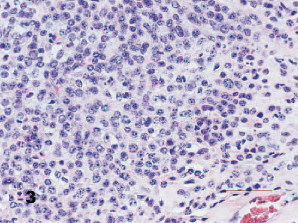
Extraosseous (caudal to right shoulder); 20-year-old grade gelding. The mass is composed of sheets of neoplastic polygonal cells within a minimal fibrovascular stroma. Neoplastic osteoblasts are associated with minimal amorphous pools of eosinophilic material (osteoid) and chondromucinous matrix. HE. Bar = 60 μm.
The final case involves a 5-year-old Appaloosa gelding with an osteosarcoma of the proximal tibia, which resulted in pronounced right hindlimb lameness. The mass was invasive and firm with a pronounced associated periosteal reaction. Histologically, the cells were fibroblastic in appearance within a minimal chondromucinous matrix. There was no treatment beyond the surgical biopsy, and the horse was lost to follow-up.
This case series presents the largest published case series of equine osteosarcomas and the first published report of an equine extraosseous osteosarcoma, to the authors' knowledge. Additionally, this case series indicates that there may be a predisposition for male and older horses, although a larger case series is warranted for further evaluation. This single case of extraosseous osteosarcoma was classified histologically as osteoblastic and demonstrated a positive outcome with surgery alone. Equine osteosarcomas are often fibroblastic (5/8) and typically contain a chondromucinous matrix (7/8). Because the majority of horses were either euthanized at the time of diagnosis or no further treatment modalities were attempted and the horse was lost to follow-up, it is difficult to assess any correlation between histologic subtype and prognosis. Additionally, metastatic disease was not identified in horses that were submitted for necropsy (2/8) or clinically in any of the horses. This suggests that the biologic behavior of equine osteosarcomas mimics that of cats, with a relatively slow rate of metastasis, although a larger case series is needed to confirm this observation. Previous case reports suggest young horses are predisposed to osteosarcoma. In this case series, the horses' ages ranged from 6 months to 20 years, with a median age of 7 years. Further investigations of equine osteosarcoma and the efficacy of various chemotherapeutic agents and alternative therapies are warranted to present additional treatment options.