
Abstract
Outbreaks of humeral fractures in dairy cows have been reported in New Zealand for several years. Gross, histologic, and histomorphometric findings in the humerus from primiparous cows with spontaneous humeral fracture were compared to age-matched control cows. Affected cows had a complete nonarticular spiral fracture of the humerus. Histologically affected humeri had a thicker growth plate with abnormal architecture, thinner cortex with increased abnormal resorption, increased resorption in the distal humerus, decreased trabecular density, abnormal trabecular architecture, presence of growth arrest lines and woven bone formation. Histomorphometry showed reduction in bone volume, trabecular perimeter, and trabecular width. Cows grazed on fodder beet had thicker growth plates with an abnormal appearance compared with cows grazed on pasture, and cows with low/marginal liver copper concentration had more resorption cavities in the distal humerus and thinner cortical bone compared with cows with adequate liver copper concentration. Decreased trabecular density (OR = 249.5), abnormal cortical resorption (OR = 54.2), presence of woven bone formation in the proximal metaphysis (OR = 37.2), and the number of resorption cavities in the distal humerus were significantly associated with a high probability of fracture. Ribs had enlargement of the costochondral junction with fractures in different stages of healing. Histology of the ribs revealed abnormal growth plate appearance, presence of fracture lines, callus tissue, fibrosis, and microfractures. Cows with humeral fracture have osteoporosis due to decreased bone formation and increased bone resorption, likely associated with inadequate feed quality and perhaps copper deficiency leading to a reduction in bone strength and fracture.
In 2008, an outbreak of spontaneous humeral fracture involving 6/200 first calving crossbreed dairy cows was reported in New Zealand. 55 Investigations into this outbreak showed that several of the affected cows had either low serum or low liver copper (Cu) concentrations, suggesting periods of Cu deficiency led to improper bone collagen crosslink formation and bone fragility, predisposing the cows to spontaneous fracture. 56 Since then, the occurrence of spontaneous humeral fracture has significantly increased (despite some farms implementing Cu supplementation to prevent deficiency) impacting animal welfare and mental health of farmers, and resulting in economic losses to the dairy industry. 12
Cases described so far have occurred mainly in first lactation cows post-calving, although occasional fractures have been seen prior to calving and in second lactation cows. 56 The clinical onset is sudden and animals develop severe forelimb lameness (also referred to as dropped elbow or dropped shoulder). 55
A small-scale study using computed tomography comparing the humeri of 10 cows with humeral fractures and 10 unaffected age-matched controls found a significant decrease in the ratio of bone volume: total volume in affected animals. 12 In the same study, histological findings in the humerus of affected animals included the presence of short cartilage spicules, thin bone trabeculae with presence of intratrabecular resorption, growth arrest lines, cortical osteoclastic resorption, and formation of additional woven bone between trabeculae. 12 Three affected animals had low liver Cu concentrations and one had low serum Cu concentration. 12 The authors concluded that the affected cows had osteoporosis, with inadequate bone deposition during crucial growth periods (leading to lower peak bone mass) and/or increased bone resorption (associated with gestation and lactation) as the main mechanisms resulting in decreased bone density. 12 Limitations of the study included that it only examined 10 cases, affected animals were from a specific region of New Zealand and there was no information regarding the animal’s diet in the months prior to the occurrence of the fracture.
Diet is important because, anecdotally, humeral fractures are common in cows grazing on a diet that is predominantly fodder beet (FB) (Beta vulgaris) before parturition (during the winter months in New Zealand). This crop has increasingly been used as a winter crop in New Zealand, despite being phosphorus and protein deficient, and has been associated with rickets/osteomalacia in lambs born to ewes that grazed on FB during gestation.13,22 However, humeral fractures also occur in cows that predominantly graze on pasture in the winter months, and it is hypothesized that these animals go through periods of protein-calorie imbalance during important growth periods leading to osteoporosis and subsequent fracture. 12
The aims of this study were (1) to determine the histological findings (qualitative) and to quantitatively evaluate histomorphometric changes in a large cohort of cows affected by spontaneous fracture of the humerus; (2) to determine whether histological and histomorphometric changes in bones from cows grazing a diet of predominantly FB over winter are different to cows that graze on a pasture-based diet over the winter; (3) to describe and compare histological changes in cows according to liver Cu concentration; and (4) to describe the histological findings in the costochondral junction (CCJ) (rib).
Materials and Methods
Study Design and Sample Collection
This was a case-control study using a convenience sample from fractured (affected) and nonfractured (control) cows. The case definition for enrolling an animal in the affected group was: a dairy cow of any breed, at least 2-year old, which had suffered a spontaneous fracture of the humerus, without any history of trauma, within 6 months of calving. Samples were provided by farmers and veterinarians who after reporting a case of spontaneous fracture of the humerus were presented with a list of samples to be collected postmortem. These included the humerus (fractured and/or contralateral), ribs (specifically including the CCJ), and a piece of liver. A pro forma submission form (containing farm and animal data, main feed offered before parturition, and relevant clinical information) was developed and sent to the case submitter for completion.
A control was defined as a 2-year-old cow (a cow with an ear tag indicating that they were born 2 years ago) of any breed that had calved recently (udder consistent with lactating) and had been culled for reasons unrelated to bone fracture of the humerus or any other bone. Control samples were obtained from a cow-rendering plant (Wallace Corporation) and from Massey University’s School of Veterinary Science postmortem service. From each control case, a sample of the humerus, the CCJ, and a piece of liver was collected postmortem. Owing to the method of sampling, no information regarding the cow diet and/or the reason for culling was available for the control animals.
Gross Evaluation
The humeri and CCJ were dissected away from surrounding soft tissues, examined grossly, and photos were taken to record the gross appearance. Subsequently, several bone slabs (~3–5 mm thick) were obtained using a band saw. Photos of the bone slabs were taken. One slab from the humerus was then cleaned with water to remove the bone marrow and allow evaluation of the trabecular bone and cortex. For each animal, a subset of bone slabs from the humerus and the CCJ were placed in 10% neutral buffered formalin solution until further processing for microscopic evaluation.
Data recorded in the gross evaluation of the fractured humerus included the location and extension of the fracture, the appearance of the cortex, the appearance of the trabecular bone, and the presence/absence of growth arrest lines.
Histological Processing and Evaluation
Sections from the humerus and CCJ were processed for histological evaluation. Bone slabs were placed in glass jars with 10% hydrochloric acid (Decalcifier hydrochloric acid, Amber Scientific Ltd). Glass jars with samples were placed on a Thermo shaker incubator at 37°C and 180 rpm (Thermo shaker MB100-4A, Hangzhou Allsheng Instruments CO., Ltd). Every 24 hours, a manual evaluation of the decalcification process was performed and the decalcifier solution was changed. When properly decalcified, bone samples were trimmed to fit a histology cassette. Samples were processed routinely for histology, embedded in paraffin, sectioned at 4–5 μm, and stained with hematoxylin and eosin (HE) for evaluation. Cases were blinded and examined by a single examiner (AWM).
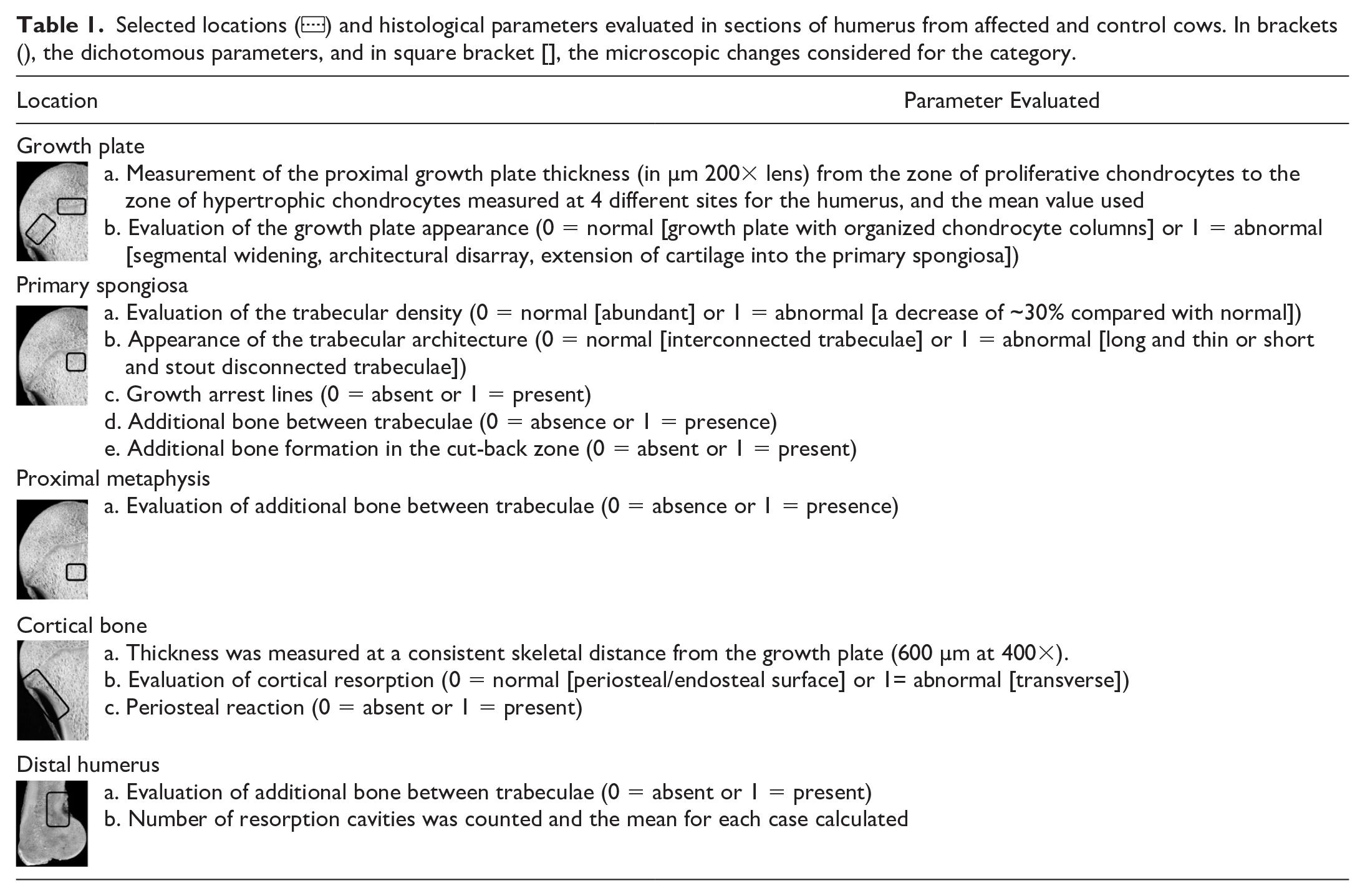
Table 1 shows the 5 locations in the humerus selected for histological evaluation and the histological features considered for evaluation. Measurements (growth plate and cortical thickness) were taken using imaging software (Olympus cellSens Standard 1.18, Olympus Corporation). Histological parameters were graded using a subjective dichotomous ordinal scale depending on whether the observed parameter was abnormal/present (1) or normal/absent (0).
Selected locations () and histological parameters evaluated in sections of humerus from affected and control cows. In brackets (), the dichotomous parameters, and in square bracket [], the microscopic changes considered for the category.
In sections of the CCJ, histological findings were evaluated and recorded. Finally, the growth plate thickness was measured at three different sites (right, center, and left sides) and the mean calculated.
Histomorphometry Processing and Evaluation
One 1 × 1 cm formalin-fixed sample of the primary spongiosa of the humerus was used for histomorphometric evaluation from a subset of 20 affected cows (10 were randomly selected from cows that grazed on FB during the winter months and 10 randomly selected from cows that grazed on pasture during the winter months) and 10 control cows.
The undecalcified bone sections were dehydrated in graded alcohol and placed first in resin infiltration solution I, then solution II for 24 hours in each (90% methyl methacrylate [MMA] and 10% dibutyl phthalate (BPO), Sigma-Aldrich). Next, samples were placed in a resin embedding solution (95% MMA/5% BPO) for 24 hours. Benzoyl peroxide (Luperox® A75, Sigma-Aldrich) was used as the polymerization agent. Samples were then cut at 6 µm using a super-microtome (Mikrotom 2050 supercut, Reichert Jung). Sections were stained with Goldner’s modified trichrome stain. For each case, bone area (B.Ar) in µm2, total area (T.Ar) in µm2, B.Ar/T.Ar, mean trabecular perimeter in µm, mean trabecular width (mean Tb.Wi) in µm, mean osteoid area (mean O.Ar) in µm2, and mean osteoid perimeter (mean O.Pm) in µm were measured. For B.Ar, T.Ar, and B.Ar/T.Ar, standard BoneJ plugins (Domander, R., Felder, A. A., & Doube, M. [2021]. BoneJ2) for ImageJ (version 1.53c, National Institute of Health) were used. Evaluation of mean trabecular perimeter, mean Tb.Wi, mean O.Ar, and mean O.Pm was measured using ImageJ software (version 1.53c, National Institute of Health). Results were compared between affected and control cases, and according to the main diet over winter (FB vs pasture).
Determination of Liver Copper Concentration
Liver samples from the affected and control groups were submitted to a commercial diagnostic laboratory (Idexx Laboratories New Zealand Ltd.) for determination of liver Cu concentration using inductively coupled mass spectrometry (NexION 2000B ICP Mass Spectrometer, PerkinElmer). Liver Cu concentration between 0 and 94 µmol/kg was considered low/marginal concentration and values > 94 µmol/kg were considered adequate liver Cu concentration.
Statistical Analysis
An independent-sample t-test was used to determine whether there were any significant differences in the values of growth plate thickness, cortical thickness, and quantity of resorption in the distal humerus between affected and control cows, between cows that grazed either FB or pasture as their main winter feed and between cows with low/marginal and adequate liver Cu concentration. Values from these parameters were first log transformed to achieve a normal distribution. If the assumption of homogeneity of variances was violated, a Welch t-test was run to determine differences between groups.
For all the dichotomous parameters, a chi-square test for homogeneity was done to compare differences in the distribution of proportions between cows in the affected and control groups, between cows in the affected group that grazed either FB or pasture as their main winter feed, and between cows with low/marginal or adequate liver Cu concentration. If the minimum sample size for each expected frequency was not met (greater than or equal to 5), then results for Fisher’s exact test were presented instead. In all tests, a P value of < .05 was considered significant.
For the comparison between cows in the affected and control groups, the parameters were put into a multivariable logistic regression model to predict the probability of cows suffering from humeral fracture. For a variable to be included as a parameter in the model, it had to have an observed count (distribution) more than 1 and the P value obtained from the t-test or chi-square test had to be significant (P < .25). 9 The model was constructed using forward selection, with variables retained at P < .05, and the model fit was assessed using the Hosmer-Lemeshow (HL) test and the Nagelkerke R 2 .
Finally, an independent-sample t-test was conducted to compare differences in the histomorphometric data between the affected and control groups, and between cows that grazed on FB compared to cows that grazed on pasture. Differences were considered significant if P < .05. All statistical analysis was done in SPSS statistics (IBM® SPSS® Statistics version 27).
Results
Study Population
A total of 80 humeri were collected from farms throughout New Zealand from cows that fitted the case definition. Out of the 80 samples of humerus, 47/80 (59%) cases also included one or several sections of CCJ, and in 66/80 (83%) cases, a piece of liver was submitted. Forty-three of the 80 (54%) affected cows were Kiwi cross cows (Holstein-Friesian × Jersey, of unknown proportions), 28/80 (35%) Holstein-Friesian, 4/80 (5%) Jersey, and 5/80 (6%) had no breed information.
Case distribution according to the predominant diet offered in the winter months showed 33/80 (41%) cases grazed on FB, 28/80 (35%) cases grazed on pasture, 14/80 (17.5%) cases were fed another type of feed, and in 5/80 (6.5%) cases information regarding main winter feed was not provided. Diets in cases classified as “other” contained a mix of feeds, including swedes (Brassica napus), maize (Zea mays) silage, oats (Avena sativa), and/or kale (Brassica oleracea).
The only reported clinical sign was nonweight-bearing lameness of the affected leg. One case reported that the cow was lame after turning around in the cow shed, another case reported bumping into an object, and in 4 cases, the cow was reported to be in estrus, artificially inseminated, and/or bulling the day prior to the fracture.
A total of 22 humeri were sourced from control animals, 17/22 (77%) controls also included one or several sections of CCJ and 22/22 (100%) included a piece of liver.
Gross Findings
In 51/80 (64%) cases, the affected humerus was included, whereas for 29/80 (36%) the nonaffected (contralateral) humerus was submitted. Gross changes described below are for the 51 humeri with fractures. Fractures in 49/51 cases (96%) were complete, nonarticular, simple, spiral fractures extending from the lateral side of the humeral head, just beneath the greater tubercle, spiraling along the diaphysis distally to end just above one of the condyles (Fig. 1a). In 2/51 cases (4%), the fracture was complete, comminuted, and transverse through the diaphysis.
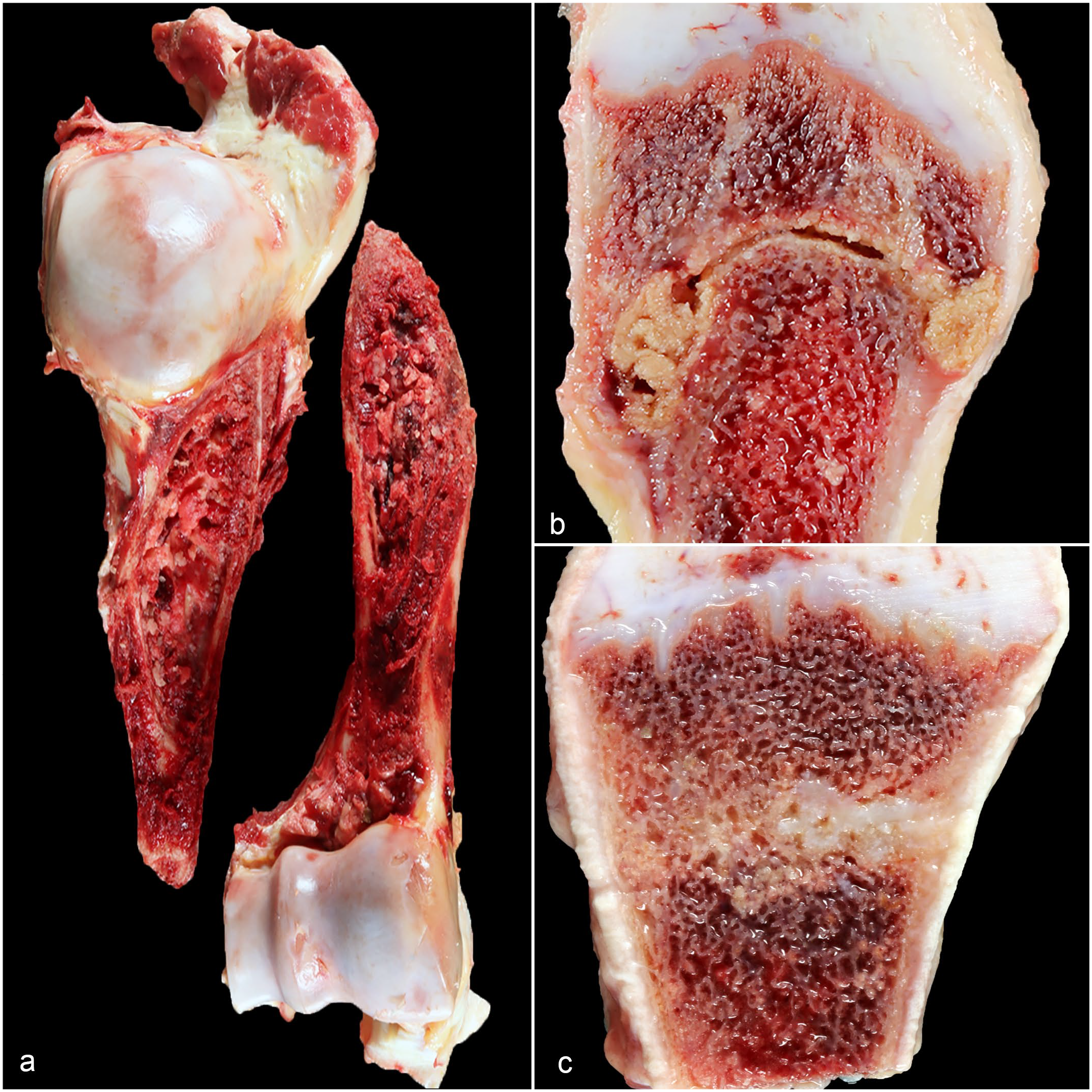
Humerus and ribs from affected cows. (a) Spiral fracture, humerus, cow. The fracture line extends from the lateral side of the humeral head (just beneath the greater tubercle) and extends distally to end just above of the condyles. (b) Costochondral junction (CCJ), cow. Cut surface of the rib and CCJ from an affected cow with a bulging, irregular cortex and a fracture line extending cortex to cortex associated with the presence of necrotic bone and hemorrhage. (c) Costochondral junction (CCJ), cow. Rib and CCJ from an affected cow show an older fracture line with the presence of cartilaginous tissue.
Evaluation of bone slabs from affected cases revealed 39/51 (76%) had a mild reduction (~30% less) in the amount of trabecular bone associated with an expansion of the bone marrow cavity toward the primary spongiosa when compared to controls (Supplemental Fig. S1). The distal humerus in 20/51 (39%) cases had evidence of increased cortical resorption identified as numerous red streak marks (Supplemental Fig. S1). No cases had macroscopically visible growth arrest lines.
Evaluation of 32/47 (68%) ribs revealed a moderate to marked enlargement of the CCJ (up to 3-fold increase compared to controls). The cut-surface in these cases revealed the presence of a fracture in the metaphysis, extending in most cases from cortex to cortex and characterized by a bulging, rounded, irregular cortex mixed with areas of necrotic bone, hemorrhage, and proliferation of tissue that appeared either edematous or fibrous and/or cartilaginous (Fig. 1b). In 9/47 (19%) cases, the CCJ did not appear enlarged, but the cut-surface revealed the presence of lesions in the metaphysis consistent with remnants of previous fractures or fractures in an advanced stage of healing, characterized by proliferation of fibrous connective tissue and/or bony or cartilaginous tissue (Fig. 1c). In 1 (2%) case, the growth plate was thickened, irregular and had small tongue-like extensions of cartilage into the primary spongiosa. In 5/47 (11%) cases, the cut-surface of the rib and CCJ appeared grossly normal.
Finally, 4/17 (24%) cows from the control group, had lesions like affected cows, consistent with a rib fracture and formation of callus tissue. In the remaining 13/17 (76%) controls, the ribs were grossly normal.
Histology Findings
Humerus
Affected vs control
The proximal humeral growth plate was significantly thicker (a difference of 35.3 µm, 95% CI, 18.2–52.4) in affected cows compared with control cows, while the humeral cortex was thicker (a difference of 153.8 µm, 95% CI, 97.5–210.1) in control cows compared to affected cows. Cows with fracture had a higher number of resorption cavities (12 more, 95% CI, 6.7–16.7) in the distal humerus compared to cows in the control group. Mean ± SD and P values are presented in Table 2.
Mean ± SD of the proximal growth plate thickness of the humerus and costochondral junction, humeral cortical thickness, and the quantity of resorption cavities in the distal humerus comparing cases in the affected vs control groups, fodder beet vs pasture groups, and low/marginal vs adequate liver copper concentration groups.
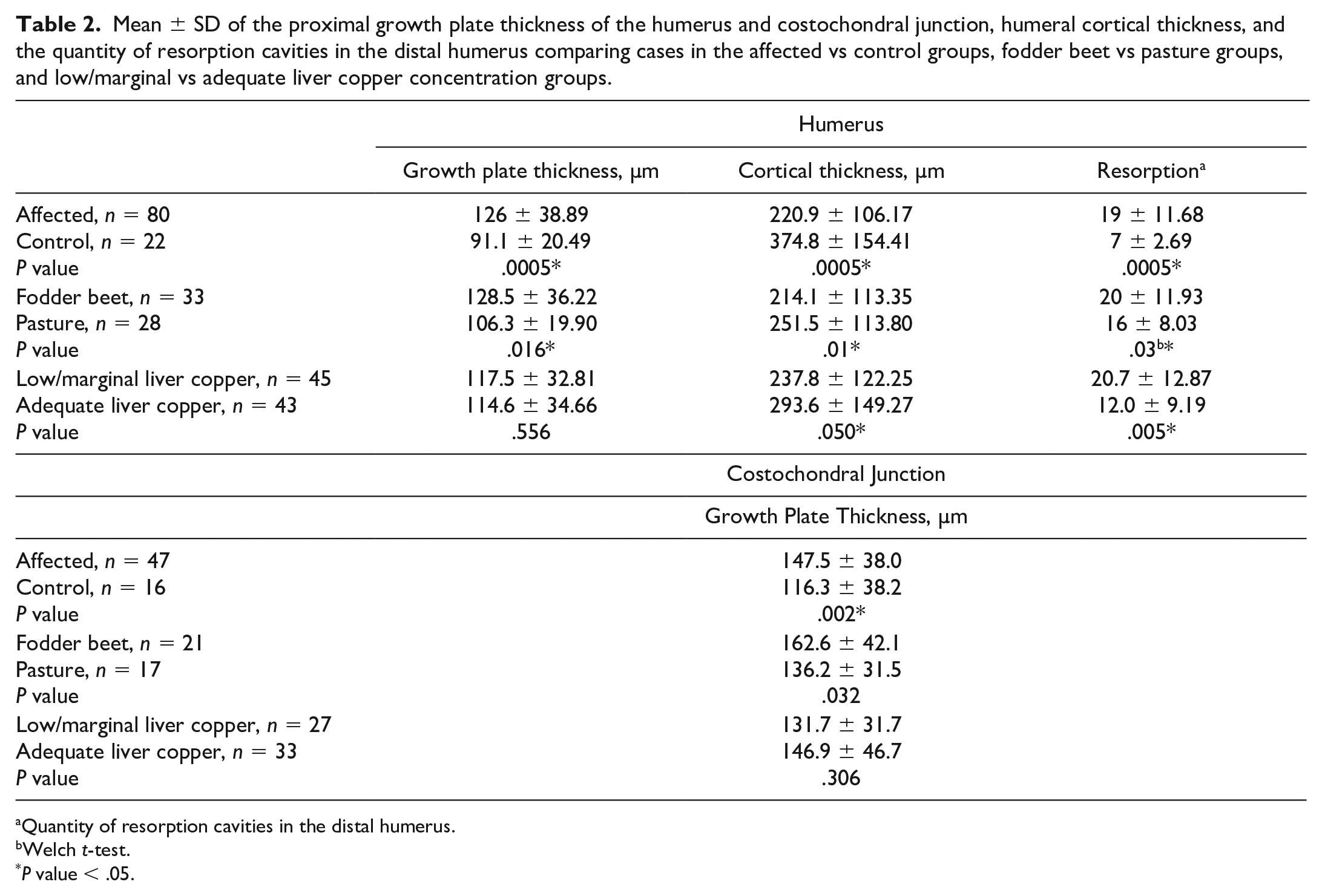
Quantity of resorption cavities in the distal humerus.
Welch t-test.
P value < .05.
In the case of the dichotomous parameters, most cows in the affected group had decreased trabecular density, abnormal trabecular architecture, abnormal growth plate appearance, additional woven bone formation in the primary spongiosa and proximal metaphysis, and 17/80 (21%) cows in the affected group had histological evidence of growth arrest lines (Fig. 2a–g). A greater number of cows in the affected group showed evidence of periosteal reactive bone formation and abnormal cortical resorption compared to cows in the control group (Fig. 2h). Finally, 24/80 (30%) had evidence of additional woven bone formation in the cut-back zone (Fig. 2i). Abnormal cortical resorption was characterized by numerous coalescing resorption cavities extending transversely through the cortex (Fig. 3a).
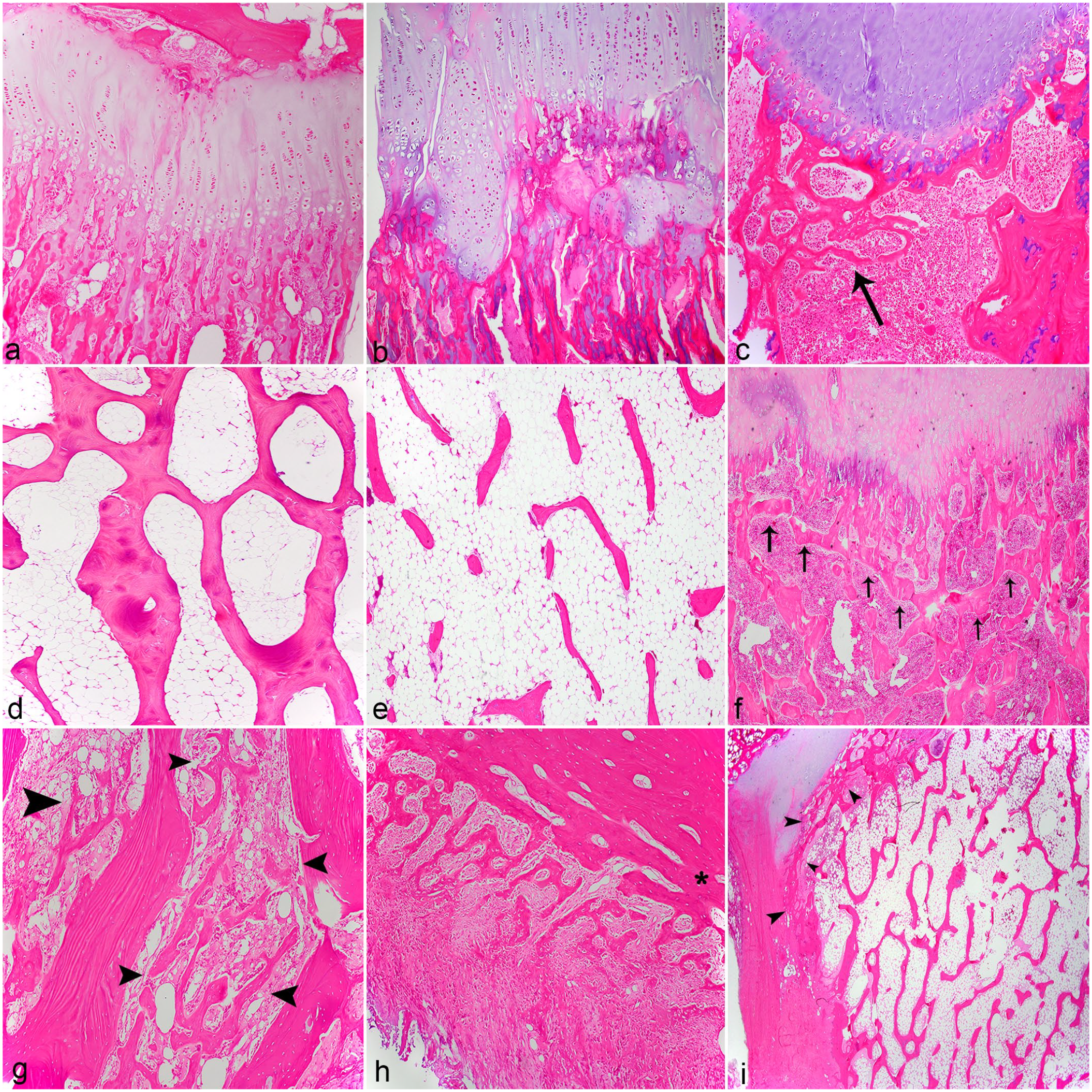
Histological appearance of humeral sections evaluated in affected and control cows. Hematoxylin and eosin (HE). (a) Growth plate and primary spongiosa, control cow. Normal growth plate architecture with thin growth plate, organized chondrocyte columns and normal appearance of the primary spongiosa. (b) Growth plate and primary spongiosa, affected cow grazed on fodder beet. Abnormal growth plate architecture with thicker, irregular growth plate, disorganized chondrocyte columns and extension of cartilage into the primary spongiosa. (c) Growth plate and primary spongiosa, affected cow grazed on pasture. Normal growth plate architecture with thin growth plate, organized chondrocyte columns but with formation of additional woven bone in the primary spongiosa (arrow). (d) Trabecular bone, control cow. Normal trabecular architecture with abundant, thick, and interconnected bone trabeculae. (e) Trabecular bone, affected cow. Abnormal trabecular architecture with fewer, thin, long, unconnected bone trabeculae. (f) Growth plate and primary spongiosa, affected cow. Transverse bone trabeculae below the growth plate (growth arrest line) (arrows). (g) Proximal metaphysis affected cow. Formation of woven bone (arrowheads) between trabeculae. (h) Cortical bone affected cow. Periosteal reactive woven bone mixed with fibrous tissue extending from the cortex (*). (i) Cut-back zone, affected cow. Additional woven bone formation in the cut-back zone (arrowheads).
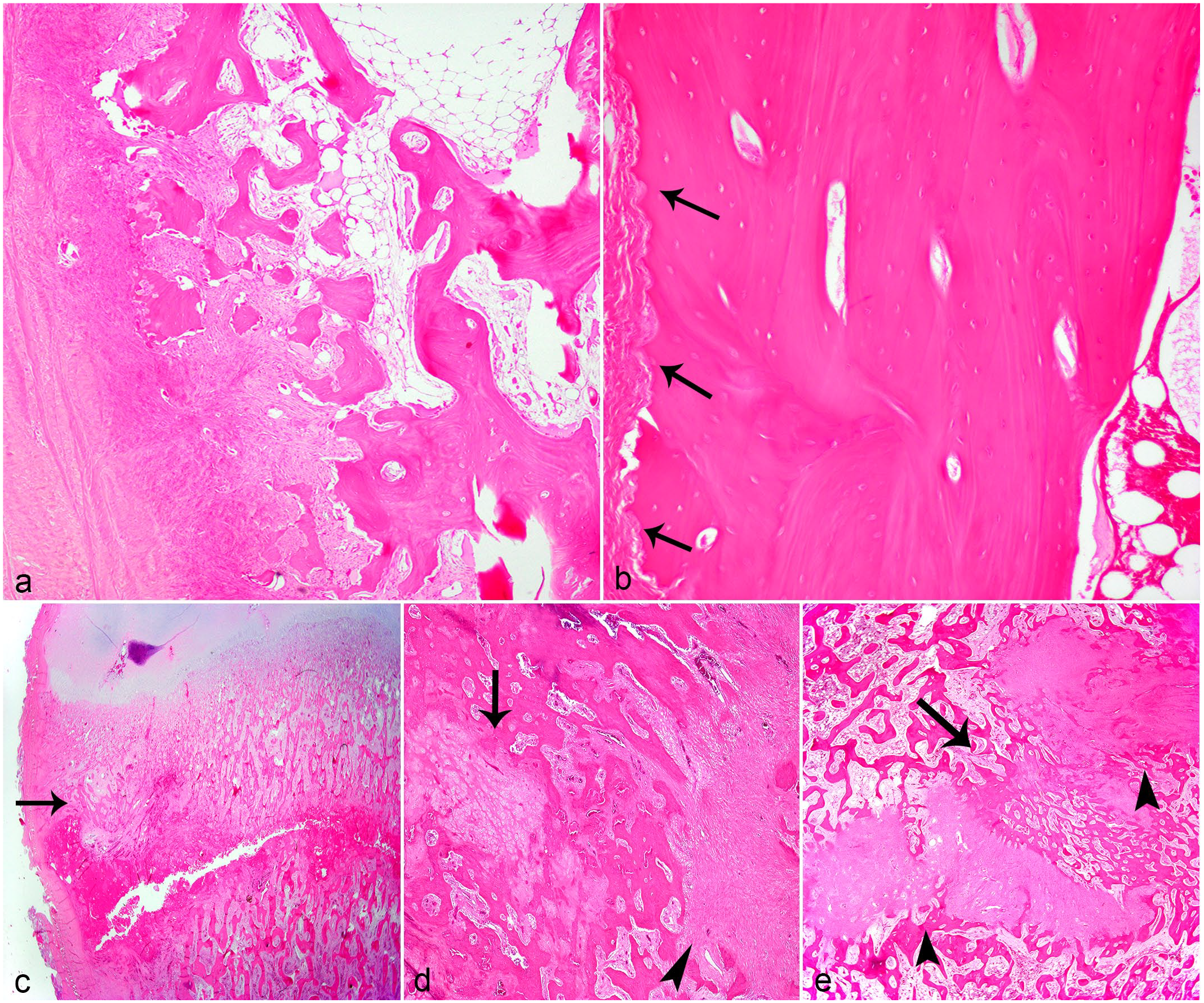
Histological appearance of humeral cortical bone, costochondral junction, and rib from affected and control cows. Hematoxylin and eosin (HE). (a) Metaphysis, humeral cortical bone, affected cow. Abnormal cortical thickness and appearance of the cortical bone from an affected cow with osteoclasts extending transversally through the cortex, leaving a cortex with a moth-eaten appearance. (b) Metaphysis, humeral cortical bone, control cow. Thick cortex, only the periosteal surface shows cortical scalloping due to resorption lacunae (arrow). (c) Costochondral junction and metaphysis, rib, affected cow. Fracture line extending from the cortex into trabecular bone associated with hemorrhage and disorganized trabecular bone (arrow). (d) Trabecular bone, rib, affected cow. Older fracture line with proliferation of fibrous connective tissue (arrowhead) and formation of soft callus tissue (arrow). (e) Trabecular bone, rib, affected cow. Irregular replacement of fracture line by a proliferation of fibrous connective tissue (arrowheads) and additional woven bone formation (arrow).
In contrast, most cows in the control group had normal trabecular density, trabecular architecture, and growth plate appearance; absence of growth arrest lines, periosteal reactive bone formation; and no additional bone formation in the primary spongiosa, proximal metaphysis, or distal humerus (Fig. 2a, d). Finally, cortical resorption was limited to the periosteal and/or endosteal side of the cortex with sparse intracortical resorption cavities (Fig. 3b). Statistical significance for each of the dichotomous bone parameters evaluated is presented in Table 3 and case distributions are presented in Supplemental Table S1. All parameters evaluated were significantly different (P < .05) when comparing cows in the affected and control groups.
Statistical significance of each of the bone parameters analyzed comparing cases in the affected and control groups, fodder beet vs pasture group and low/marginal vs adequate liver copper concentration group.
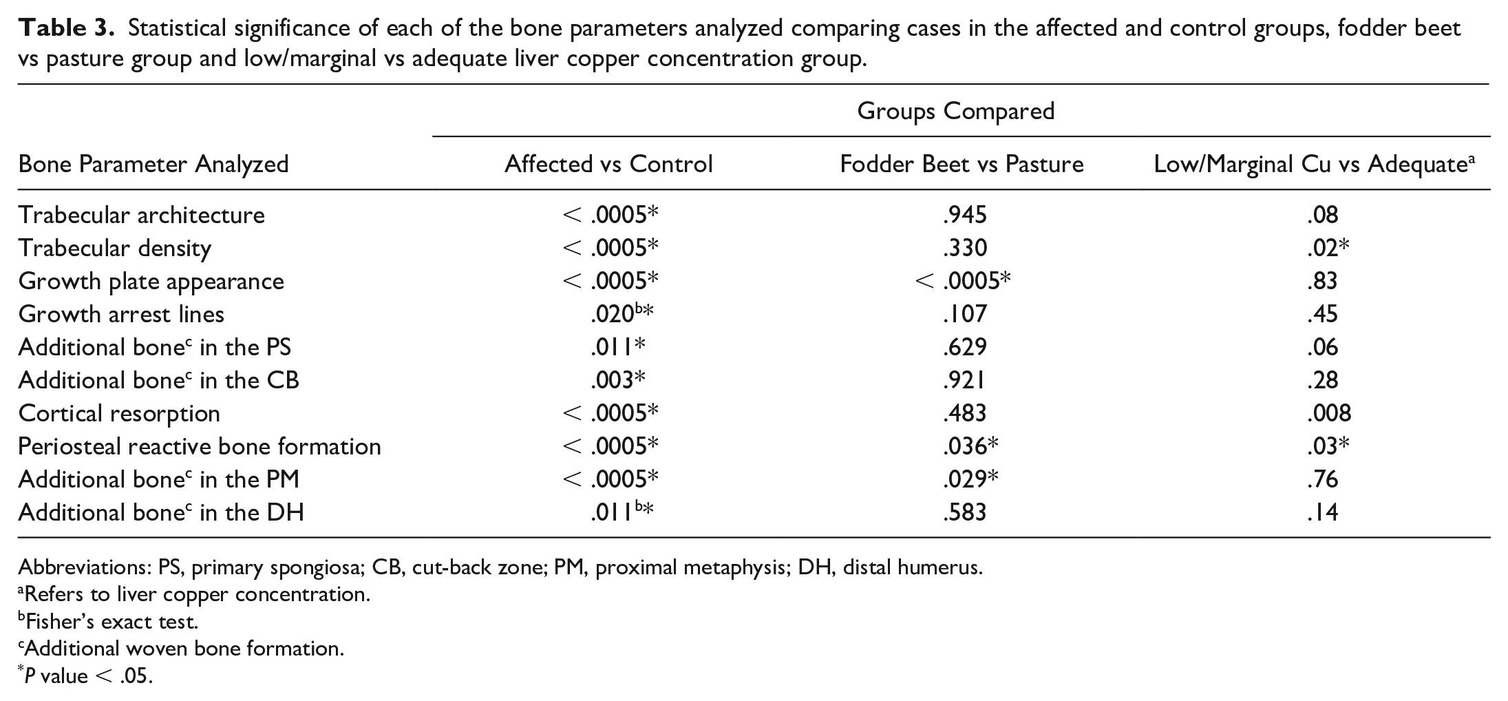
Abbreviations: PS, primary spongiosa; CB, cut-back zone; PM, proximal metaphysis; DH, distal humerus.
Refers to liver copper concentration.
Fisher’s exact test.
Additional woven bone formation.
P value < .05.
FB vs pasture
The growth plate was thicker (a difference of 22.3 µm, 95% CI, 6.91–37.63) and growth plate appearance was significantly abnormal in cows grazing FB compared to cows grazing pasture (Fig. 2b, c). Cortical thickness and quantity of cortical resorption in the distal humerus were not significantly different between groups. Mean ± SD and P values are presented in Table 2. Cows that grazed on pasture had significantly more woven bone formation in the primary spongiosa and greater formation of periosteal reactive bone compared to cows that grazed on FB. Statistical significance for each of the dichotomous bone parameter evaluated is presented in Table 3 and case distribution is presented in Supplemental Table S1.
Low/marginal vs adequate liver Cu concentration
A significantly greater proportion of affected cows 41/66 (62%) had low/marginal liver Cu concentrations compared to control cows 4/22 (18%). When analyzing the quantity of resorption cavities in the distal humerus, regardless of the fracture status, the mean number of resorption cavities was greater (9 more, 95% CI, 3.8–13.5) in cows with low/marginal liver Cu concentration compared with cows with adequate liver Cu concentrations. Also, cortical bone was thicker (a difference of 55.73 µm, 95% CI, 2–113.4) in cows with adequate liver Cu concentration compared to cows with low/marginal liver Cu concentration. There were no significant differences in growth plate thickness between groups. Mean ± SD and P values are presented in Table 2.
For the dichotomous parameters, significantly more cows with low/marginal liver Cu concentration had decreased trabecular density (34/45, 76%), abnormal cortical resorption (36/45, 80%), and formation of periosteal reactive bone (31/45, 69%) compared to cows with adequate liver Cu concentrations. There were no significant associations between all other dichotomous bone parameters analyzed. Statistical significance for each of the dichotomous bone parameter evaluated is presented in Table 3 and case distribution is presented in Supplemental Table S1.
Ribs
The main microscopic changes seen in the rib sections from affected cows were an abnormal growth plate appearance characterized by an irregular growth plate with extension of hypertrophic chondrocytes into the primary spongiosa forming tongues and/or islands of cells. Furthermore, all sections of rib evaluated had incomplete or complete fracture lines extending cortex-to-cortex. Fractures had a variety of changes ranging from presence of necrotic bone mixed with congestion, hemorrhage, and edema to callus tissue formation (Fig. 3c). In a few cases, attempts at callus tissue formation were observed, characterized by a proliferation of cartilaginous tissue (soft callus) and/or woven bone (hard callus) (Fig. 3d). In most cases, fracture lines were filled with a proliferation of fibrous connective tissue associated with an irregular proliferation of woven bone at the periphery on the fracture line (Fig. 3e). Nonetheless, in most cases, where callus tissue was identified, there was also proliferation of fibrous connective tissue interspersed with cartilage and woven bone indicative of an abnormal repair process. Other findings included the presence of numerous microfractures, thickening of the periosteum by proliferation of fibrous connective tissue, identification of granulation tissue, marked trabecular resorption, and an irregular bulging cortex.
Lesions in the ribs from control cows showed 5/22 (23%) cases had small (2–5 cells thick) nests of chondrocytes in the primary spongiosa. In 4 cases, where grossly a fracture line was observed, these were histologically characterized by proliferation of fibrous connective tissue and multifocal areas with soft callus and hard callus proliferation. Lesions in control animals also had areas of hemorrhage, formation of additional bone between trabeculae and thickened periosteum.
The rib growth plate was thicker (a difference of 31.1 µm, 95% CI, 9.1–53.1) in cows in the affected group compared with cows in the control group. Finally, the growth plate of the rib was thicker (a difference of 26.4 µm, 95% CI, 1.4–51.4) in cows that grazed FB in winter compared to cows that grazed pasture. Mean ± SD and P values are presented in Table 2.
Histomorphometry Findings
Comparison between the affected and control groups showed cows in the control group had greater B.Ar (P < .005), higher B.Ar/T.Ar (P < .005), longer mean trabecular perimeter (P = .027), and mean Tb.Wi (P = .03) values. O.Ar (P = .24) and O.Pm (P = .18) were not significantly different between groups. There were no statistically significant differences in any of the histomorphometric parameters evaluated when comparing FB and pasture as main winter feed. Mean ± SD and P values are presented in Table 4.
Comparison of histomorphometric results between affected vs control and fodder beet vs pasture.
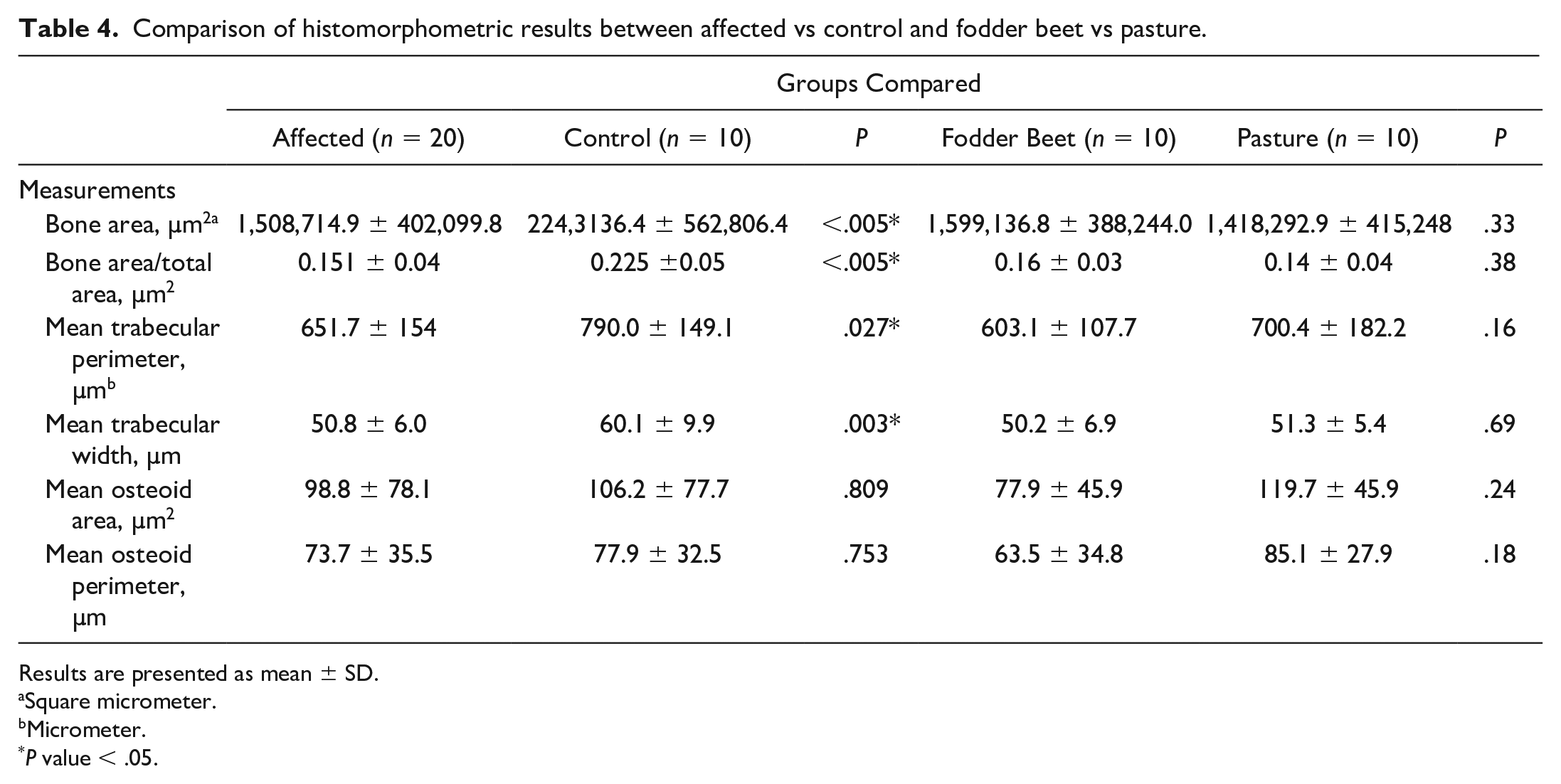
Results are presented as mean ± SD.
Square micrometer.
Micrometer.
P value < .05.
Logistic Regression Analysis
The following 9 bone parameters were tested in the logistic regression model: trabecular architecture, trabecular density, growth plate appearance, formation of additional bone in the primary spongiosa and proximal metaphysis, type of cortical resorption, growth plate thickness, cortical thickness, and amount of resorption in the distal humerus. Four were retained in the final model: decreased trabecular density, abnormal cortical resorption, presence of additional woven bone formation in the proximal metaphysis, and the number of resorption cavities in the distal humerus. The logistic regression model was statistically significant, χ2(4) = 85.152, P < .0005. The HL test was not statistically significant (P = .95) indicating the model is a good fit. The model explained 88.6% (Nagelkerke R 2 ) of the variance in humeral fracture and correctly classified 94.9% of cases. Sensitivity was 96.1% and specificity was 80.9%. Positive predictive value was 97.3% and negative predictive value was 86.9%. The odds of an animal with decreased trabecular density having a fracture were 249.4 times higher than the odds of an animal with normal trabecular density having a fracture. Abnormal cortical resorption was also associated with an increased likelihood of humeral fracture (odds ratio, 54.2) as was the presence of additional bone in the proximal metaphysis of the proximal humerus (odds ratio, 37.2).
Discussion
Humeral fractures are infrequent in ruminants due to the musculoskeletal configuration around the humerus and the need for very high forces to fracture the humerus. 42 The fact that the fractures in affected cows in this study are spontaneous and numerous cows can be affected at the same time within a single herd implies a significant reduction in bone strength/quality as the main mechanism leading to the appearance of fracture. Furthermore, the gross and histological changes present in sections of ribs from a large proportion of cows with humeral fractures, showing fractures of different ages and stages of healing, indicate that the issues with bone strength and quality are systemic and have been compromised for some time.
The histological and histomorphometric results from this study reveal that cows with humeral fracture have osteoporosis characterized by reduced trabecular density, abnormal trabecular architecture, reduced cortical thickness with increased abnormal cortical resorption, and the presence of growth arrest lines. We believe that these characteristics are the main factors that contributed to decreased bone strength ultimately resulting in humeral fracture.
Osteoporosis is characterized by a reduction in bone mass, bone mineral content, and bone matrix, which severely compromises bone quality and strength and increases the risk of bone fracture.36,41,46 Histological changes of osteoporosis are characterized by decreased trabecular number, trabecular thickness, cortical thickness, and increased trabecular separation and cortical porosity, which are remarkably similar to the findings presented in this study of cows with humeral fracture.19,51
Mechanisms leading to osteoporosis include failure to achieve peak bone mass (failure of bone formation), high bone turnover (excessive bone resorption), and/or low bone turnover (normal osteoclastic activity with reduced osteoblastic activity).10,41 Considering the histological and histomorphometric findings in this study, both failure of bone formation and excessive bone resorption have contributed to the osteoporosis observed in affected cows.
When considering bone formation, the impact of nutrition on bone formation/growth (achievement of peak bone mass) and strength, especially during the early years of life, is important. Failure to achieve peak bone mass, and low protein/caloric intake during adolescence leads to osteoporosis and increased fracture risk in humans.5,6 Similarly, prepubertal nutrition has an important effect not only on mature body weights in dairy cows, but also on milk yield and bone growth.21,23,24,53 In cows, liveweight is important because it determines bone size and is a major determinant of the strain stress index (a proxy for bone strength) in the humerus of dairy cows.23,24 Furthermore, although calves born from dams with high milk production tend to be lighter, if heifers reach pre-mating target liveweights (before 12–14 months of age), there are no significant differences in skeletal (frame) size in later months, further emphasizing the importance of nutrition in early life.26,49
Analysis of growth patterns in New Zealand dairy cows shows there are normally 2 periods of reduced growth rate, the first is ~9 months of age (coinciding with the period of lowest pasture quantity and quality in winter), and the second between 22 and 24 months of age, corresponding to the second winter. 27 This is significant because when growth of the metacarpus is compared with the humerus in growing dairy cows, the humerus keeps growing during the second year of life making it more susceptible to additional growth checks. 24
Several microscopic changes in bone can provide information regarding nutrition and bone growth. First, the presence of growth arrest lines is important. Growth arrest lines are transverse bone trabeculae that appear histologically when the activity of the growth plate stops for a period of time and is suggestive of starvation and/or malnutrition for a period of time.10,39 The presence of these growth arrest lines in cases of humeral fracture previously described led to the suggestion that protein/calorie malnutrition was one of the factors contributing to reduced bone strength and osteoporosis. 12 While only 21% of cows with humeral fracture in the present study had growth arrest lines, remodeling can cause resorption and disappearance of growth arrest lines. 32 Increased remodeling was observed in many sections of humeral fractures in the current study and could have masked the identification of greater numbers of growth arrest lines.
Second, the histological appearance of the growth plate can also provide valuable information on patterns of growth in long bones.10,50 The significant difference in growth plate thickness (both in the humerus and CCJ) and growth plate appearance in affected cows compared to control cows in this study may indicate a mismatch between the actual bone mass and the expected bone mass. Catch-up growth (referred to as compensatory growth) is a phenomenon that occurs after a period of nutrient restriction or other inhibitory factor whereby there is inhibition of chondrocyte senescence leading to longer chondrocyte columns and a thicker growth plate compared to age-matched controls, such as was seen in physes from cows with humeral fractures.18,20
The other mechanism that can lead to the appearance of osteoporosis and that needs to be considered is increased bone resorption. In dairy cows, increased osteoclastic bone resorption is observed physiologically during gestation and this increases up to 3-fold during lactation with the majority of this resorption occurring in trabecular bone.29,43 Furthermore, primiparous cows and cows with higher milk yield have more active bone resorption than multiparous cows and cows with lower milk yield, as assessed by plasma and urine bone biochemical markers.33,34 Excessive bone resorption can lead to fewer and thinner trabeculae (observed in 81% of affected cases), with loss of trabecular connectivity (observed in 78% of affected cases), compromising bone architecture and strength leading to fracture. 45 These frequently observed trabecular changes, plus the higher probability of breaking a bone with decreased trabecular density, suggest increased resorption is an important factor impacting bone strength in these cows. Decreased trabecular density, trabecular connectivity, and thickness affect the biomechanical strength of bone interfering with the capacity of trabecular bone to withstand load. 38
As a result of the reduction in the quantity and quality of trabecular and cortical bone, additional bone may be formed between trabeculae, particularly in areas with marked stress and strain.17,52 In affected cows, additional woven bone formation was observed in the primary spongiosa (66% of cases), cut-back zone (30% of cases), proximal metaphysis (75% of cases), and in the distal humerus (24% of cases). This formation of “extra” bone in affected cases indicates that numerous areas of the humerus from affected cows are under mechanical stress. 44 Additional woven bone between trabeculae appears histologically around 7 days after trauma in small animals and around 2 weeks in humans. 44 However, this new bone is not readily available for resorption and this could mean that osteoclastic activity increases along the already small and poorly formed pre-existing trabeculae to supply Ca for lactation. 44 Finally, formation of periosteal reactive bone is also a mechanism to increase bone strength at sites of maximum stress-strain, and for fracture repair, as was observed in 75% of samples from cows in the affected group.3,14,52
In the humeral cortex of affected cows, there was a significant reduction in cortical thickness and a significant increase in resorption activity that was abnormal. Cortical bone thickness is determined by the rate of endosteal bone resorption (not by a lag in bone formation) and is key for bone strength.2,45 Contrary to trabecular bone, only minimal resorption is observed in cortical bone before and after gestation. 43 Clumping and enlargement of resorption canals in the cortex causes a reduction in cortical thickness and focal areas of weaker bone leading to fracture, which is seen in humans with nontraumatic intracapsular femoral neck fractures. 31 Abnormal cortical microstructure affects maximum stress distribution facilitating propagation of microcracks that can lead to bone fracture. 1 A similar pathophysiological process is likely occurring in the humeral cortex of affected cows with enlargement of resorption cavities severely compromising cortical bone thickness and strength.
In addition, increased cortical resorption was observed in the distal humeral sections in affected cows and this is an important observation considering the distal humerus has been described as the biomechanically weaker area of the bovine humerus. 7 Bouza-Rodriguez and Miramontes-Sequeiros 7 performed a biomechanical analysis on a 3-dimensional model of the bovine humerus and found that when basic stimulations (compression, bending, and torsion) were applied to the model, the maximum stress was reached in the distal metaphysis. In addition, this study found that the lower diameter of bone in the distal humerus may also contribute in making this zone weaker, and that the increased cortical thickness in this area is a type of bone adaptation, trying to decrease the maximum stress. 7 The increased resorption observed in the distal humerus in affected cows combined with the observation that this is a potential weak point suggests the distal humeral metaphysis is the origin of the fracture line, contradicting a previous hypothesis that identified the cut-back zone as a weak area and the zone where the fracture most likely started. 12
Finally, the binomial logistic regression model used in this study showed that decreased trabecular density, abnormal cortical resorption, formation of additional bone in the metaphysis, and the number of resorption cavities in the distal humerus strongly predicted the presence of humeral fracture in cows, further supporting these parameters as important in the determination of bone strength and risk factors for fracture.
FB is an alternative crop that has been increasingly fed to pregnant, nonlactating dairy cows in parts of New Zealand. 37 Recently, health and welfare issues in animals grazing this crop have been reported, including poor liveweights gains and nutritional congenital rickets in sheep.13,15,22,54 Health issues are associated with the high sugar content of FB, which is also relatively low in protein, fiber, and minerals (phosphorus, Ca, and Mg).15,22 Cows grazing FB in this study had a significantly thicker and abnormal growth plate appearance, compared to cows grazing pasture, which may indicate the presence of rickets in animals that had FB as their main winter feed, accelerating the onset of fractures. However, there was lack of a significant difference in O.Ar and O.Pm as evaluated by undecalcified bone sections. This suggests there is not an apparent lag in the mineralization of bones between the groups evaluated (affected vs control and FB vs pasture), which does not support the diagnosis of rickets in animals on FB.
The presence of significantly more additional woven bone formation and periosteal reactive bone in pasture-grazed cows imply bone from these cows may be under greater mechanical stress compared with cows that grazed FB.8,11
Another important factor that was studied in this group of cows was the relationship between liver Cu concentration and humeral fractures. Copper deficiency results in inadequate formation of crosslinks in collagen and elastin molecules leading to decreased mechanical strength and potentially bone fracture. 28 Decreased lysyl oxidase activity (due to Cu deficiency) leads not only to inadequate collagen crosslink formation, but also to a reduction in bone formation by osteoblasts. 16
Experimentally, Cu-deficient calves and lambs have a thickened and disorganized growth plates, decreased osteoblasts and increased osteoclasts, along with a decreased number of bone trabeculae and loss of trabecular connectivity.30,47,48 Other bone-related clinical signs in ruminants grazing Cu-deficient pasture include poor growth and weight gain, lameness, enlargement of joints, increased brittleness of bones, and increased incidence of spontaneous fractures.25,28,35,47 In addition, several studies have described a relationship between Cu and osteoclasts differentiation and activity. One study found a significant inhibition of resorption in bone cultures when treated with copper sulfate (associated with activation of the enzyme prostaglandin endoperoxide reductase). 57 Induction of a hypoxic microenvironment that may inhibit bone resorption is also described with Cu, and finally, a significant reduction in bone resorption was observed in cell cultures and was associated with a significant reduction in glutathione (a protective antioxidant) level in the cells.4,40
In the cows in this study, the cortex was significantly thinner and there was significantly more resorption in the distal humerus of cows with low/marginal liver Cu concentrations compared to cows with adequate liver Cu concentrations, suggesting Cu deficiency could have resulted in increased osteoclastic activity. Low/marginal liver Cu concentrations were also associated with decreased trabecular density, formation of additional bone, abnormal cortical resorption, and presence of periosteal reactive bone formation. These findings imply a likely correlation between decreased bone strength and increased fracture risk in cows with low/marginal liver Cu concentration. The activity of lysyl oxidase associated with the formation of collagen crosslinks and the relationship between the activity of osteoblasts and osteoclast with Cu concentration in tissues requires investigation.
Conclusion
Qualitative (histology) and quantitative (histomorphometry) analysis of bone sections supports a diagnosis of osteoporosis as the main disease occurring in these cows. Periods of inadequate feed quality and perhaps Cu deficiency have led to a lag in bone formation with cows failing to achieve peak bone mass at a critical point in their production cycle (ie, the start of lactation), resulting in a reduction in trabecular number, width, and connectivity. In addition, there is increased cortical bone resorption leading to a marked reduction in cortical thickness and bone strength. Spontaneous fractures of the humerus are a direct consequence of these changes.
Moreover, the influence of FB feeding during important growth periods and depletion of Cu concentration on bone quality, structure, and the occurrence of bone fractures in animals needs to be further investigated.
Supplemental Material
sj-pdf-1-vet-10.1177_03009858221122500 – Supplemental material for Osteoporosis is the cause of spontaneous humeral fracture in dairy cows from New Zealand
Supplemental material, sj-pdf-1-vet-10.1177_03009858221122500 for Osteoporosis is the cause of spontaneous humeral fracture in dairy cows from New Zealand by Alvaro Wehrle-Martinez, Kevin Lawrence, Penny J. Back, Chris W. Rogers, Michaela Gibson and Keren E. Dittmer in Veterinary Pathology
Footnotes
Acknowledgements
We thank the farmers and veterinarians that collected samples, and Matthew Perrot, Evelyn Lupton, and Petru Daniels for their technical support.
Declaration of Conflicting Interests
The author(s) declared no potential conflicts of interest with respect to the research, authorship, and/or publication of this article.
Funding
The author(s) disclosed receipt of the following financial support for the research, authorship, and/or publication of this article: The author received funding from the Massey University School of Veterinary Science Postgraduate Research Fund and the Wairarapa Veterinary Association research grant toward this research.
Supplemental Material for this article is available online.
References
Supplementary Material
Please find the following supplemental material available below.
For Open Access articles published under a Creative Commons License, all supplemental material carries the same license as the article it is associated with.
For non-Open Access articles published, all supplemental material carries a non-exclusive license, and permission requests for re-use of supplemental material or any part of supplemental material shall be sent directly to the copyright owner as specified in the copyright notice associated with the article.