
Abstract
With increasing numbers of pet rabbits living out their natural lifespan, rabbit oncology is stepping more and more into the limelight. On the other hand, rabbit tumors are less covered in recent editions of textbooks of veterinary pathology than before. We present 1238 cases with neoplastic and non-neoplastic masses in rabbit tissue, submitted from 2008 to 2019, supplemented by a review of the literature on neoplasms in rabbits. Cutaneous masses comprised 47% of submissions. Trichoblastoma was by far the most common skin neoplasm, and nodular suppurative panniculitis was the second most frequent skin nodule in this series. Epithelial as well as mesenchymal cutaneous neoplasms can be virally induced in rabbits (eg, Shope papilloma, myxomatosis) but were infrequent in the current cases. Mammary neoplasms comprised 21% of submitted masses and 94% of these had histologic features of malignancy. Tumors of the female reproductive tract were responsible for 9% of biopsies and were predominantly uterine adenocarcinoma. Polypoid proliferation of rectal mucosa was the most common lesion in the alimentary tract. A broad spectrum of other neoplasms was described, including sarcomas at vaccination sites and ocular posttraumatic sarcomas, comparable to lesions described in cats.
Rabbits bred for meat and fur production or for research usually do not grow old, whereas pet rabbits are often allowed to live out their physiologic life span. As the patient population grows older, the incidence of neoplastic diseases increases and up-to-date medical care is requested for this species with increasing frequency. This has led to a considerable change in the focus of rabbit medicine. Here we present data on samples from privately owned pet rabbits submitted for routine histopathology to the IDEXX Surgical Pathology Laboratory in Ludwigsburg, Germany, and consider these findings in reference to the published literature. Cases originated from Northern, Central, and Eastern Europe, consisting of 1238 neoplastic and non-neoplastic masses received from 2008 to 2019 without prior selection, including 858 neoplasms.
The age at presentation was 5.2 ± 2.5 years (mean ± standard deviation) for all masses (range: 2 months to 18 years). Rabbits with neoplastic masses were older (5.7 ± 2.2 years, Fig. 1), and this can be regarded as middle-aged given the usual life span of approximately 9 years. However, age differences were statistically not significant. While skin tumors occurred earlier in life (mean: 5.0 years of age), mammary (mean: 5.7 years) and testicular tumors (mean: 6.8 years) were more frequent in older animals. Polypoid proliferation of rectal mucosa (so-called rectal papilloma) is a condition of older rabbits as described below.
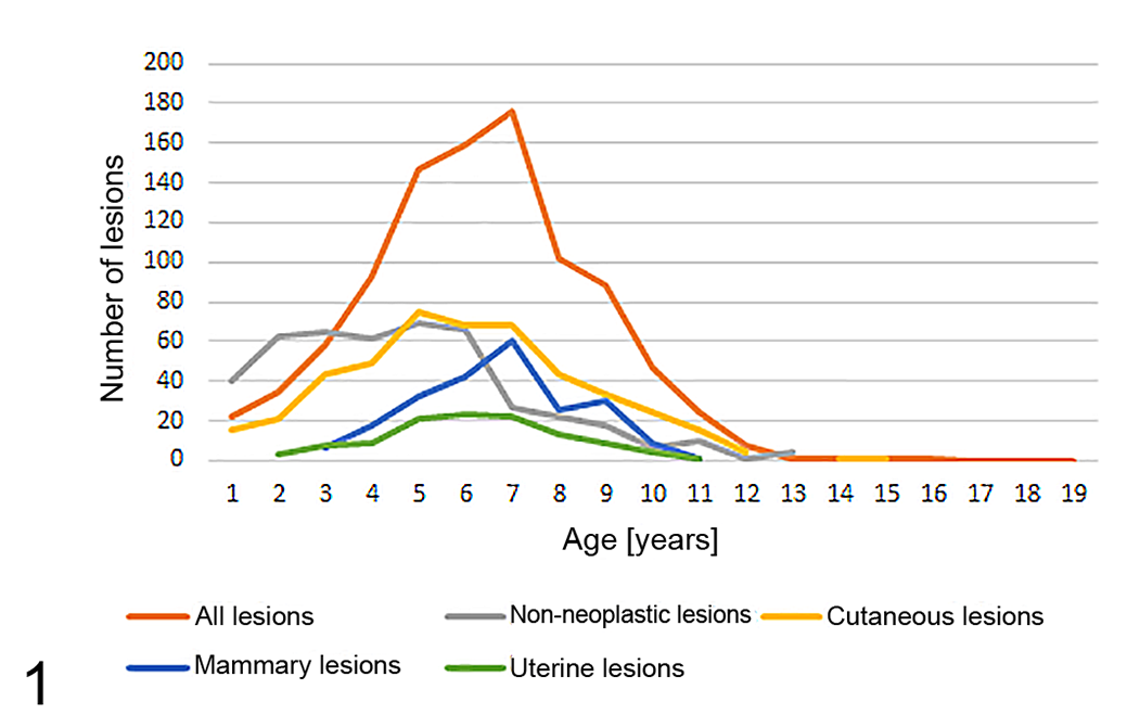
Age distribution of European pet rabbits with neoplastic and non-neoplastic masses of various body systems.
Both sexes were equally affected by neoplasia (Table 1), with the exception of lesions in genital organs and mammary gland. Of 226 mammary lesions in which the sex was known, only 1 adenocarcinoma occurred in a male. This is similar to feline 98 and canine 9 mammary tumors but contrasts with the situation in guinea pigs, in which a high proportion of mammary neoplasms occur in males. 3
Number of Pet Rabbits of Each Sex in the Study.
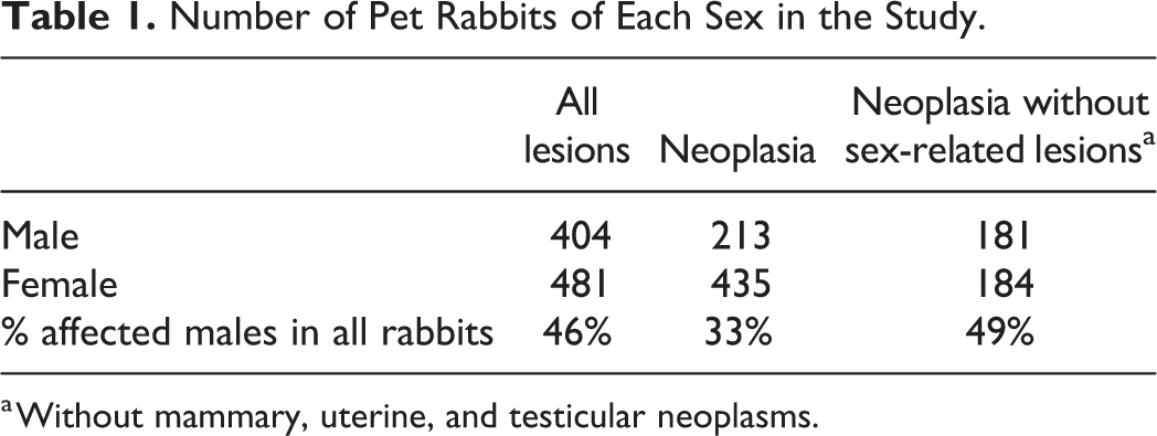
a Without mammary, uterine, and testicular neoplasms.
Published reports 100 and our own data indicate that the condition of disseminated collagenous hamartomas (described in more detail below) occurs nearly exclusively in males (25/28 with known sex in our study). A tendency for skin tumors, especially sarcomas, to occur more in males was also described in a survey of American pet rabbits 99 and became evident in the analysis of our data (Table 2). Furthermore, in the current study population, melanomas occurred mostly in males (14/16, 88%).
Cutaneous Masses in 582 Pet Rabbits.
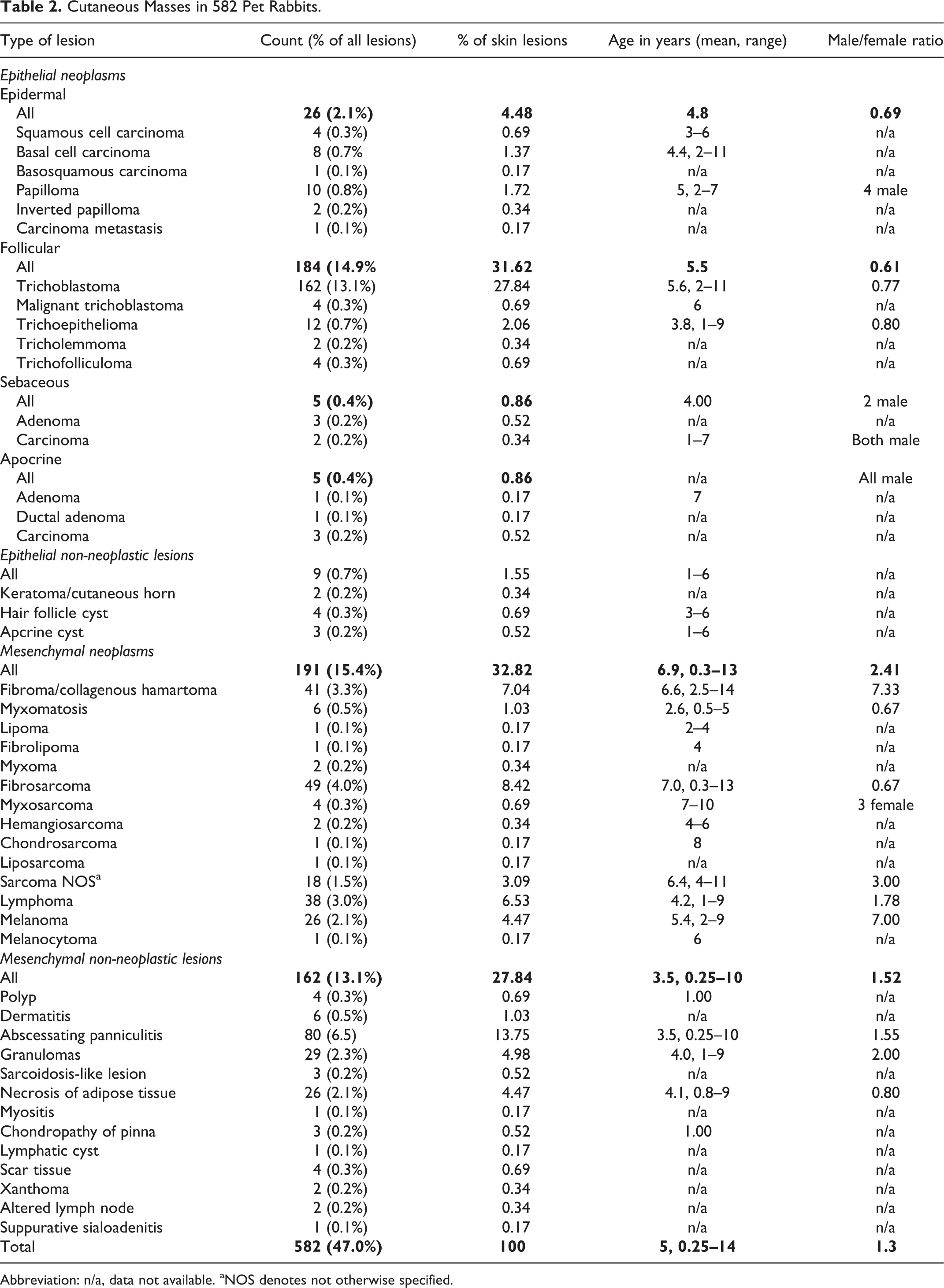
Abbreviation: n/a, data not available. aNOS denotes not otherwise specified.
In the present study, some neoplasms such as fibrosarcoma occurred in aging rabbits, whereas benign epithelial tumors were found in younger rabbits. Since female rabbits have a high risk of mammary and uterine adenocarcinoma, higher longevity of male rabbits might bias epidemiologic studies. Breed predisposition could not be determined because the breed was unspecified or nonspecific (eg, dwarf rabbit) in most submissions.
Skin
Cutaneous lesions represented the major group of submissions with 582/1238 (47%) cases in this study population (Supplemental Table S1 lists all of the lesions and their frequency). Of these, 411 (71%) were neoplasms including 220 epithelial and 191 mesenchymal neoplasms (Table 2). The remaining 171 cases represented inflammatory and degenerative conditions including 109 (18%) cases of panniculitis (Fig. 2). Panniculitis may be overrepresented among suspected tumors in rabbits because rabbit pus is of caseous consistency and may macroscopically resemble tumor necrosis.
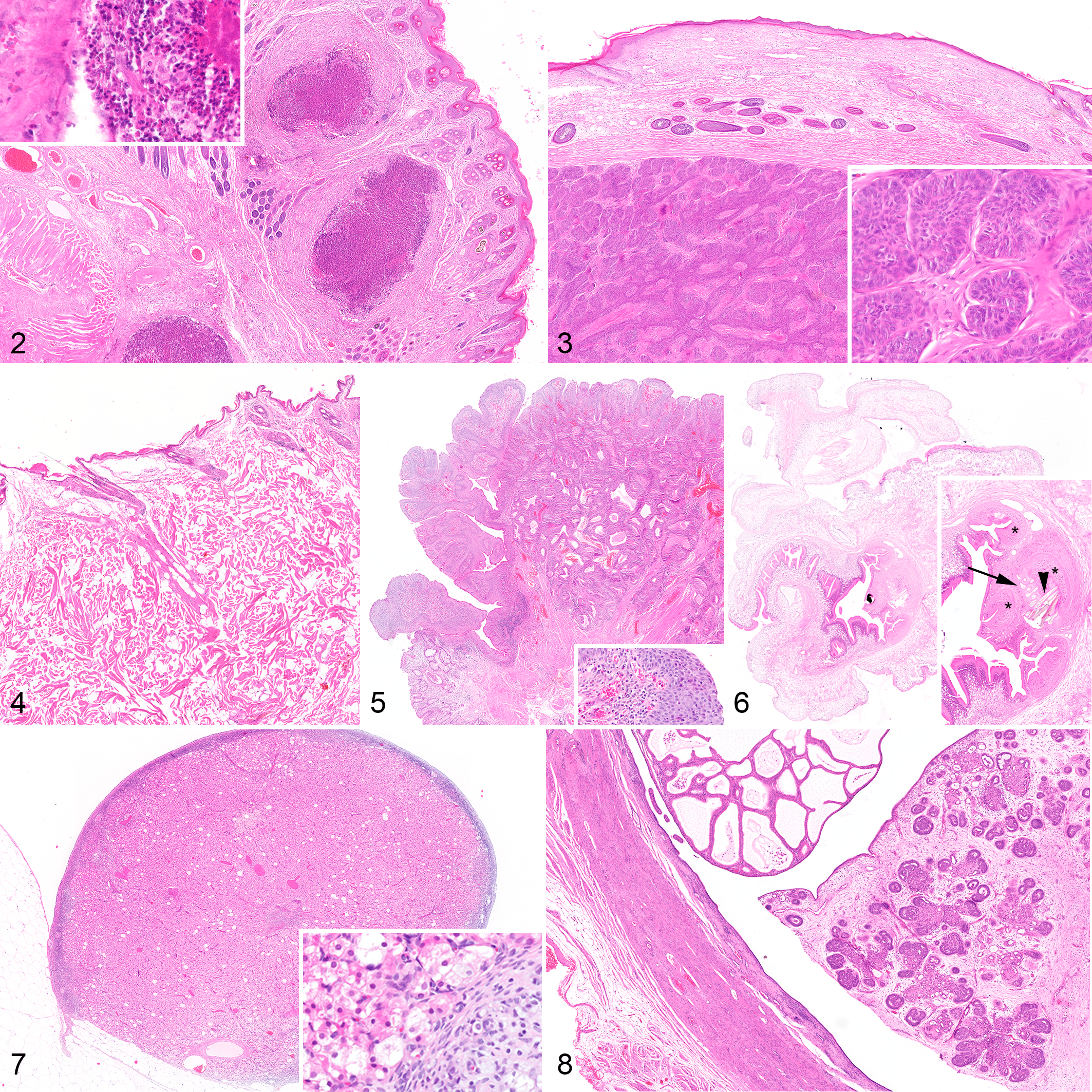
Subcutaneous abscess, rabbit. The mass is composed of cavitations filled with caseous debris with Splendore-Hoeppli material. Inset: Abscess capsule with pyogenic membrane, containing numerous heterophils. Hematoxylin and eosin (HE).
Skin: Epithelial Tumors
Next to mammary carcinomas, trichoblastomas were the most frequent neoplasms in the rabbit, comprising 166/1238 (18%) of all neoplastic and non-neoplastic masses. About 35% were situated on the trunk, 20% on the neck, and the remaining in about equal proportions on head, hindlimb, and forelimb. As this distribution parallels the size of these areas, this might be designated as a random pattern, contrasting with the situation in the dog with a predisposition for the head and especially the ear base. Histologically, trichoblastoma in rabbits usually resembled trabecular-type trichoblastoma in dogs (Fig. 3), although 4/166 trichoblastomas (2.4%) had infiltrative growth as evidence of malignancy in our case series. Tumor cells in trichoblastomas in rabbits reportedly express cytokeratin 14 and 17. 58
Other follicular masses were uncommon (26/1238, 2%) and those of adnexal glands were rare (1%, 13/1238). The latter might be explained by the fact that apocrine glands in rabbits can hardly ever be found outside of glabrous areas and the region at the ventral part of the mandible, also termed the mandibular gland. 94 Consequently, most apocrine lesions in our series (3 out of 5 with known location) were situated at the chin.
Papillomas occurred on the pinna (n = 9) and on the nose (n = 1). The predominant occurrence on the pinna may suggest a viral cause as for equine aural plaque. One neoplasm exhibited a plaque-like morphology resembling this lesion in horses. Viral cytopathic effects were scant and inclusion bodies could not be detected. Foci with spongiosis and vacuolation were regularly present, but koilocytes were only noted in one neoplasm from the nose.
Actinic damage might be another predisposing factor, but gradual transition from dysplasia to carcinoma, as occurs in cats, 37 was not observed. A predominance on the head and pinna was also reported in pet rabbits from the United States of America. 99 Shope papilloma was diagnosed there, but the cottontail rabbit (Sylvilagus floridanus) is the reservoir for the causative virus and is not present in Europe, so that whether that causes the lesions in European rabbits is uncertain. Immunohistochemistry or polymerase chain reaction for papillomavirus in European rabbit papillomas require further investigation. Malignant transformation towards squamous cell carcinoma has been described for Shope papilloma 81,86 but was not observed in papillomas of our European case series.
Of the 4 squamous cell carcinomas, 3 were present in the hindleg around the tarsal joint, but numbers are too low to speculate on a correlation with the high prevalence of “sore hocks.”
Skin: Mesenchymal Tumors
Rabbits develop a specific multinodular condition characterized by well-differentiated collagenous nodules and plaques all over the body (Fig. 4), which is termed fibroma, collagenous hamartoma, 106 or scleroderma. 57 In the current case series, 8 of 12 cases of these disseminated collagenous hamartomas occurred in males and only one in a female (sexes of further cases were not given). In one case, testicular interstitial cell tumors were found simultaneously. Also, orchidectomy led to partial resolution of skin lesions in a rabbit with bilateral interstitial cell tumors and increased serum testosterone level prior to surgery. 57 Therefore, a hormonal pathogenesis has been suspected. It is uncertain whether this is a specific condition, or a stereotypic response toward metabolic imbalances. Over time, male rabbits may normally develop a thickened dermis on the back, suggesting a hormonal influence on collagen fiber synthesis in the rabbit. 95
Detecting a viral cause of neoplasms in rabbits was a major paradigm change in medicine in the 1930s. 87 Leporipoxvirus infection may cause fibrous proliferation, best known in Europe with myxomatosis, while Shope fibroma is induced by a different but related leporipoxvirus and is more frequent in North America. Theoretically, Shope fibroma virus might cause sporadic outbreaks if carried by travelling rabbit handlers into Europe. However, given the lack of the virus reservoir in Europe (wild rabbits of the Sylvilagus genus), viral spread can be excluded. A third related and highly aggressive poxvirus has been isolated from Shope fibroma tumors and designated malignant rabbit fibroma virus. 91 It caused multiple myxosarcomas and rapid death due to opportunistic secondary infection. Correlation with a transplantable sarcoma from a rabbit at an injection site of Shope fibroma virus in the 1930s is uncertain. 2 Neither virus was reported to cause natural disease and both disappeared from the literature in recent decades. Viral mesenchymal neoplasms with histologic morphology of Shope fibroma were not represented in our caseload.
The majority of epithelial skin lesions in the rabbit were benign (206/229, 90%), whereas the majority of mesenchymal neoplasms are malignant in our case series (139/191, 73%) and in the literature. 99 Fibrosarcoma is a disease of the aging rabbit (mean age 7 years, range 0.3–13 years). It has been postulated that these neoplasms might be vaccine-induced, similar to the cat, after aluminum particles were detected within a fibrosarcoma. 75
Of 49 fibrosarcomas, 28 (57%) occurred on the trunk and hindlimb in our series. The head and especially the lips were overrepresented with 20% of fibrosarcomas.
Skin: Melanomas
Of 411 cutaneous neoplasms in this series, 27 (6.6%) were of melanocytic origin. Most exhibited a spindle cell morphology with prominent pleomorphism, predominantly oval nuclei with coarse chromatin and moderately conspicuous nucleoli, a moderate amount of cytoplasm with variable numbers of brown pigment granules, and infiltrative growth. Interestingly, 15 of 26 melanomas with a known location were situated on the head, and at least 7 of these were on the pinna, and the pattern was similar in a US survey. 99 Prominent UV-exposure in this sparsely haired area might play a role. Melan-A, PNL-2, S100, and HMB-45 have been detected with immunohistochemistry in malignant melanomas of 2 rabbits. 93
Skin: Tumor-Like Lesions
Panniculitis most frequently affects the thorax and neck. These lesions present as multiple cavitations surrounded by heterophilic granulocytes and a variable amount of granulation tissue. A traumatic cause is likely and correlation with malocclusion is to be considered especially in the head and neck area. In contrast, panniculitis occurring in the limbs is usually diffuse and superficial rather than nodular. It causes a characteristic ulcerative and suppurative pododermatitis of the hind legs (“sore hocks”), and biopsies of such lesions are not submitted as potential neoplasia.
Especially in the neck region, nodular subcutaneous lesions occur in the adipose tissue, consisting of necrotic foci with moderate histiocytic inflammation. Eight of 24 cases of nodular necrosis with known location occurred in the dewlap, a skin fold most prominent in female rabbits but also occurring in obese bucks. A possible cause is blunt trauma.
Hematopoietic System
Cases of lymphoma in our series included 38 arising in the skin, 2 in lymph node, 1 in intestine, 1 in liver, and 1 in spleen. Systemic disease with involvement of multiple organs was not present in the submissions in this case series. Thymic lymphoma is also reported. 76
Cutaneous lymphomas are far more common in the European pet rabbit population (9.2% of skin neoplasms in this series) than that reported in the United States of America (0.5% 99 and 1.4% 66 ). A screening study did not find herpesviral or retroviral causes. 79 Cutaneous lymphoma may occur at any age. 109 Most cases are diffuse large B-cell lymphoma or T-cell-rich B-cell lymphoma. 79 Tumor cells are frequently of intermediate size, without epitheliotropism, and extend deeply into the subcutis and musculature. Mitotic count is variable. T-cell-rich B-cell lymphoma may be challenging because of its innocuous appearance with low mitotic count and mixed cellular composition. Poor demarcation and a lack of different zones or nodular structure might help differentiate it from inflammation.
Our series had only one case of thymoma, although this is reportedly the most common mediastinal neoplasm in the rabbit. 60 Most are benign, but malignant forms have been described. 101 A hemophagocytic histiocytic sarcoma with disseminated growth has been described in a 7-year-old rabbit 46 with immunolabeling for vimentin, CD204, Iba-1, and lysozyme, but not for CD163. Myeloid leukemia was also reported in a rabbit. 70
Until recently, immunohistochemistry in rabbits was limited by an origin of the majority of primary antibodies in the rabbit. Development of increasing numbers of monoclonal antibodies will probably lead to rapid progress in immunophenotyping of tumors in rabbits.
Musculoskeletal System
Musculoskeletal masses in our series were exclusively inflammatory and degenerative lesions, especially nodular inflammation of the distal limbs, extending to tendons and bones. No neoplasms of this system were included in our series.
Osteosarcoma has been previously reported in rabbits. 40,44,107,108,111 The age of affected rabbits ranged from 1 to 9 years with a bimodal distribution, with 5 rabbits below 3 years of age and 9 older than 6 years. Of 14 reported cases, 8 were situated in flat bones, 1,42,48,67,82,102,107,111 4 in the limbs, 40,45,59,106 and 1 each in soft tissue of the lip 78 and the forelimb. 111 The most common location was the head (6 cases) and especially the mandible (3 cases). Irradiation of the mandible in experimental rabbits resulted in osteosarcoma in 17 of 90 treated animals within a period of only 8 months, 16 and 4 of 9 rabbits developed osteosarcoma within 1 year after irradiation of the hindlimbs. 53
Rhabdomyosarcomas have been described 74,99 on the abdomen and the hind limb, respectively. Diagnosis was confirmed by immunohistochemistry for desmin in both cases.
Alimentary System
Oral Cavity
Twenty-eight of 49 (57%) masses in the oral cavity in our series were non-neoplastic and this is explained by the high frequency of dental abscesses in rabbits. Eight other nodular masses of the head and neck had sialadenitis, most likely representing extension of periodontal inflammation to the salivary glands.
Our series included 21 oral neoplasms: 9 papillomas, 4 sarcomas, 3 ameloblastomas, 2 adenocarcinomas, 2 odontogenic fibromas, and 1 squamous cell carcinoma. Rabbits with oral neoplasia were older (5.7 years, statistically similar to 5.1 years for all oral lesions) with a roughly equal sex distribution (7 males, 5 females, further sexes unknown). It is notable that in rabbits, oral papillomatosis is not restricted to young animals but may occur at any age (mean age 4.5 years, range 2–9 years in our case series). Single reported cases of oral neoplasms include osteosarcoma (see above), fibrosarcoma, 15 keratinizing ameloblastoma, 41 ameloblastic fibroma, 104 ossifying fibroma, 110 salivary gland adenocarcinoma, 10 cementoma, 71 and complex odontoma. 71
Gastrointestinal Tract
Gastrointestinal neoplasms in our series comprised 1 leiomyoma in the stomach, 1 lymphoma in the intestine, 1 tubular adenocarcinoma in the colon, and 1 papillary adenocarcinoma in the cecum. Others have reported gastric adenocarcinoma and intestinal leiomyosarcoma, 36 and lymphoma of the stomach, 79 small intestine, 30,38 and cecum. 45 A cecal polyp caused intussusception in an aged rabbit. 77
We identified 36 anorectal polyps. Ages ranged from 1 to 11 years with a mean of 5.5 years and affecting 18 males and 15 females (sexes unknown for other cases). These lesions consisted of a polypoid expansion of the rectal mucosa at the junction between stratified squamous and glandular epithelium (ie, the anorectal transition). Nonkeratinizing stratified squamous epithelium predominated, but both parts were present in most cases. The surface was usually ulcerated and severely infiltrated with heterophils. Mass formation resulted predominantly from granulation tissue in the lamina propria (Fig. 5) and variable edema. We did not detect viral cytopathogenic effects. Clinically, this condition is referred to as rectal papilloma. 95 Histologically, however, the lesions suggest reactive proliferation due to chronic inflammation consistent with an inflammatory polyp rather than neoplasia. The opening of rectal glands in this area 28 might be a predisposing factor. In contrast, experimentally sensitized rabbits fed carrageenan developed diffuse and dysplastic, non-papillomatous lesions. 55
Liver and Pancreas
One case each of pancreatic acinar adenocarcinoma, hepatic lymphoma, hepatocellular carcinoma, and cholangiocarcinoma were in our series. Other reported neoplasms include bile duct adenoma 36 and biliary cystadenoma. 21 Biliary neoplasms have been described as comparatively common in the rabbits from 4 to 8 years of age. 92
Abdominal Cavity
In 15 cases in our series, intraabdominal nodules were formed of necrotic adipose tissue. Trauma is a possible cause, because of nodular presentation in contrast to the diffuse pattern expected with saponification by pancreatic lipase. Mineralization was frequent, which is also common with abdominal fat necrosis in cats and dogs. 85 Intraabdominal neoplasia was not present in our series, but mesothelioma has been described in a 3.5-year-old rabbit. 63 Two intraabdominal masses in our series were caused by cestodal larvae (Fig. 6).
Respiratory System
Neither primary neoplastic nor non-neoplastic masses of the respiratory system were present in our submissions. In the literature, nasal adenocarcinoma, 62 anaplastic sarcoma 23 and osteochodroma of the larynx, 13 and adenocarcinoma of the trachea have been reported. 13
Neoplasms of the lung are rarely described. Histiocytic sarcoma with frequent systemic spread to the lung has been described. 11 Pulmonary carcinoma has been reported. 7,23,27,83 Additionally, small fibropapillomas have been described in a population of rabbits, all of which were older than 2 years, with a mean age of 4 years, and without Shope papilloma virus exposure. 20 Lesions were characterized by epithelial papilliform proliferation with a prominent fibrous stalk.
Urinary Tract
Neoplasms of the urinary tract appear to be rare in the rabbit. In this case series, submissions from the urinary tract contained only degenerative and inflammatory lesions, and one inflammatory polyp of the urinary bladder from a 3-year-old male rabbit.
Renal neoplasms have been described in rabbits, especially nephroblastomas (also known as embryonic nephroma and Wilms’ tumor). We only found 1 case report each of renal adenocarcinoma 49 and nephroblastoma 39 in the literature in the last 50 years. Metastases were not detected. In an older compilation of case reports on rabbit neoplasms, embryonal nephroma was described with the second highest frequency next to uterine carcinoma. 26 However, this does not equal a high prevalence, because an uncommon neoplasm has a higher likelihood of publication than a well-known one.
Transitional cell carcinoma of the renal pelvis and ureter in combination with adrenal gland carcinoma has been described in a 9-year-old rabbit. 80 Transitional cell carcinoma of the bladder has been reported in a 6-year-old rabbit. 18
Female Genital Tract
In 1938, the high incidence of uterine neoplasms in the rabbit was reported. 35 In a further study presenting necropsy data from 849 female rabbits, the same authors reported 142 uterine adenocarcinomas (16.7%). 33 In our series of biopsy material, uterine adenocarcinoma was the third most common neoplasm (84/1238, 6.8%), after mammary carcinoma (250/1238, 20.2%) and trichoblastoma (166/1238, 13.4%). This discrepancy might be explained by differences between frequency of occurrence and detection or between clinical presentation and actual submission for pathologic examination. Rabbits with uterine neoplasia were 5.6 ± 1.7 years of age (mean ± SD).
Of the 135 uterine masses in our case series, uterine adenocarcinoma was the most frequent with 84 cases (62%), followed by 13 cases (10%) of leiomyoma, 8 (6%) of leiomyosarcoma, and 1 teratoma. Ten of our cases had concurrent uterine adenocarcinoma and either leiomyoma or leiomyosarcoma in the uterus.
Rabbits have high numbers of polygonal finely vacuolated eosinophilic interstitial cells in the ovary. These can easily be mistaken for luteoma (Fig. 7), but we could not convincingly demonstrate luteoma in any submission. According to examinations of the last century, this so-called interstitial organ develops from degrading follicles. 72 They are a continual source of progesterone, which might explain the high prevalence of neoplasia in the endometrium and mammary gland in aging does.
The mean age of rabbits with uterine adenocarcinoma was 5.6 years (range 3–9 years). In an investigation on uterine disorders in 59 pet rabbits, simultaneous occurrence of endometrial hyperplasia and adenocarcinoma was observed in 11 rabbits and the mean age of hyperplasia was 1.6 years lower than that for adenocarcinoma. 103 Diffuse endometrial hyperplasia is a common finding in rabbits. 103 In cases of uterine adenocarcinoma, the non-neoplastic endometrium usually exhibits diffuse hyperplasia, and multifocal progression to malignancy at different locations is common. All this is indicative of stepwise carcinogenesis and supports a hypothesis of a hormonal background. In an experimental study with repeated biopsies of uterine neoplasms, progression from acinary patterns via papillary and cystic structures to disorganized architecture was observed in the majority of neoplasms. 34 Further evidence was provided in a study of spontaneous uterine lesions in 88 pet rabbits showing the neoplastic endometrium had higher expression of estrogen and progesterone receptors, particularly in tubular/solid adenocarcinomas, compared with papillary adenocarcinoma, hyperplasia, or normal endometrium. This supports a hypothesis of a hormonal background in carcinogenesis in certain types of tumors in rabbits. However, this study failed to detect differences of clinical outcome. 4
Intraluminal growth with formation of papillary structures and nests is the most frequent stage at the time of submission in this series (Fig. 8). Invasive growth is found with increasing frequency in larger neoplasms. Mitotic rate is low to moderately elevated, usually about 2 to 8 per 10 hpf (2.37 mm2) without obvious correlation to the growth pattern. In larger neoplasms, necrotic areas also occur, and resultant hemorrhagic discharge is often the reason for clinical detection.
Pulmonary metastases were present in nearly 80% of rabbits with uterine adenocarcinoma in a compilation of necropsy findings, 14 although it should be noted that aggressive neoplasms may be overrepresented at necropsy. Uterine adenocarcinoma must be regarded as a serious threat to longevity in female rabbits, but follow-up investigations are necessary to determine lethality of the lesion without bias.
Uterine leiomyosarcoma is more frequent in rabbits than in other species (38% of all 21 uterine smooth muscle neoplasms vs less than 10% in the dog). 96 This might reflect a tendency to detect these neoplasms late in their progression, as tumors are commonly large at the time of submission. An extreme case weighing 500 g in our series had extensively infiltrated into the abdominal cavity. Myxosarcoma with similarly invasive growth has been described in a 4-year-old rabbit. 73
Diagnosis of deciduosarcoma was made in a rabbit, appearing as a spindle cell tumor with large epithelial cells, karyomegaly, and anisocytosis. 19 Two cases of malignant mixed Müllerian tumor (also known as carcinosarcoma) have been described. 31,103
Ovarian neoplasms of rabbits are rarely reported. Individual cases of granulosa cell tumor, 47 hemangioma, 36 adenoma, 12 adenocarcinoma, 103 and choriocarcinoma 50 have been described in the ovary, and one adenoma in the uterine tube. 17
Male Genital Tract
Our series included 32 testicular neoplasms in 31 rabbits. The 3 main types of testicular tumors found in other domestic animals were also detected in the rabbit; that is, Sertoli cell tumor, interstitial cell tumor, and seminoma. Mean age was higher in rabbits with interstitial cell tumors and seminomas than for those with other neoplastic conditions. However, overall low case numbers preclude statistical analysis of this difference. The neoplasms were usually in an advanced stage at the time of submission, having replaced major parts of the testicular parenchyma. Compared to those of dogs, testicular neoplasms of rabbits had a higher percentage of neoplasms with histomorphologic evidence of malignancy, specifically invasive growth in all tumor types as well as increased pleomorphism in interstitial tumors (Table 3). 97 In this study, invasion of blood vessels was present with all 3 types of neoplasms. Metastases have been described in a seminoma. 6 Neoplastic cryptorchid testicles may also result in testicular torsion. 112
Testicular Neoplasms in Pet Rabbits.
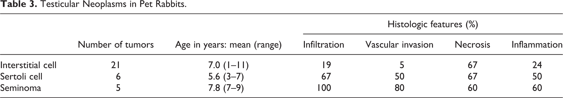
In recent publications, interstitial cell tumors in rabbits have been designated as granular cell tumors due to occurrence of PAS-positive cytoplasmic granules in these neoplasms otherwise identical to interstitial cell tumors in other species. 105 It is questionable whether this neoplasm is in fact different from interstitial cell tumor, or instead there is species-specific staining behavior.
Further neoplasms of the male genital tract were restricted to one scrotal melanoma in our series. The literature presents a testicular teratoma. 69
Mammary Gland
The mammary gland is the second most frequent location of neoplasms in rabbits in our series, after the skin. Unlike the dog, all mammary complexes have an identical risk for the development of neoplasia in the rabbit. In our study, distribution between cranial, central, and caudal gland were 41%, 19%, and 41% (for 341 neoplastic nodules), and right and left were nearly even with 49% and 51%, respectively (331 nodules). Development from hyperplastic lesions over time was already suspected in 1939. 32 Influence of hormonal stimulation is highly likely, but correlation between tumor type and expression of hormonal receptors could not be detected using immunohistochemistry. 22
Most mammary neoplasms in rabbits were simple and histologically malignant in our series (250/265, 94%). Malignant tumors are represented by tubular, papillary, tubulopapillary, solid, adenosquamous, comedo type, complex, ductal, cribriform, anaplastic, spindle-cell carcinomas, and malignant myoepitheliomas. Benign tumors—papillomas, adenomas, and rarely complex adenomas—are uncommon. Mammary neoplasms are exhaustively described in the literature, 8,84,89 but with the limitation of unknown significance of different morphologic patterns for prognosis.
In our series, follow-up was available for only 22 rabbits with mammary tumors (Table 4). There is no obvious correlation between the histologic type and the survival time, not even a tendency of longer survival with better differentiated neoplasms. Of rabbits with mammary adenocarcinoma, 45% survived for longer than one year. This is an unexpectedly high percentage considering the usually obvious evidence of malignancy. Four cases surviving 1 year or longer were euthanized or died with lung metastasis despite there being no reported further neoplasms in the mammary gland, but postmortem examination apparently did not occur. This suggests either slow growth of metastases or a tendency for first diagnosis at a late stage of disease in rabbits in comparison with the situation in dogs and cats. However, metastasis may be widespread, including the uvea. 29
Survival Times for Different Types of Mammary Tumors in Pet Rabbits. The Data Show the Number of Cases.a
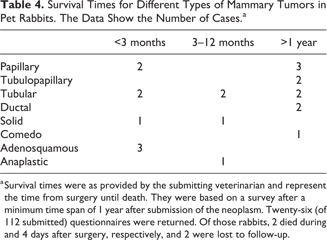
a Survival times were as provided by the submitting veterinarian and represent the time from surgery until death. They were based on a survey after a minimum time span of 1 year after submission of the neoplasm. Twenty-six (of 112 submitted) questionnaires were returned. Of those rabbits, 2 died during and 4 days after surgery, respectively, and 2 were lost to follow-up.
Endocrine System
There were no endocrine tumors in our series. In the literature, adenoma 5 and carcinoma of the adrenal gland 43 were described, the latter after administration of a dye. Hyperplasia of the adrenal gland is reportedly frequent. 92 Neoplasia and hyperplasia of the adrenal cortex may induce hyperandrogenism and aggressive behavior. 61 Similar to ferrets, lack of feedback inhibition due to spaying has been suspected as possible pathogenesis. 5 Other reported endocrine neoplasms are prolactin-secreting acidophilic adenoma of the pituitary gland, with hyperplasia and neoplasia of the mammary gland, 64,88 as well as teratoma of the pituitary gland, 65 and carcinoma of the thyroid gland. 25
Nervous System and Sensory Organs
Four intraocular neoplasms were present in our series. One adenocarcinoma in a 7-year-old rabbit was suspicious for iridociliary adenocarcinoma and infiltrated strongly into the surrounding soft tissue of the orbit. One iridal lymphoma occurred in a 7-year-old rabbit. A round cell tumor and a spindle cell tumor in 2- and 3-year-old rabbits formed metastases in the skin of the head and neck, respectively; these were both suspicious for amelanotic melanoma, but immunohistochemistry was not performed.
Eight epithelial neoplasms were present in the conjunctiva with a broad spectrum between benign lesions, in situ carcinoma, and infiltrative carcinoma. Four rabbits aged between 4 and 9 years presented with melanoma of the eyelid, and all neoplasms had evidence of malignancy, specifically infiltration and increased cellular pleomorphism.
Intraocular sarcomas were reported in combination with lens rupture 24 and presumably occurred posttraumatically as for the feline neoplasm. 68 B-cell lymphoma has also been observed in 3 rabbits following trauma. 51 Teratoma at the optic chiasm has been described. 52
Malignant peripheral nerve sheath tumors apparently occur with some frequency, 99 but neoplasms of the central nervous system of rabbits are rarely reported. One schwannoma extended from the spinal roots to the brachial plexus. 90 Ependymoma was described 54 and various glial tumors could be experimentally induced in young rabbits. 56
Supplemental Material
Supplemental Material, sj-pdf-1-vet-10.1177_03009858211002190 - Not Just Uterine Adenocarcinoma—Neoplastic and Non-Neoplastic Masses in Domestic Pet Rabbits (Oryctolagus cuniculus): A Review
Supplemental Material, sj-pdf-1-vet-10.1177_03009858211002190 for Not Just Uterine Adenocarcinoma—Neoplastic and Non-Neoplastic Masses in Domestic Pet Rabbits (Oryctolagus cuniculus): A Review by Berit Baum in Veterinary Pathology
Footnotes
Acknowledgements
The author would like to thank Drs Nora Berghoff, Jaco van der Lugt, Melissa Behr, and Janice Harvey for their valuable input; Dr Nikola Pantchev for parasitologic consultation; and Dr Annika Lehmbecker for help with figures.
Declaration of Conflicting Interests
The author declared no potential conflicts of interest with respect to the research, authorship, and/or publication of this study.
Funding
The author received no financial support for the research, authorship, and/or publication of this article.
Supplemental material for this article is available online.
References
Supplementary Material
Please find the following supplemental material available below.
For Open Access articles published under a Creative Commons License, all supplemental material carries the same license as the article it is associated with.
For non-Open Access articles published, all supplemental material carries a non-exclusive license, and permission requests for re-use of supplemental material or any part of supplemental material shall be sent directly to the copyright owner as specified in the copyright notice associated with the article.