
Abstract
Prevalence and age distribution of tumors is largely unknown in pet rabbits. Currently available studies focused on specific organ systems or specific tumor types and never covered a comparative examination of all tumor types. Previous studies on laboratory rabbits suggested a low tumor prevalence but were mostly limited to young adult animals. In the present study, all tumor types and several tumor-like lesions of all organ systems were analyzed retrospectively in archived pet rabbit samples of all ages. Cases included necropsy cases (n = 2,014) or postmortem tissue samples (n = 102) as well as surgical biopsies (n = 854). All lesions suspicious of neoplasia were reevaluated by histopathology and, when indicated, by immunohistochemistry. Necropsy cases had a tumor prevalence of 14.4% in both sexes or 19.8% in female intact rabbits of all age groups, and up to 47.2% or 66.7%, respectively, in rabbits older than 6 years. Overall, the most common tumor types were uterine adenocarcinoma (prevalence in female intact animals: 13.1%), lymphoma (prevalence: 2.8%), and thymoma (prevalence: 2.1%). Lymphoma, the most common tumor of rabbits ≤24 months of age, were of B-cell immunophenotype in 96% of cases and most commonly located in the lymph nodes (57%), gastrointestinal tract (54%), kidneys (48%), spleen (42%), and liver (41%). Tumors accounted for 81.1% of surgical biopsies and mostly comprised cutaneous, mammary, and uterine tumors. In conclusion, tumor types and prevalence varied significantly with respect to age, revealing some differences from previous studies on laboratory rabbits.
Keywords
Current knowledge on spontaneous neoplasms in pet rabbits (Oryctolagus cuniculus) is sparse. Although several tumor types have been reported in a large number of individual case reports, studies comprising large case numbers exist exclusively concerning specific tumor types—such as cutaneous lymphomas 32 —or organ systems—such as the female genital tract, 8 skin, 23,40 mammary gland, 4,35 or oral cavity. 27 To the authors’ knowledge, a comprehensive study covering all tumor types and all organ systems and ages of pet rabbits has not been conducted to date. Therefore, prevalence and age distribution of most tumor types is largely unknown.
However, individual studies have determined the prevalence of spontaneous tumors in laboratory rabbits. The overall tumor prevalence determined in these studies ranged between <0.5% and 2.7%, suggesting that neoplasia is rare. 5,30,41,43 The highest prevalence described by Weisbroth 43 is of 2.7% with 16 neoplasms in 599 laboratory rabbits, of which 9 occurred in 107 rabbits older than 2 years, rendering a prevalence of 8.4% for this age group. The largest case number is described by Von Löliger 41 with 19 tumors discovered in a total of 915 laboratory rabbits and 585 breeding rabbits (652 young animals and 602 sexually mature animals) and suggests a prevalence of 1.3%. Further studies focusing on neoplasia in a large case number of laboratory animals, such as Bell and Henrici 5 (2 tumors in >400 rabbits) and Polson 30 (7 tumors in 560 rabbits), do not state any age-related information. Therefore, aforementioned studies cannot be easily translated to pet rabbits, as the age of analysis usually differs significantly between laboratory and pet animals. Only few studies on uterine adenocarcinomas in female laboratory rabbits include older animals and suggest a much higher prevalence—between 13.7% and 24.4%—for this tumor type. 2,16,17,20 Furthermore, Weisbroth 43 suggested that rabbits of more than 2 years of age are more prone to neoplasia than younger animals. Hence, it seems likely that the prevalence of various tumors in pet rabbits is much higher compared to the previous general studies on laboratory rabbits due to the higher life expectancy, although the prevalence of uterine adenocarcinoma in pet rabbits may be similar to the above studies of aged laboratory rabbits. Furthermore, the relative frequency of the various tumor types of laboratory rabbits may not be applicable to old rabbits as different tumors may occur in different age groups. For example, one study determined lymphoma as the most common tumor type (12 of total 19 tumors) in 1500 breeding and laboratory rabbits, 41 while Bell and Henrici 5 only found 2 renal tumors in more than 400 rabbits. Uterine adenocarcinoma—which is generally considered the most common tumor type in rabbits 43 —is indeed very rare in young rabbits of <2.5 years. However, its prevalence consistently increases with age to a prevalence of up to 75% in rabbits older than 6 years 8,16,20 and therefore clearly exceeds the prevalence of lymphoma and renal tumors reported by Von Löliger 41 and Bell and Henrici, 5 respectively, for this age group.
The present study aims at a comprehensive analysis of all tumor types in all organ systems in pet rabbits for the first time. We reevaluated all cases of pet rabbits with masses suspicious of neoplasia and tumor-like lesions submitted between 1995 and 2019. In the following, tumor prevalence; organ distribution of primary, metastatic, and hematopoietic tumors; as well as sex and age distribution were evaluated systematically.
Materials and Methods
Case Selection
In this study, all spontaneous neoplastic masses and tumor-like lesions were retrospectively analyzed from all necropsy and organ samples from pet rabbits (Oryctolagus cuniculus) submitted between January 1995 and December 2019 to the Institute of Veterinary Pathology, Freie Universität Berlin. All tumor samples were obtained during routine pathological examination. Use of these samples was approved by the local governmental authorities (State Office of Health and Social Affairs of Berlin, Approval ID: StN 011/20). Samples were divided into 3 sample types: (1) full postmortem examination performed by a pathologist (necropsy case), (2) postmortem tissue sampling by the referring veterinarian (tissue sample), and (3) intravital tissue removal during surgery (surgical biopsy). Age, sex, and breed information was obtained from submission forms and necropsy reports. Cases lacking archived paraffin-embedded tissue for histological reevaluation were excluded. Tumors of bone marrow was not evaluated in this study, as bone marrow was not systematically examined in all cases. A subset of cases included in this study has been published previously. 6 –8,18,44
Histopathological and Immunohistochemical Examination
All lesions suggestive of neoplasia were reexamined in order to facilitate uniform histological evaluation. Tumor types were diagnosed according to current general guidelines of domestic animal histopathology 14,26,46 and detailed specifications for rabbits when available. 6,31,40 Special stains and/or immunohistochemistry (IHC) was performed when required. Methods for IHC are summarized in Supplemental Table S1. Overall, all soft-tissue sarcomas were immunolabeled with antibodies against smooth muscle actin (SMA) and stained with Masson’s trichrome for collagenous connective tissue. For testicular tumors, IHC with S100 antibodies and a periodic acid-Schiff reaction (PAS) was performed as diagnostic criterion for granulosa cell tumors according to Reineking et al. 31 Furthermore, all thymomas were confirmed by IHC for pan-cytokeratin (CK), lymphomas by Pax-5 (B-cell marker) and CD3 (T-cell marker), histiocytic sarcomas by CD204 (also known as macrophage scavenger receptor 1), mesotheliomas by CK and vimentin, and melanomas by melan-A (MART-1). Further immunohistochemical labeling were performed for individual cases with uncertain histological criteria. Respective tumor tissues from rabbit reference cases or rabbit tissues devoid of any histological alterations were employed as positive or negative controls, respectively, for all immunohistochemical procedures. All melanomas with abundant melanin pigment were used as positive controls for melan-A IHC after partial bleaching.
Data Analysis
Tumor prevalence was determined exclusively for the necropsy cases as full pathological examinations were performed for this group only. For the purpose of this study, tumor prevalence is defined as the number of necropsied rabbits with neoplasia divided by the total number of rabbits necropsied (or a subset of these based on age or sex). Prevalence of tumors of the female and male reproductive tract were exclusively determined for intact animals of the respective sex. All 3 sample types (necropsy case, tissue samples, surgical biopsies) were used for relative comparison of tumor types present within a specific organ system and determination of mean age, median age, and age range of the tumor types. Age distributions of tumor prevalence were determined exclusively in the necropsy cases with age intervals set to 6 months. Due to uneven case numbers within the different age intervals, different numbers of intervals were assigned to 6 age groups (Suppl. Figs. S1, S2): (1) 0 to 6 months; (2) 7 to 12 months; (3) 13 to 24 months; (4) 25 to 48 months; (5) 49 to 72 months; and (6) 73 to 180 months.
Neoplastic changes of each sample type were divided into primary tumors of a specific organ, tumor metastasis, and round-cell tumors. For the purpose of the present study, tumor metastasis was defined as the spread of a non-round-cell tumor into another organ. Involvement of multiple organs by round-cell tumors (lymphoma and histiocytic sarcoma) is referred to as “disseminated tumor.” Tumor-like lesions were analyzed separately.
Statistical analysis was performed using R version 4.0.1 (R Foundation Vienna). Prevalence is each reported with a 95% confidence interval (CIs), determined by the Wilson method with continuity correction. For statistical analysis, a negative binomial regression was used to calculate prevalence ratios (PR) and P values for tumor occurrence. Prevalence ratios are mathematically identical to risk ratios. Since our study cannot measure risk as all animal characteristics were recorded exclusively postmortem, PR was reported to avoid misinterpretation. Details to PR are provided in Martinez et al. 25 A significance threshold of .05 was used.
Raw data can be made available upon reasonable request.
Results
Necropsy Cases
Necropsy findings from 2014 pet rabbits were evaluated. Age and sex distribution of the entire population is presented in Supplemental Tables S2, S3, and S4 and Figures S1 and S2. Neoplastic disease was diagnosed in 290 rabbits (prevalence: 14.4%, CI: 12.9% to 16.1%) with a total of 330 different neoplasms. Prevalence of multiple tumors (n = 35) increased with age (P <.001) and the observed average number of tumors did not exceed a theoretical model (Suppl. Fig. S3). The most commonly affected organ systems were the female reproductive tract, hematopoietic organs, skin, mammary gland, testes, and the digestive system (Table 1). Histologic criteria of malignancy were evident in 228 of 330 (69.1%) tumor cases from 212 of 290 rabbits (73.1%; prevalence: 10.5%, CI: 9.2% to 12.0%). Mean age of rabbits with neoplasia was 69.2 months (median age: 72 months; range: 2–180) and tumor prevalence increased with age group from 0.9% (CI: 0.3% to 2.8%) in rabbits ≤6 months to 47.2% (CI: 40.7% to 53.8%) in rabbits >6 years of age (Fig. 1, Suppl. Table S5). The PR increased by a factor of 51.4 between the youngest and oldest age groups (Suppl. Table S6). Similarly, prevalence continuously increased with age for malignant neoplasms, non-round-cell tumors, metastatic neoplasia, and neoplasia in female rabbits (Figs. 1, 2, Suppl. Tables S5, S7). Overall tumor prevalence was 19.7% (CI: 17.1% to 22.5%) in female intact rabbits, 22.4% (CI: 12.8% to 36.2%) in female neutered rabbits, 8.4% (CI: 6.5% to 10.9%) in male intact rabbits, and 14.2% (CI: 10.3% to 19.2%) in male castrated rabbits. Compared to male intact rabbits, the age-adjusted PR for female intact rabbits was 2.33 (P < .001), for female neutered rabbits 1.39 (P = .349), and for male castrated rabbits 1.40 (P = .130, Suppl. Table S8). Breed information was available for 328 rabbits (16.3%) with a total of 25 pure and 8 mixed breeds. Lop (n = 140 cases) and Lionhead rabbits (n = 73 cases) had no significant (P = .352 and P = .224, respectively) predisposition for neoplasia (PR with adjustment of age and sex: 0.79 and 0.66, respectively). Tumor predisposition was not calculated for other breeds as they included ≤19 cases.
Overview of Primary Tumor Prevalence in the Different Organ Systems and Hematopoietic Tumors in Full Postmortem Examinations (Necropsy Samples) of Pet Rabbits.
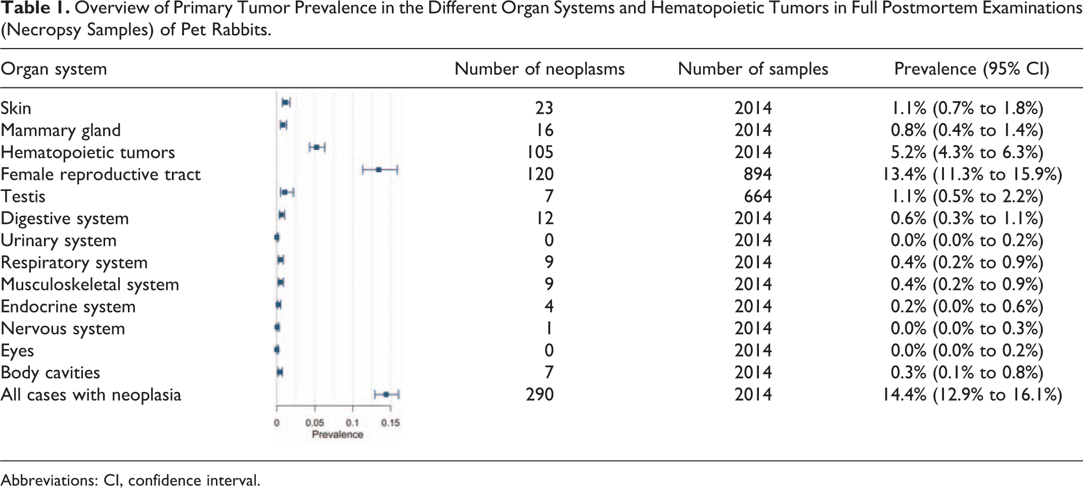
Abbreviations: CI, confidence interval.
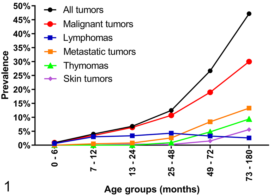
Age distribution of tumor prevalence in necropsy cases (full postmortem examination performed by a pathologist) of rabbits of all sexes with tumors (n = 290/2014). Six different age groups are compared. Metastatic tumors include only non-round-cell tumors. Prevalence is defined as the number of rabbits affected divided by all rabbit necropsy submissions within the respective age group.
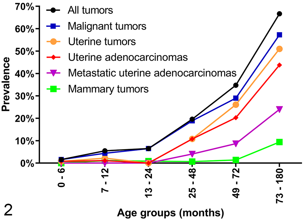
Age distribution of tumor prevalence in necropsy cases (full postmortem examination performed by a pathologist) of female intact rabbits with tumors (n = 177/894). Six age groups of variable age intervals are compared. Prevalence is defined as the number of rabbits affected divided by all rabbit necropsy submissions within the respective age group.
Non-round-cell tumors were found in 229 necropsy cases (prevalence: 11.4%, CI: 10.0% to 12.9%) with a mean age of 74.5 months (median age: 72 months; range: 5–180). Metastatic spread, especially to the lungs (Suppl. Table S19), was present in 74 non-round-cell neoplasms from 73 rabbits (32.3%; prevalence: 3.6%, CI: 2.8% to 4.6%) with higher PR for older rabbits (Suppl. Table S10). Most metastatic tumors (90.4%, Suppl. Table S11) occurred in female intact rabbits with a prevalence of 7.4% (CI: 5.8–9.4), which was higher compared to male intact (age-adjusted PR: 11.30, P < .001), male castrated (age-adjusted PR: 22.90, P = .010), and female neutered rabbits (age-adjusted PR: 7.41, P = .195). Round-cell tumors were found in 64 rabbits (prevalence: 3.2%, CI: 2.4% to 4.1%) with a mean age of 47.2 months (median age: 44 months; range 2–128). They were found to be disseminated in 58 cases (91%) and localized in 6 cases (9%).
Postmortem Tissue Samples
Tissue samples were received from 102 cases with a total of 264 organ tissue samples (Suppl. Table S12). Sex and age distribution is presented in Supplemental Table S2 and S3. One tumor type was found in 28 rabbits, 3 rabbits had 2 tumor types, and 1 rabbit had 3 tumor types (n = 32/102, CI: 23.0% to 41.2%). Histological examination revealed morphologic criteria of malignancy in 27/37 cases (73%). Mean age of affected rabbits was 73.2 months (median age: 76 months; range: 6–155) and 50% of the affected rabbits with known sex were intact females (Suppl. Table S13). Non-round-cell tumors were found in 22 rabbits with 27 different tumors identified and metastatic disease was diagnosed in 9 of those rabbits (41%) and 10 of those tumors (37%, Suppl. Table S14). Round-cell-tumors were diagnosed in 10 cases of a mean age of 58.1 months (median age: 72 months; range: 6–119) and disseminated disease was confirmed in 7 cases (70%).
Surgical Biopsies
Surgical biopsies of 854 pet rabbits (Suppl. Tables S2, S3) were evaluated and included 1013 organ samples (Suppl. Table S12). A total of 600 tumors were identified in 574 rabbits (proportion: 67.2%, CI: 64.0% to 70.4%) with a mean age of 61.8 months (median age: 60 months; range: 7–120). Affected rabbits were female intact in 55.8% of the cases of known sex (Suppl. Table S15). Neoplasia was most commonly found in the skin, mammary gland, and female reproductive tract (Table 2).
Overview of the Proportion of Primary Tumors in the Different Organ Systems and Hematopoietic Tumorsa in Surgical Biopsies of Pet Rabbits.
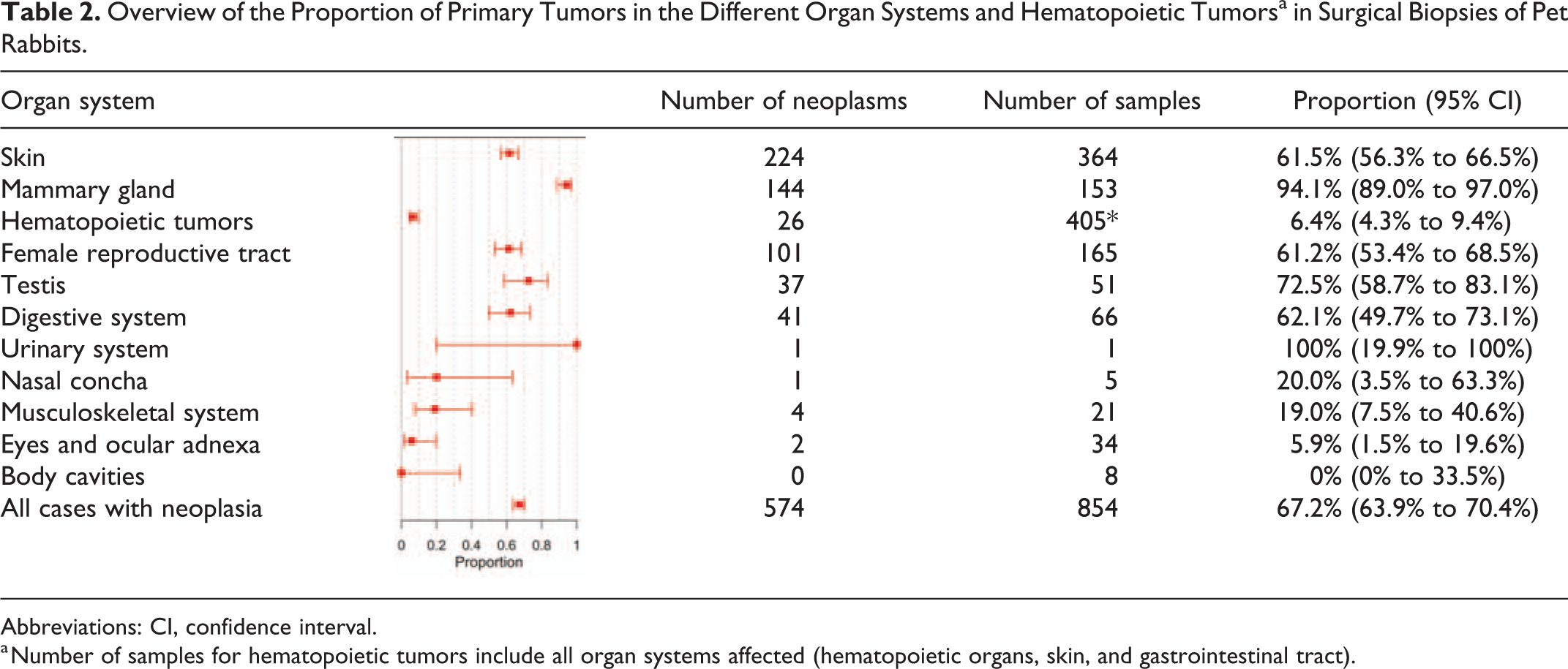
Abbreviations: CI, confidence interval.
a Number of samples for hematopoietic tumors include all organ systems affected (hematopoietic organs, skin, and gastrointestinal tract).
Tumors of the Skin (Including Subcutis)
Primary, nonhematopoietic tumors of the skin were found in 23 necropsy cases (prevalence: 1.1%, CI: 0.7% to 1.8%), in 2/2 tissue samples, and in 224/364 surgical skin biopsies (proportion: 61.5%, CI: 56.3% to 66.5%). Mean age of affected rabbits was 66.1 months. Of the necropsy cases, tumor prevalence continuously increased from 0% in the youngest age group to 5.6% in the oldest age group (Fig. 1, Suppl. Table S5). Rabbits of the oldest age group had a 19.24 times higher relative PR (P = .023) than rabbits between 25 and 48 months of age. Of the 17 types of cutaneous neoplasms identified in 249 cases of the 3 sample types (Suppl. Table S16), trichoblastoma was the most frequently occurring type (n = 86; Fig. 3), followed by soft-tissue-sarcoma (n = 56), lipoma (n = 44; 3 infiltrative), melanoma (n = 13; 1 amelanotic), and Meibomian gland adenoma/hyperplasia (n = 12). Tumors of all sample types occurred with similar frequency in female and male rabbits except for Meibomian gland adenoma/hyperplasia, which was found predominately in males (Suppl. Table 17). Anatomical localizations of cutaneous tumors are presented in Supplemental Table S18. Tumor-like lesions included collagenous hamartoma (n = 13), apocrine cysts (n = 2), infundibular cysts (n = 2), and follicular hamartoma (n = 1; Suppl. Table 19).
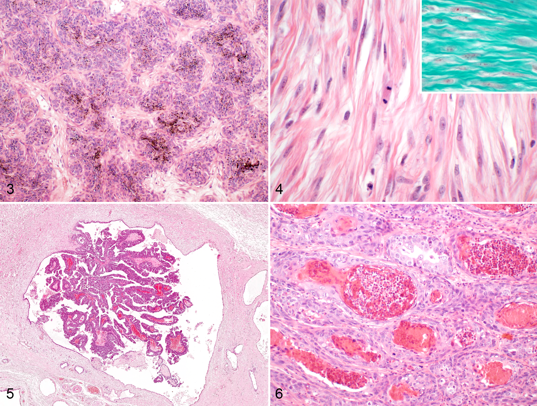
Trichoblastoma, abdominal skin, rabbit. There are lobules of basaloid cells and brown melanin pigment. Hematoxylin and eosin (HE).
Tumors of the Mammary Gland
Tumors of the mammary gland were found in 16 necropsy cases (prevalence: 0.8%, CI: 0.4% to 1.3%), 1/1 tissue samples, and 144/153 surgical biopsies (proportion: 94.1%, CI: 89.0% to 97.0%). For female intact rabbits, tumor prevalence was 1.7% (CI: 1.0% to 2.8%) with a continuous age-related increase to up to 9.4% (CI: 4.9% to 17.1%) in animals >6 years of age (Fig. 2, Suppl. Table S7). In all sample types combined (Suppl. Table S20), the most frequently occurring tumor types were simple carcinoma (46.0%), intraductal papillary adenoma (19.3%, Fig. 5), comedocarcinoma (7.5%, Fig. 6), simple adenoma (7.5%), and intraductal papillary carcinoma (5.0%). Mean age was 65.5 months (range: 6–156), and sex was predominately female intact (Suppl. Table S17). Of the 12 malignant mammary tumors of necropsy cases and tissue samples, metastasis was found in 5 cases (41.7%) with spread into the lungs (n = 5), liver (n = 2), regional lymph node (n = 1), and spleen (n = 1). Tumor-like lesions were found in 12 cases of female intact rabbits with multilocular cysts of the mammary gland.
Tumors of Hematopoietic Organs
Lymphoma was identified in 56 necropsy cases (prevalence: 2.8%, CI: 2.1% to 3.7%), 7 tissue samples, and 25 surgical biopsies (Suppl. Table S21). Lymphomas had highest prevalence in 25- to 48-month-old rabbits (Fig. 1, Suppl. Table S5), which was not significantly different compared to the youngest (PR: 7.07, P = .098) and oldest age group (PR: 0.59, P = .888). There was no significant difference in the PR of the different sexes (P = .889). Lymphomas of the necropsy cases were disseminated in 52 cases (93%; 48 B-cell and 4 T-cell lymphoma) and localized to the intestine (n = 3) or spleen (n = 1) in 4 cases (7%; all B-cell lymphoma). Examination of tissue samples confirmed disseminated disease in 5 of 7 cases (71%). Masses were commonly identified in lymph nodes, gastrointestinal tract, kidneys, spleen, and liver (Suppl. Tables S9, S14). Most cases were of B-cell immunotype (n = 84, 96%; inset Fig. 7), while only 4 necropsy cases were consistent with T-cell immunotype (4%) based on IHC. B-cell lymphomas were infiltrated with few (n = 44; 52%), moderate (n = 30; 36%) or high (n = 10; 12%) numbers of (non-neoplastic) T-cells. Cell size was always large in B-cell lymphomas and small to intermediate in T-cell lymphomas. Cutaneous lymphomas exhibited epitheliotropism in 1/33 cases.
Histiocytic sarcoma was found in 8 necropsy cases (prevalence: 0.4%, CI: 0.2% to 0.8%), 3 tissue samples, and 1 surgical skin biopsy. Of the necropsy cases, 6 were disseminated (75%) and 2 were localized to the lung (25%), while disseminated spread was evident in 2 of 3 tissue samples (Suppl. Tables S9, S14).
Thymoma (Fig. 8) was identified in 42 necropsy cases (prevalence: 2.1%, CI: 1.5% to 2.9%) and in 6 tissue samples. Prevalence of thymomas continuously increased with the age groups up to 9.4% (CI: 6.2% to 14.0%) in rabbits >6 years (Fig. 1, Suppl. Table S5). Comparison of the sixth to fourth age group revealed a PR of 10.86 (P < .001). Neutered male (PR: 4.27, P < .001) and female (PR: 4.05, P = .278) rabbits each had a higher prevalence compared to intact rabbits of respective sex.
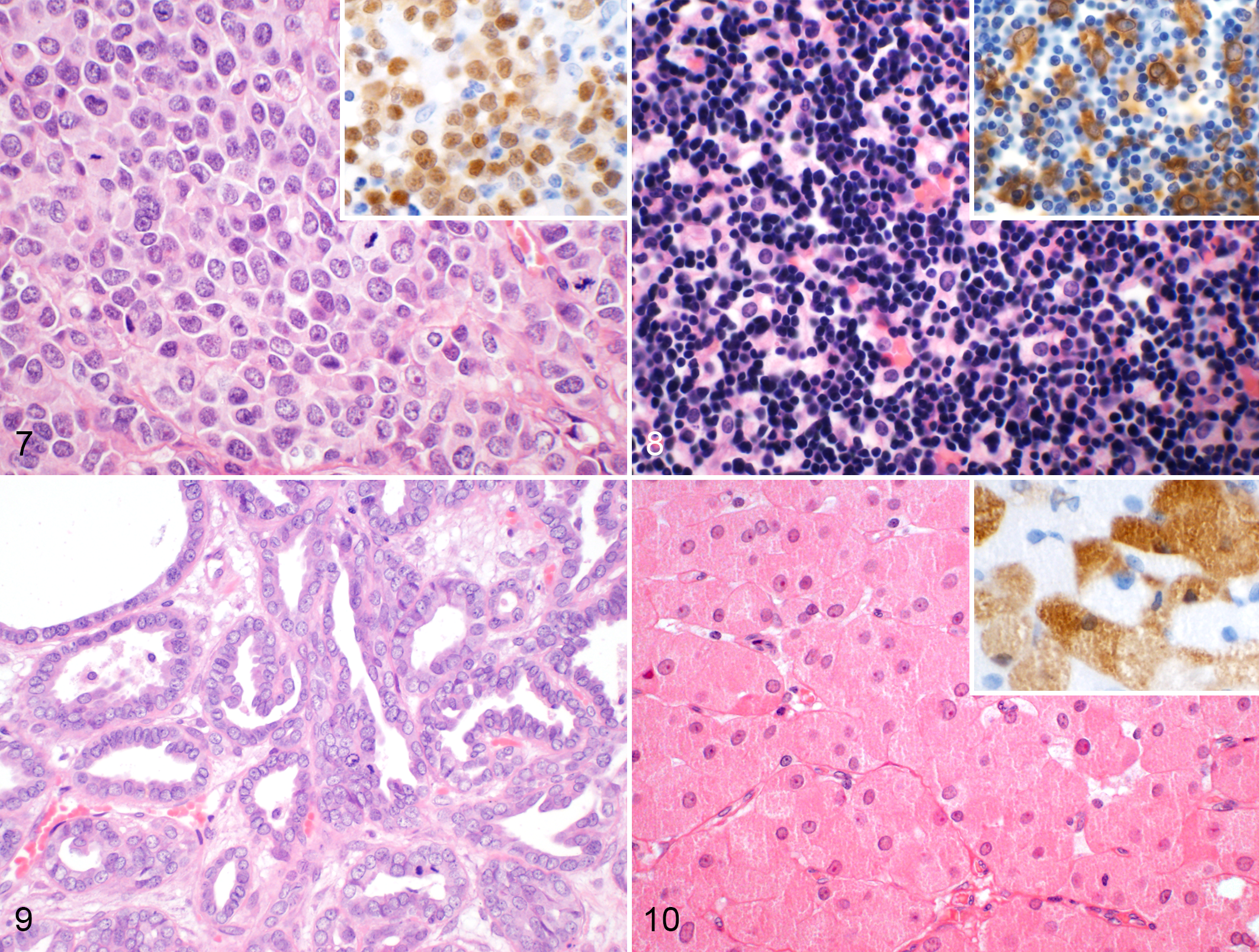
Diffuse, large B-cell lymphoma, mesenteric lymph node, rabbit. Large, centroblastic round cells are present and contain 3 mitotic figures. HE. Inset: Neoplastic cells have nuclear immunolabeling for Pax-5 (B-cell marker).
A splenic hematoma (tumor-like lesion) was diagnosed in one necropsy case.
Tumors of the Female Genital System
Primary ovarian neoplasia was identified in 2/894 necropsy cases (prevalence: 0.2%, CI: <0.1% to 0.8%) and 4 of 142 (proportion: 2.8%, CI: 1.0% to 7.2%) ovarian biopsies (Suppl. Table S22). A total of 238 uterine tumors were found in 117/894 necropsy cases (prevalence: 13.1%, CI: 10.9% to 15.6%), 9/14 tissue samples (proportion: 64%, CI: 38% to 84%), and 97 of 159 (proportion: 61.0%, CI: 53.0% to 68.4%) uterine biopsies. Of all sample types, the most frequent tumor type was uterine adenocarcinoma which comprised 75.6% of all uterine tumors (Fig. 9) followed by leiomyoma with 14.3% (Suppl. Table S23). Metastasis of uterine adenocarcinoma was found in 46/98 necropsy cases (47%, CI: 37% to 57%) and in 5/8 tissue samples (63%; Suppl. Tables S9, S14). Tumor prevalence continuously increased with age for all uterine neoplasia (PR between the sixth and third age groups: 4.8, P < .001). There was no significant difference between the PR of uterine adenocarcinoma for the Lop (P = .968) and Lionhead (P = .582) breeds compared to other breeds. Concerning the necropsy cases, uterine adenocarcinomas (n = 98) were accompanied by additional tumors in 21 cases (21.4%), including 9 mammary tumors. The PR for mammary neoplasia with concurrent uterine adenocarcinoma was 13.25 (P < .001) and decreased with adjustment for age by 72% to a PR of 3.71 (P = .035). One case of vaginal leiomyosarcoma was diagnosed at necropsy (prevalence: 0.1%).
Tumors of the Male Genital System
A total of 47 testicular tumors were diagnosed in 7/664 necropsy cases (prevalence: 1.1%, CI: 0.5% to 2.2%) and 37/51 biopsy cases (proportion: 73%, CI: 58% to 84%; 3 cases with bilateral tumors). Granular cell tumor was the most frequently occurring tumor type (Fig. 10, Suppl. Table S24). Cryptorchidism was diagnosed in 4/664 postmortem cases (0.6%, CI: 0.2% to 1.6%; age reported in 2 cases: 36 and 132 months), 2 of which had granular cell tumors (50%; age reported in one case: 132 months). Furthermore, cryptorchidism was reported in 7/37 testicular biopsies with neoplasms (median age: 84 months; range: 6–108), which included 4 granular cell tumors, 2 teratomas, 1 Sertoli cell tumor, and 2/14 testicular biopsies without neoplasia (both rabbits 24 months old).
Tumors of the Digestive System
Tumors of the oral cavity were found in 4 necropsy cases (prevalence: 0.2%, CI: <0.1% to 0.5%) and 10/14 surgical biopsies (Suppl. Table S25). Adenocarcinoma (n = 2) and adenoma (n = 1) of the salivary gland were identified in 2 necropsy cases (prevalence <0.1, CI: <0.1% to 0.4%) and in 1/10 surgical biopsies. In the intestine, anorectal papillomas were found in 1 necropsy case (prevalence <0.1%, CI: <0.1% to 0.3%) and 30/37 intestinal surgical biopsies. Median age of these rabbits was 55 months (age range: 36–108). Intestinal adenocarcinomas were identified in 1 necropsy case (prevalence <0.1%) and 1 tissue sample.
Neoplasia of the liver including gallbladder was identified in 5 necropsy cases (prevalence: 0.3%, CI: <0.1% to 0.6%; Suppl. Table S26). Tumor-like lesions included focal, multilocular cysts lined mostly by cuboidal epithelium and separated by moderate amounts of fibrous connective tissue in 6 necropsy cases (prevalence: 0.3%, CI: 0.1% to 0.7%). Rabbits with cystic lesions had a median age of 94.5 months (range: 56–95).
One primary pancreatic adenocarcinoma (prevalence <0.1%, CI: <0.1% to 0.3%) with metastasis to the peritoneum, liver, gall bladder, and kidneys was found in a necropsy case. Tumor-like lesions included one necropsy case of pancreatic nodular hyperplasia.
Tumors of the Urinary System
A renal nephroblastoma was diagnosed in a tissue sample of a 23-month-old, female rabbit. In the urinary bladder, a transitional cell carcinoma was identified in one surgical biopsy. Tumor-like lesions included one polyp in the urinary bladder of a necropsy case.
Tumors of the Respiratory Tract
In the upper respiratory tract, 2 tumors were identified in the necropsy cases (prevalence <0.1, CI: <0.1% to 0.4%) and in one tissue and surgical biopsy sample each (Suppl. Table S27). Primary tumors of the lungs (papillary adenoma, n = 6; adenocarcinoma, n = 2) were diagnosed in 7 necropsy cases (prevalence: 0.3%, CI: 0.1% to 0.8%) and one tissue sample with a median age of 106.5 months (range: 57–180).
Tumors of the Musculoskeletal System
Osteosarcomas of the bone tissue were identified in 5 necropsy cases without distant metastasis (prevalence: 0.3%, CI: <0.1% to 0.6%) and in 3/11 surgical biopsies. Median age was 54 months (range: 18–87). Location of the neoplasms were the mandibles (n = 4), nasal bones with invasion into the nasal cavity (n = 2), maxilla (n = 1), and spine (n = 1).
Neoplasms primarily located in the skeletal muscle were identified in 4 necropsy cases (prevalence: 0.2%, CI: <0.1% to 0.6%) and one surgical biopsy and were consistent with anaplastic sarcoma with giant cells in 3 cases and fibrosarcoma in 2 cases. Of the 4 necropsy cases, all had metastatic spread to the lungs and in one case to further organs.
Tumors of the Endocrine Glands, Nervous System, and Eyes
Tumors of the endocrine glands were found in 4 necropsy cases (prevalence: 0.2%, CI: <0.1% to 0.6%; Suppl. Table S28). One glioblastoma was found in the brain of a 16-month-old, female intact rabbit at necropsy (prevalence <0.1%, CI: <0.1% to 0.3%). Primary tumors of the eye were diagnosed in 2/28 surgical biopsies (7%) as uveal melanoma.
Tumors of the Body Cavities
Seven primary tumors of the body cavities were found at necropsy. Mesotheliomas were present in 4 rabbits (prevalence: 0.2%, CI: <0.1% to 0.6%) with a median age of 62.5 months (range: 48–69). Of these, 2 were located in the peritoneum, 1 in the pleura, and 1 in the pericardium. Furthermore, 3 sarcomas (IHC: CK-negative, vimentin-positive) were found in 3 rabbits (prevalence: 0.1%, CI: <0.1% to 0.5%) in the pelvic (n = 2) or pleural cavity (n = 1).
Tumors of Unknown Primary Origin
In 8 necropsy cases, the tumor type was not readily assessable (Suppl. Table S29). One papillary adenocarcinoma (IHC: CK-positive, TTF-1-negative, thyreoglobulin-negative) was located in the neck region. The remaining 4 carcinomas and 3 sarcomas were found in multiple organs in terms of a metastatic tumor of inconclusive primary location. Furthermore, tissue samples with primary location was not submitted for 2 cases with pulmonary metastasis and the affected organ could not be determined in 3 surgical biopsies or abdominal masses.
Discussion
The present study is the first to describe tumor prevalence in all organs of pet rabbits. In all age groups combined, neoplasia was found in 14.4% of the necropsied pet rabbits, which is much higher than the prevalence previously reported for laboratory rabbits. 5,30,41,43 More than two thirds of the tumors at necropsy had histologic features of malignancy and approximately a quarter of all tumor cases or one third of the morphologically malignant tumor cases had distributed into multiple organs as metastatic or disseminated tumors. Greene and Strauss 17 report a similar rate of metastasis, that is, in 30.3% of the cases, of malignant tumors in laboratory rabbits. The most frequently occurring tumor types of the present necropsy cases were uterine adenocarcinoma, (disseminated) lymphoma, and thymoma. Tumors of the skin, mammary gland, and testis occurred occasionally. Lymphomas were the most frequently occurring tumor type in young rabbits (≤24 months of age). A further tumor type that seems to occur predominately in young to middle-aged rabbits—however, with much lower prevalence—is nephroblastoma (embryonal nephroma). 5,19,41,43 The prevalence of uterine adenocarcinoma, thymoma, skin tumors, and mammary tumors increased with age, of which uterine adenocarcinoma was the most frequently occurring tumor type in older rabbits. Due to the high prevalence of uterine adenocarcinoma in female intact rabbits, this cohort seems to have a higher prevalence of metastatic neoplasia compared to other sexes. Interestingly, female neutered rabbits had the highest overall tumor prevalence. However, this may be explained by the fact that this cohort was of much higher median age in comparison to the other sexes. Subsequently, the prevalence ratio with adjustment for age confirmed that female intact rabbits have a higher probability to develop neoplasia, consistent with the findings of a previous study on laboratory rabbits. 17 The higher overall age of female neutered rabbits in the present study should, however, not be considered suggestive of neutering leading to a longer life expectancy. In this regard, the results may be biased as preventative ovario(hyster)ectomy is generally not performed prior to maturity. Furthermore, rabbits receiving such measures of disease prevention may be more likely to receive thorough diagnostic workup as geriatric patients which may also include postmortem examination. Further studies are necessary to show the effect of neutering on life expectancy in female rabbits and whether this measure of prevention of uterine neoplasia and other uterine disorders 8 is beneficial regarding overall welfare.
Some rabbits had multiple tumors; however, the prevalence of multiple tumors did not exceed the calculated value which is furthermore similar to the findings reported by Greene and Strauss. 17 However, a correlation of uterine adenocarcinoma and mammary tumors with the concurrent hormonal stimulation had been suspected. 17 In the present study, we found a high prevalence rate of concurrent mammary neoplasia and uterine adenocarcinoma, which was, however, highly dependent on high prevalences of both tumor types in older rabbits. Interestingly, a recent study has determined that mammary carcinomas mostly do not express estrogen and progesterone receptors, based on IHC, suggesting a hormone-independent tumor growth in rabbits. 12 Also, uterine adenocarcinomas often lack immunoreactivity for those receptors, depending on the growth pattern. 1
The greatest limitation of the present study was the retrospective manner of analysis. Therefore, all potential neoplastic and tumor-like masses were reexamined histologically. However, due to the retrospective nature of the study, it cannot be excluded that some neoplastic masses were not identified or reported by the initial diagnostic service. Also, postmortem tissue samples referred by a veterinarian were analyzed separately in this study, as it cannot be guaranteed that all neoplastic masses present were submitted for histological examination. A further limitation lies in the submission for routine diagnostic examination itself. To the authors’ experience, pet owners are more likely to submit an animal to necropsy in cases of unexpected death, that is, in which the cause of death may not be obvious to the owner, such as rabbit hemorrhagic disease. On the contrary, neoplasia is usually a chronic disease that may be suspected by a thorough clinical examination, particularly in cases of neoplasia of the skin and mammary gland and these cases may be less likely submitted to necropsy, leading to a potential underrepresentation. These limitations therefore suggest that neoplasia may potentially be of higher true prevalence in pet rabbits than determined in the present necropsy cases. Nonetheless, as we have reported all tumors regardless of being a clinically relevant or an incidental finding, we expect the organ distribution of neoplasms to be largely representative for the pet rabbit population. It is, however, unknown whether the age distribution of the necropsy cases reflect the actual mean life expectancy or was biased by the owner’s probability to request necropsy, particularly in cases involving young rabbits. The risk to develop neoplasia correlates with the rabbits’ age, as seen in this study for several tumor types and for uterine adenocarcinomas in previous studies involving laboratory rabbits. 16,20 Therefore, tumor prevalence of the different age groups is especially interesting. We highlight that the tumor frequency of surgical biopsies cannot be compared to the tumor frequencies of respective organ systems. Frequency of tumor types is certainly influenced by the relative number of submitted organs. In the present study, the most frequently submitted, and, hence, overrepresented tissues of surgical biopsies were skin, female reproductive tract, and mammary gland. Concomitantly, the tumor numbers regarding these organs may also be overrepresented with frequencies which partially stand in contrast to those of necropsy cases. Regardless, surgical biopsies may add valuable information regarding the different tumor types that may occur in particular organ systems and the median age of animals affected.
It is currently unknown whether the various pet rabbit breeds are prone to distinct inheritance of susceptibility to neoplastic disease. For laboratory rabbits, it has been suspected that uterine adenocarcinoma occurs at variable rate in different breeds based on tumor prevalence from 4 to 60 evaluated animals per breed. 16 In the present study, breeds of the rabbits were often not stated or indicated as “dwarf” rabbits, which have a poorly defined genetic background. Therefore, only the 2 most common breeds—dwarf Lop and Lionhead—were evaluated and revealed no obvious tumor predisposition.
Hematopoietic tumors occur occasionally in rabbits and include lymphoma, thymoma, and histiocytic sarcoma. Although lymphoma seems to be a relatively common tumor in rabbits, only few studies 32,41 and some case reports or series are currently available. 22,24,29,45 According to von Lölinger, 41 lymphoma was the most common tumor type in a population of laboratory and breeding rabbits, which were affected at 10 to 30 months of age. In the present study, however, lymphoma occurred in all age groups—including very young rabbits, in which other tumor types occurred rarely. Therefore, lymphoma was the most frequently occurring tumor type in rabbits ≤2 years. The youngest rabbit affected in the present study was 2 months old, while a rabbit of 7 weeks of age had been previously reported. 45 Similar to cutaneous lymphomas, 32 most lymphomas were diffuse large B-cell lymphomas or T-cell-rich B-cell lymphomas regardless of the affected organ system. Only very few cases in the present study and in some previous reports have been diagnosed as T-cell lymphoma. 29,38,45 However, a B-cell marker was lacking in the study by White et al, 45 while Toth et al 38 used an unusual B-cell marker, that is, mouse polyclonal anti-rabbit IgM. Therefore, the differential diagnosis of a T-cell-rich B-cell lymphoma cannot be conclusively excluded. The marker most commonly applied for B-cells in rabbits is CD79a. 15,22,29,32 In the present study, Pax-5 was used, which yielded satisfying staining in both neoplastic tissue and positive controls from normal hematopoietic organs, that is, lymph node, spleen, and caecum. The rather uniform B-cell immunotype of lymphomas in rabbits, which was confirmed in this study, may suggest standardizable treatment options for rabbits. However, information on survival time 32 and therapeutic approaches 39 is very sparse in rabbits and further studies are required. In addition to prevalence and age distribution, lymphoma distribution in internal organs had not been systematically investigated to date. In 12 cases described by Von Löliger, 41 a predominant involvement of the kidneys and lymph nodes was mentioned but further information on organ-specific tumor distribution is lacking. Our necropsy cases commonly showed widely disseminated masses, suggestive of later stages of disease. Hence, it is difficult to determine the primary organs from which the lymphomas had originated. We find it, however, very interesting that the gastrointestinal tract was the second most frequently affected organ beside the lymph nodes and, hence, suspect a high prevalence of gastrointestinal lymphomas, especially of the cecum, similar to the case description by Isikawa et al. 22
Skin was the third most commonly affected organ system in the present necropsy cases and the organ or tissue most frequently affected by neoplasia in surgical biopsy submissions. Frequencies of diagnosed tumor types were similar to previous studies. 23,40 Consistent with a previous case report, 47 IHC with antibodies against melan-A was also useful to identify amelanocytic melanoma in our study. Basal cell tumors—especially trichoblastoma—seem to be the most frequently occurring type of skin tumor and are mostly benign. 23 Histological criteria of malignancy or metastasis are only rarely reported in basal cell tumors of rabbits. 9,23 In the present biopsy cases, we found some basal cell carcinomas, a morphologically malignant trichoepithelioma, and confirmed metastatic spread of a basal cell carcinoma to the regional lymph node. In contrast to studies from the United States, 23,40 Shope fibroma and Shope (squamous) papillomas were not present in our study from Germany. Consistent with previous studies, 23,40 collagenous hamartomas were the most frequently occurring tumor-like lesion in the present study and also occurred almost exclusively in male rabbits. A hormonal stimulation of collagenous hamartoma and mesenchymal skin tumors in male rabbits had been proposed. 40 In contrast to the findings by von Bomhard et al, 40 mesenchymal tumors, including myxosarcomas, were found with similar frequency in male and female rabbits in the present study.
Mammary tumors were found occasionally in the present study with highest prevalence in old female rabbits. Male rabbits were rarely affected, consistent with previous studies which described a single male in more than 300 cases of mammary tumors. 4,12,23,35 Although simple tumors are most common, rabbits also have relatively high frequencies of intraductal and comedo-type tumors as well as occasional lipid-rich and adenosquamous tumors. Malignant myoepithelioma had been reported previously only in a single case report. 13 The 2 present findings were suggested based on histological and immunohistochemical examination with antibodies against CK and SMA. Unfortunately, further immunohistochemical markers such as p63 and calponin were not available. 46 Some cystic mammary glands were identified as tumor-like lesions in the present study. However, it cannot be excluded that these represent ductal ectasias secondary to intraductal tumors not present in the tissue sections examined, as proposed by Baum and Hewicker-Trautwein. 4 Criteria of malignancy have been quantified for different mammary tumor types in rabbits showing that morphologically malignant tumor types are common. 4,12,35 However, as survival data have not been examined along with these criteria, the cutoff values for malignancy determination or a histological grading system are currently unavailable for rabbits. Therefore, we used established cutoff values for canine and feline tumors. 46 Further studies are necessary to develop a specific histological grading system for mammary neoplasia in rabbits. Indeed, numerous mammary carcinomas diagnosed at necropsy in the present study had metastasized—especially to the lung—suggesting a high risk for malignant behavior.
The male genital system, and particularly the testes, were among the 5 most common organ systems with neoplasia in the present necropsy (prevalence: 1.1%) and biopsy cases (4.3% of all biopsy submissions). Webb et al 42 report testicular tumors in 1.9% of all rabbit submissions to 2 pathological institutions. Up until recently, interstitial cell tumors had been considered the most frequently occurring tumor type. 3,21,37 However, 2 recent studies have identified granular cell tumors to be more common and describe these tumors to be difficult to distinguish from interstitial cell tumors without the use of special stains, especially PAS reaction. 31,42 More specifically, according to Reineking et al, 31 granular cell tumors are positive for PAS, whereas interstitial cell tumors are negative. By this means of differentiation, 31,42 the present study confirms that interstitial cell tumors, Sertoli cell tumors, seminomas, and teratoma are less common than granular cell tumors in rabbits. Metastasis of testicular tumors has been reported in a single case report of seminoma 3 and was not detected in the present study. Granular cell tumors—and possibly other testicular tumors—have a good prognosis in rabbits. 42 Previous literature on the association of testicular tumors and cryptorchid status is not available. To date, there is only one report on cryptorchidism associated with a teratoma. 33 In the present study, we found testicular tumors in 2 of 4 necropsy cases and in 7 of 9 biopsies of undescended testes. This finding should be considered as first evidence that the risk for developing testicular tumors might be higher in cryptorchid rabbits; however, further studies are necessary to confirm this assumption.
The most common non-hematopoietic tumors of the digestive system seem to be anorectal polyps and tumors of the oral cavity. Although anorectal polyps were reported in a review article, 39 primary literature on this tumor type is not available to date. A viral etiology is currently unknown and should be examined in future studies. In the oral cavity, various tumor types may occur and—consistent with results from a previous study 27 —odontogenic tumors seem to be relatively common in rabbits. Also, biliary tumors—especially adenomas—were occasionally identified in the present study, in previous studies of laboratory rabbits, 17,43 as well as in case descriptions of pet rabbits. 34 However, biliary cystic lesions were more common than proliferative masses. Cystic lesions had characteristics of previous descriptions of a biliary hamartoma (developmental abnormality) 36 and biliary cystadenoma. 11,28,34 As had been discussed by Cullen, 10 differentiation of these 2 disorders is neither straightforward nor essential as the clinical consequence is the same for these 2 benign masses. Cullen 10 also states that most hepatic cystic lesions in domestic animals are more likely biliary developmental disorders than neoplasms. As proliferative epithelium was not evident, we chose to classify these lesions as tumor-like processes although present exclusively in rabbits of more than 4.5 years of age. However, we highlight that these cystic lesions may be the same entity as the biliary cystadenoma in rabbits reported previously.
In conclusion, we found a tumor prevalence of 14.4% in all necropsied pet rabbits, most of which were morphologically malignant. Prevalence increased relative to age up to 47.2% in rabbits older than 6 years. The most frequently occurring tumor type was the uterine adenocarcinoma, hence rendering female rabbits to be more commonly affected by neoplasia. Lymphoma was found most frequently in young rabbits, was commonly located in the lymph nodes, gastrointestinal tract, kidneys, spleen, and liver, and was mostly of B-cell immunotype. Thymoma was frequently present in rabbits of old age and was always benign in the present study. Primary, non-hematopoietic tumors were occasionally found in the skin, mammary gland, testis, and digestive system and seem to be rare in the remaining organ systems in rabbits.
Supplemental Material
Supplemental Material, sj-pdf-1-vet-10.1177_0300985820973460 - Neoplasia and Tumor-Like Lesions in Pet Rabbits (Oryctolagus cuniculus): A Retrospective Analysis of Cases Between 1995 and 2019
Supplemental Material, sj-pdf-1-vet-10.1177_0300985820973460 for Neoplasia and Tumor-Like Lesions in Pet Rabbits (Oryctolagus cuniculus): A Retrospective Analysis of Cases Between 1995 and 2019 by Christof A. Bertram, Beate Bertram, Alexander Bartel, Anja Ewringmann, Marco A. Fragoso-Garcia, Nancy A. Erickson, Kerstin Müller and Robert Klopfleisch in Veterinary Pathology
Footnotes
Acknowledgements
We thank Nicole Huth, Cornelia Zieger, Charlene Lamprecht, and Michaela Scholz for technical assistance.
Declaration of Conflicting Interests
The author(s) declared no potential conflicts of interest with respect to the research, authorship, and/or publication of this article.
Funding
The author(s) received no financial support for the research, authorship, and/or publication of this article.
Supplemental material for this article is available online.
References
Supplementary Material
Please find the following supplemental material available below.
For Open Access articles published under a Creative Commons License, all supplemental material carries the same license as the article it is associated with.
For non-Open Access articles published, all supplemental material carries a non-exclusive license, and permission requests for re-use of supplemental material or any part of supplemental material shall be sent directly to the copyright owner as specified in the copyright notice associated with the article.