
Abstract
Orofacial masses or swellings are a common presenting complaint in lagomorphs. Similar gross appearances of the masses can complicate clinical interpretation, and histologic review often provides the final diagnosis. Underlying causes vary from infectious to neoplastic. Although inflammatory changes are most commonly reported, various neoplasms occur, although the prevalence of specific tumor types is relatively unknown. We reviewed retrospectively 120 cases (87.5% biopsy, 12.5% autopsy) of neoplastic and non-neoplastic orofacial masses received from January 2000–February 2023 at 2 institutions: University of Guelph, Canada (Animal Health Laboratory and Department of Pathobiology), and Finn Pathologists, United Kingdom. All final diagnoses were achieved through histologic assessment. We included masses or mass-like swellings from the oral cavity, including the mandible and maxilla, and surrounding skin and soft tissues of the oral cavity and jaw. Submissions included pet and commercial (meat and fur) rabbits. Neoplastic lesions were most common (60%), including trichoblastomas, papillomas, melanocytic neoplasms, sarcomas, round-cell tumors, carcinomas (including squamous cell carcinoma), lipomas, odontogenic neoplasms, polyps, osteoma, neuroma, peripheral keratinizing ameloblastoma, and apocrine adenoma. Inflammatory diagnoses (30%) included abscesses, osteomyelitis, dermatitis, and sialadenitis. Other diagnoses (7%) included cysts, as well as hyperplastic skin and proliferative bone lesions. Three cases had no definitive diagnosis. The importance of histologic assessment in diagnosing orofacial “masses” in rabbits is highlighted, given that the most common diagnostic category overall was neoplasia.
Domestic rabbits (Oryctolagus cuniculus) can be a relatively stoic species and can fail to show clinical signs of disease, which can be a challenge to pet owners, veterinarians, and pathologists. Nevertheless, cutaneous lesions, or changes involving the head or mouth, tend to be visible grossly and can thereby be more easily identified as a potential problem. Clinical signs, such as drooling, difficulty prehending food, and weight loss, can be dramatic and easily noted by owners, contributing to the discovery of oral lesions. Masses involving the skin and soft tissues of these regions may be more commonly observed visually and or palpated by owners upon handling of the animal, and result in presentation to a veterinarian. Hence, sample submissions from these areas are commonly received in diagnostic laboratories for assessment. Although inflammatory masses are commonly reported in rabbits, particularly in the orofacial region, neoplasia, including neoplasms with an ectopic presentation, can also occur, complicating interpretation of the gross picture and demonstrating that neoplasia is a critical differential diagnosis for any mass-type lesion.2,4,5,24,27,37
Although rabbits are a common domestic species, both for commercial use and as pets, knowledge regarding spontaneous neoplasms and prevalence of tumor types is relatively limited. 5 Individual case reports of orofacial masses exist for several tumor types in rabbits; however, studies of larger case numbers are usually focused on specific entities or organ systems2,5,6,7,17,19,20,22,30,37 or report unspecified neoplasia as a cause of death. 27 Various tissue types in the head and neck region, including bone, teeth, and soft tissue components, can all be involved in neoplastic transformation, resulting in disparate neoplastic presentations for similar grossly appearing masses. As well, masses typically regarded as inflammatory, such as abscesses, may mask an underlying neoplasm.8,28 Recognizing and being aware of the potential tumor types observed in these regions can have important prognostic and therapeutic implications for this species.
We reviewed biopsy and postmortem reports of orofacial masses and swellings in rabbits and summarized the more common findings in the tissues of the head and neck. We highlight the difficulty for both clinicians and pathologists when assessing masses or mass-like structures in rabbits, solely based on gross findings, and demonstrate the importance of histopathology in making a definitive diagnosis.
Case selection
We reviewed retrospectively submission forms and final reports from January 2000 to February 2023, for domestic rabbits (either commercial or pet rabbits; Oryctolagus cuniculus) from 3 diagnostic services at 2 institutions (Animal Health Laboratory and Department of Pathobiology, University of Guelph, Canada [institution 1]; Finn Pathologists, United Kingdom [institution 2]). Submissions from experimental rabbits were not routinely received at either institution; none was available for inclusion in this review. Inclusion required histologic assessment of a facial mass or swelling in the skin and soft tissues of the face, head, and neck, including base of ears, dewlap, lips, and oral cavity. Neural tissue (brain or spinal cord), nasal sinuses, and enucleated eyes were not included in the submissions reviewed, and cytologic examination was not reported. The minimum data collected included lesion location and histologic diagnosis, and included age, sex, and breed when available. For categorization purposes, neoplasms were defined as masses resulting from new, abnormal growth of a tissue. 25 Those lesions categorized as inflammatory included responses to either known infectious organisms or unknown organisms or cause. Lesions categorized as “other” included hyperplastic or cystic mass-like lesions. Statistical analyses were performed between the neoplastic and inflammatory groups for age and sex demographics using unpaired t-tests and one-way ANOVA as appropriate.
Results
A total of 120 submissions met the inclusion criteria for our review, comprising 80 cases (67%) from Finn Pathologists and 40 cases (33%) from the University of Guelph. Samples submitted included those taken surgically by the submitter, following postmortem examination by the submitting veterinarian, or performed at the diagnostic laboratory itself. We reviewed 15 autopsy samples (12%) and 105 biopsy samples (88%). Of the 15 autopsy submissions, 7 of 15 (47%) final diagnoses were neoplastic, 5 of 15 (33%) were inflammatory, and 1 of 15 (7%) fell into the “other” category. Two cases (13%) in the autopsy category resulted in no definitive diagnoses. Of the 105 biopsy sample submissions, 65 (62%) were neoplastic, 32 (30%) were inflammatory, 7 (7%) were “other,” and 1 (1%) had no definitive diagnosis.
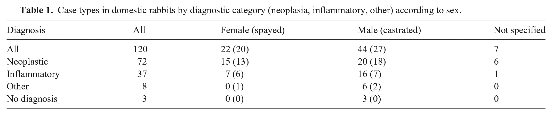
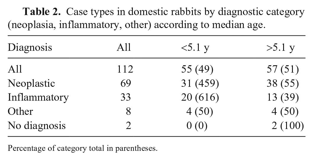
Demographic data were summarized when provided (Tables 1, 2; Suppl. Table 1). Age was available for 112 animals with a median age at diagnosis of 5.1-y-old (range: 0.3–14.0-y-old). Information on the sex of the submitted animal was available for 113 cases, which included 42 females (20 intact, 22 spayed) and 71 males (44 intact, 27 castrated; Table 1). Various domestic breed types were submitted (Suppl. Table 1). The most numerous breed submitted was “lop,” not further specified (n = 11), although breed information was not specified for 41 individuals. A more descriptive location was provided for 119 of 120 submissions; the most common locations included the mandible or maxilla (jaw; 24 of 120), followed by dewlap (16 of 120), chin (10 of 120), oral cavity (10 of 120), and lip or neck (9 of 120).
Case types in domestic rabbits by diagnostic category (neoplasia, inflammatory, other) according to sex.
Case types in domestic rabbits by diagnostic category (neoplasia, inflammatory, other) according to median age.
Percentage of category total in parentheses.
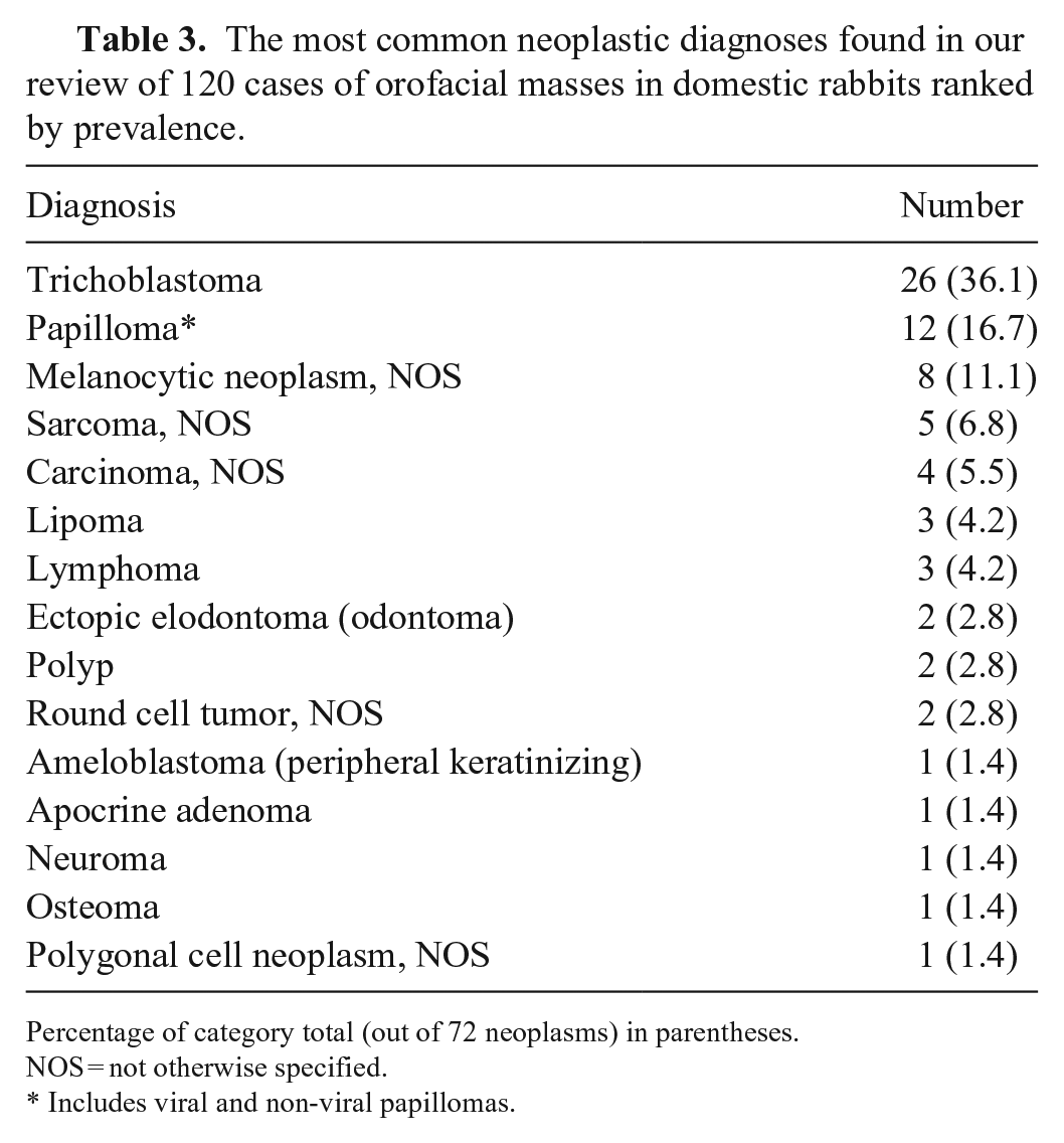
A diagnosis of neoplasia was made for 72 of 120 cases, providing an overall prevalence of 60.0% for neoplasms in the orofacial region. Of the neoplastic histologic diagnoses included, 24 different diagnoses were made. These included: apocrine adenoma, peripheral keratinizing ameloblastoma, carcinoma (adenocarcinoma, squamous cell carcinoma, unspecified carcinoma), ectopic elodontoma (odontoma), laryngeal and oral polyps, lipoma, lymphoma (epitheliotropic, multicentric, unspecified), melanocytic neoplasms (amelanotic melanoma, melanoma), neuroma, osteoma, papilloma (unspecified, viral), sarcomas (rhabdomyosarcoma, giant-cell sarcoma, myxosarcoma, neurofibrosarcoma, soft-tissue sarcoma), trichoblastoma, and round-cell tumors as well as polygonal-cell neoplasms that were not further specified (Table 3).
The most common neoplastic diagnoses found in our review of 120 cases of orofacial masses in domestic rabbits ranked by prevalence.
Percentage of category total (out of 72 neoplasms) in parentheses.
NOS = not otherwise specified.
Includes viral and non-viral papillomas.
Neoplastic diagnoses were made in 28 females, 38 males, and 6 cases with sex not specified, and were largely commensurate with the number of submissions per sex category with no statistically significant difference across the groups (Table 1). The median age at diagnosis was statistically significantly greater for those with neoplastic masses (median age: 6.0 y; range: 2.0–14.0 y) compared to inflammatory masses (median age: 3.6 y, range: 0.3–9.0; p < 0.01). Aside from the jaw, neoplasia was the most common histologic diagnosis per location, with the dewlap (9 of 72) and oral cavity (8 of 72) representing the most common sites (Suppl. Tables 2A, 2B). There was no notable association of type of neoplasm with any particular anatomic location, except for trichoblastomas in the cheek (5 of 6 diagnoses). Although the oral cavity was a common location for tumors, none was of odontogenic origin, and the 2 elodontomas (odontomas in species with continuously erupting incisors) were ectopic in the lip.
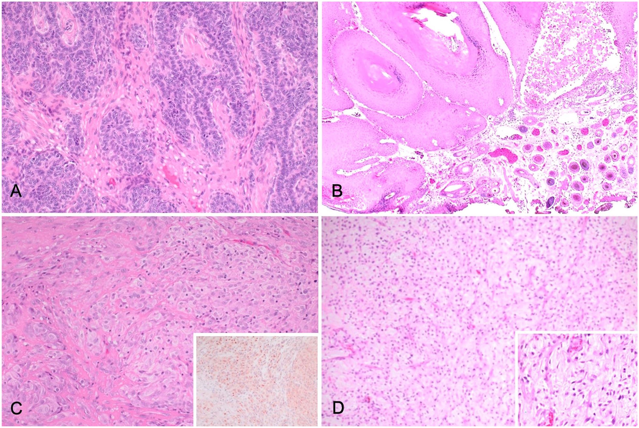
Trichoblastomas were the most common neoplastic diagnosis (26 of 72, 36.1%; Fig. 1A), followed by papillomas (12 of 72, 17%; of which 3 of 12 were viral and 9 of 12 were non-viral; Fig. 1B), melanocytic neoplasms (melanoma [Fig. 1C] and amelanotic melanoma; 8 of 72, 11%), and sarcomas (including myxosarcoma [Fig. 1D; 5 of 72, 6.8%] and carcinomas [4 of 72, 5.5%]). Papillomas were largely observed in areas associated with haired skin, except for 1 on the “lip,” 1 on the tongue, 2 on the third eyelids, 1 below the left nostril, and 1 associated with the gingiva of teeth 408–409. Those identified as viral papillomas were cases in which characteristic intracytoplasmic viral inclusion bodies and cytoplasmic clearing with eccentric nuclei (koilocytes) were observed. Trichoblastomas were diagnosed in the haired skin of the nose, neck, jaw, head, face, dewlap, chin, cheek, and base of the ear, representing a common skin mass. This mass was diagnosed in both sexes and across a large age range.
The most common neoplastic diagnoses found in our review of 120 cases of orofacial masses in domestic rabbits.
The remaining diagnoses comprised inflammatory lesions, such as abscesses, sialadenitis, myxomatosis, granulation tissue, inflammation secondary to fat necrosis, botryomycosis, and pyoderma and/or dermatitis (37 of 120, 31%; Table 4). Of the 8 cases with “other” diagnoses, 7 were reported as hyperplasia, and 1 as a cyst.
The most common inflammatory diagnoses found in our review of 120 cases of orofacial masses in domestic rabbits ranked by prevalence.
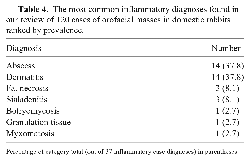
Percentage of category total (out of 37 inflammatory case diagnoses) in parentheses.
Three submissions had no definitive diagnosis following histopathology (Table 1). For these 3 cases, all were male rabbits, and the tissue submitted was identified as having been taken from the region of the jaw. The results for these 3 cases (although specified as not definitive) included possible osteoma, possible odontogenic tumor or fibrosis, and insufficient sampling. The most numerous inflammatory mass-like effects were caused by abscesses or dermatitis and/or pyoderma (14 of 120; 12% each), and the most numerous non-inflammatory or non-neoplastic diagnosis was hyperplasia (7 of 120 cases; 6%), which was most commonly reported as hyperplasia of the apocrine gland (Suppl. Table 3).
Inflammatory diagnoses were most common in intact males (13% of all diagnoses and 16 of 36 inflammatory diagnoses [44%]). Other diagnoses (8 of 120; 7%) and no definitive diagnoses (3 of 120; 2%) were encountered less commonly (Table 1). Most samples from the jaw (maxilla or mandible; 14 of 24) and lip (5 of 9) were diagnosed as inflammatory lesions, with most of the submissions listed as “jaw” being from the skin associated with the jaw; these were diagnosed as abscesses (11 of 14, 79%; Suppl. Table 4). Only one report of a jaw-associated abscess specified osteomyelitis as part of the final diagnosis.
Discussion
In our case series, the most common diagnosis for a mass lesion from the orofacial region was neoplasia, which is similar to other case series for rabbits that examined results of autopsy and biopsy samples.2,5 Interestingly, but perhaps not surprisingly, given the variety of tissue types present in the orofacial region, we found a variety of diagnoses of neoplasia. Most neoplasms were described in areas in which that tissue is normally found (i.e., melanocytic neoplasms in association with epidermis, osteoma with bone of the jaw, etc.), except for elodontomas (odontoma; 2 of 120), which were described as being within an ectopic location for both cases (both considered to have been taken from the lip). 37
Consistent with the literature on common neoplastic diagnoses in rabbits, the neoplasm diagnosed most commonly in our review was trichoblastoma.11,12 This is not surprising considering that our region of interest included haired skin; trichoblastoma represents disordered growth of the hair follicle commonly reported in rabbits in a variety of locations, although most often observed in submissions from the head and neck.18,32
Papillomas were, also unsurprisingly, the second most commonly reported lesion in the orofacial region in this species. 32 This is presumably the result of viral replication of the rabbit oral papillomavirus (Papillomaviridae, Kappapapillomavirus 1) occurring in the oral mucosa or by cottontail rabbit (Shope) papillomavirus (Kappapapillomavirus 2) in the haired skin, although non-viral squamous papillomas are also reported commonly in rabbits.15,21,32,34 Of the 12 papillomas diagnosed in our case review, only 3 were diagnosed as viral papillomas (with the method of diagnosis uncertain). The 3 viral papillomas were observed on the tongue, under the chin, and the “right jaw” (haired skin presumed, based on this location description).
The next most common neoplastic diagnoses were melanocytic neoplasms, followed by sarcoma and carcinoma. Of these, melanocytic neoplasms and carcinomas were observed both in the oral cavity (an amelanotic melanoma from the “gum,” a melanoma from the “palate,” an oral squamous cell carcinoma rostral to the incisors, and an oral adenocarcinoma of indeterminate origin from the “upper left gingiva”) and in association with samples submitted from the skin (ear, chin, eyelid, neck, face, and head for the melanocytic neoplasms, and base of ear and eyelid for carcinomas). In contrast, sarcomas were only reported in cases submitted from sites associated with skin (dewlap, face, and neck).
Determining the cellular origin of a neoplasm is complicated in any species, 26 and a similar difficulty was observed in some of the submissions in our review. For some of the diagnoses made, the pathologist was unable to definitively differentiate the cell of origin for the observed neoplasm. For these results, the neoplastic diagnosis was based on the morphologic appearance of the neoplastic cells. These cases included diagnoses of polygonal-cell neoplasm, round-cell tumor, or sarcoma. Although the reasons for not pursuing further testing in these cases remain unknown, the most likely possibilities for not pursuing immunohistochemistry for further differentiation include cost and, potentially, lack of validation or unavailability of particular immunohistochemical stains at the institution. Rabbits are used frequently for raising antibodies, 33 which increases the challenge of finding an immunohistochemical stain that could be used for diagnostic purposes in this species. A limitation of our review is its retrospective nature.
Rabbits are a species recognized for the frequency of dental disorders and oral or tooth root abscessation (often subsequent to malocclusion and trauma, with subsequent odontogenic dysplasia).4,9,13,14,23 Hence, abscessation was not unsurprisingly a common reason for tissue submission in our series. Interestingly, although in diagnoses of neoplasia (or “new growths”), most bone or jaw and/or dental disorders were reactive or inflammatory (hyperplasia). Unfortunately, as most samples received were biopsy specimens, the tissues examined in the reviewed reports were largely soft-tissue–associated structures; no distinctly dental-associated submissions were received in the biopsy services from either institution. The few samples involving bone that were assessed as part of our review were from autopsy cases wherein distinct subcutaneous abscesses associated with osteomyelitis (1 case), osseous proliferation associated with the dental arcade (1 case), or an osteoma (1 case) were observed.
Although neoplastic diagnoses were relatively common in the oral cavity in our review, interestingly, neoplasms of dental origin were uncommon. The only neoplasm of odontogenic origin developed in what was considered an ectopic location for that tissue.29,37 Neoplasms of odontogenic origin are believed to arise from germinal tissues of the tooth and hence can recapitulate any of the tooth-forming tissues. It has been thought that odontogenic neoplasms were generally anatomically confined to the jaw, wherein tooth germinal tissue is anatomically located. However, neoplasms arising from odontogenic epithelium have been reported arising from other oral or oral-associated locations, such as the gingival mucosa or cheek tissue of rabbits, and the facial skin of cats.16,24,37 The cause for this ectopia is not certain; both ectopic germinal rests and phenotypic mimicry have been suggested as potential reasons but, in part given the relative rarity of these neoplasms, not yet proven. 24
Other neoplasms diagnosed in the oral cavity in our review included those of epithelial or round-cell origin (carcinoma, melanocytic neoplasms, undifferentiated round-cell tumor, polygonal-cell neoplasm, polyp, and papilloma). We found that neoplasia of the soft tissues of the oral cavity, or possible oral involvement with a more systemic neoplastic disorder, was more common than neoplasms of dental or hard tissues.
Rabbits are recognized for their frequently encapsulated abscesses.9,10,14 Because rabbits lack lysosomal enzymes that digest dead cells to a liquid form, purulent material is often dense and more solid in texture, making abscesses difficult to drain, which often results in encapsulation or walling off of the necrotic material.1,31 Not surprisingly, abscesses can appear grossly as a “mass,” and even when surgically removed or incised, can be difficult to differentiate from necrotic foci in neoplastic growths. Abscesses in our review arose primarily from the jaw and associated tissues (chin, dewlap), although other locations around the head have been reported. 14 The second most common inflammatory lesion noted in our review was dermatitis, and, perhaps unsurprisingly, was primarily associated with locations in the orofacial region where pooling of fluids (saliva, nasolacrimal secretions) could be observed.3,35,36 The reasons for the inflammatory lesions in our review were not definitively identified, but based on location, are suspected to again potentially reflect disorders of dentition.
Interestingly, most inflammatory diagnoses in rabbits appeared to occur in intact males (16 of 36 inflammatory diagnoses, inclusive of female, spayed females, castrated males, and unspecified rabbits). The reason is uncertain, although potentially may reflect secondary bacterial infection in areas of conspecific trauma (e.g., bite wounds as a result of cohabitation or breeding activities). 3 Unfortunately, a lack of history, unavailable animal management data, and unknown purpose of use, means that our hypothesis is purely speculative.
Our review highlights the importance of histopathology in the investigation of masses from the orofacial region in rabbits. Given the specific ways in which rabbits process inflammation, inflammatory masses in the orofacial region can be difficult to differentiate grossly from neoplastic growths. Our review also highlights the potential for ectopic neoplasms (ectopic elodontoma [odontoma]) to occur in these animals. Not all masses in the orofacial region are inflammatory, and not all are neoplastic; although location and signalment may demonstrate trends, histologic examination is invaluable in allowing differentiation.
Supplemental Material
sj-pdf-1-vdi-10.1177_10406387241234326 – Supplemental material for Orofacial masses in domestic rabbits: a retrospective review of 120 cases from 2 institutions, 2000–2023
Supplemental material, sj-pdf-1-vdi-10.1177_10406387241234326 for Orofacial masses in domestic rabbits: a retrospective review of 120 cases from 2 institutions, 2000–2023 by Emily Rätsep, Latasha Ludwig and Melanie Dobromylskyj in Journal of Veterinary Diagnostic Investigation
Footnotes
Acknowledgements
We thank Geoffrey A. Wood at the University of Guelph, and Sophie Bowen-Carpenter and Ken Smith at the Royal Veterinary College, for their support in this collaboration, as well as the technicians and original reporting pathologists involved in these cases.
Declaration of conflicting interests
The authors declared no potential conflicts of interest with respect to the research, authorship, and/or publication of this article.
Funding
The authors received no financial support for the research, authorship and/or publication of this article.
Supplemental material
Supplemental material for this article is available online.
References
Supplementary Material
Please find the following supplemental material available below.
For Open Access articles published under a Creative Commons License, all supplemental material carries the same license as the article it is associated with.
For non-Open Access articles published, all supplemental material carries a non-exclusive license, and permission requests for re-use of supplemental material or any part of supplemental material shall be sent directly to the copyright owner as specified in the copyright notice associated with the article.