
Abstract
Twenty-seven mammary tumors from 18 male (3 intact, 15 neutered) dogs were collected. The average age at diagnosis was 9.2 years (range, 2–14 years). Seven of the dogs were Cocker Spaniels. Five dogs had multiple mammary tumors. All tumors were benign. Twenty-six were simple adenomas with mixed acinar and papillary patterns. The acinar pattern was predominant in 17 cases. One adenoma was complex with a prominent myoepithelial component. The myoepithelial component of 25 of the 25 tumors was immunohistochemically positive for calponin and p63. In the cases for which relevant clinical information was available, there was no reported history of obesity, testicular tumors, or sex hormone therapy. Surgery was the only reported treatment for these tumors. Only 1 dog was reported to have developed an additional mammary tumor. None of the dogs for which case outcome was known died or was euthanatized as a result of mammary tumor. Although uncommon, mammary tumors do occur in male dogs. Although mammary tumors may be quite cellular, the presence of an intact myoepithelium, which can be demonstrated with immunohistochemistry for calponin and p63, indicates benignancy, as the clinical behavior documents.
Mammary tumors are among the most common canine neoplasms, but the vast majority of these tumors occur in intact female dogs.1,3,5,7–11,15 The percentage of canine mammary gland tumors that occur in males ranges from 0 to 2.7% (average <1%).3,5,9–11,15 The types of mammary tumors in male dogs are often not reported. Of those reported (for supplemental materials, please visit http://vet.sagepub.com/supplemental), 23 have been classified as benign and 28 as malignant.5,7–10,12,13,17 Few reports include histologic descriptions,9,12,17 immunohistochemical results, 13 or follow-up information.9,10,13 The purpose of this article is to describe the histologic, immunohistochemical, and clinical features of mammary tumors in 18 male dogs and compare those results with the information in the literature.
Materials and Methods
Two veterinary pathologists (F. Y. S. and T. P. Lipscomb) collected all mammary tumors from male dogs that they received working for the veterinary diagnostic laboratory of Marshfield Laboratories between November 2001 and November 2009. The histologic specimens were reviewed by all authors.
All tissues were fixed in 10% neutral-buffered formalin, embedded in paraffin by conventional methods and processed routinely, sectioned at 4–6 μm, and stained with hematoxylin and eosin (HE).
Immunohistochemistry was performed using Ventana Autostainers (Benchmark or Benchmark XT platforms, Ventana Medical Systems, Inc, Oro Vally, AZ) with diaminobenzidine (DAB), for calponin, or 3-amino-9-ethylcarbazole (AEC), for p63, as chromogens and Harris’ hematoxylin counterstain. Antibodies included those for calponin (1:1600, monoclonal: CALP) and p63 (1:100, monoclonal: 63P02). Nonimmune mouse and rabbit serum (1:10 000) replaced the antibody for negative controls. Canine brain, adrenal gland, thyroid gland, small intestine, haired skin, lymph node, and skeletal muscle served as external controls.
Clinical information was obtained from the biopsy submission forms and from follow-up questionnaires and telephone conversations with the attending clinician or veterinary technician. Information obtained included age when neutered (if neutered), exact location of tumors, duration of tumor prior to surgery, age at presentation, presence or absence of recurrence or metastasis, date of last examination, pertinent medical history (history of testicular disease, hormonal therapy, obesity), and cause of death/euthanasia (if applicable).
Results
Selected clinical and pathological data for 27 mammary tumors from 18 dogs are summarized in Table 1 . Grossly, the tumors were described by the histology technicians, who processed them as soft to firm, tan to off-white, pedunculated, sessile or subcutaneous masses that ranged from 0.5 to 3.0 cm in greatest dimension. Of the 20 tumors that were specified as to mammary gland, 12 involved the fourth or fifth glands, 5 involved the first or second glands, and 3 involved 1 of the third glands. The duration of the lesions prior to surgery was recorded in 10 cases and ranged from several days to years.
Summary of Clinical and Histologic Findings of 27 Mammary Tumors From Male Dogs
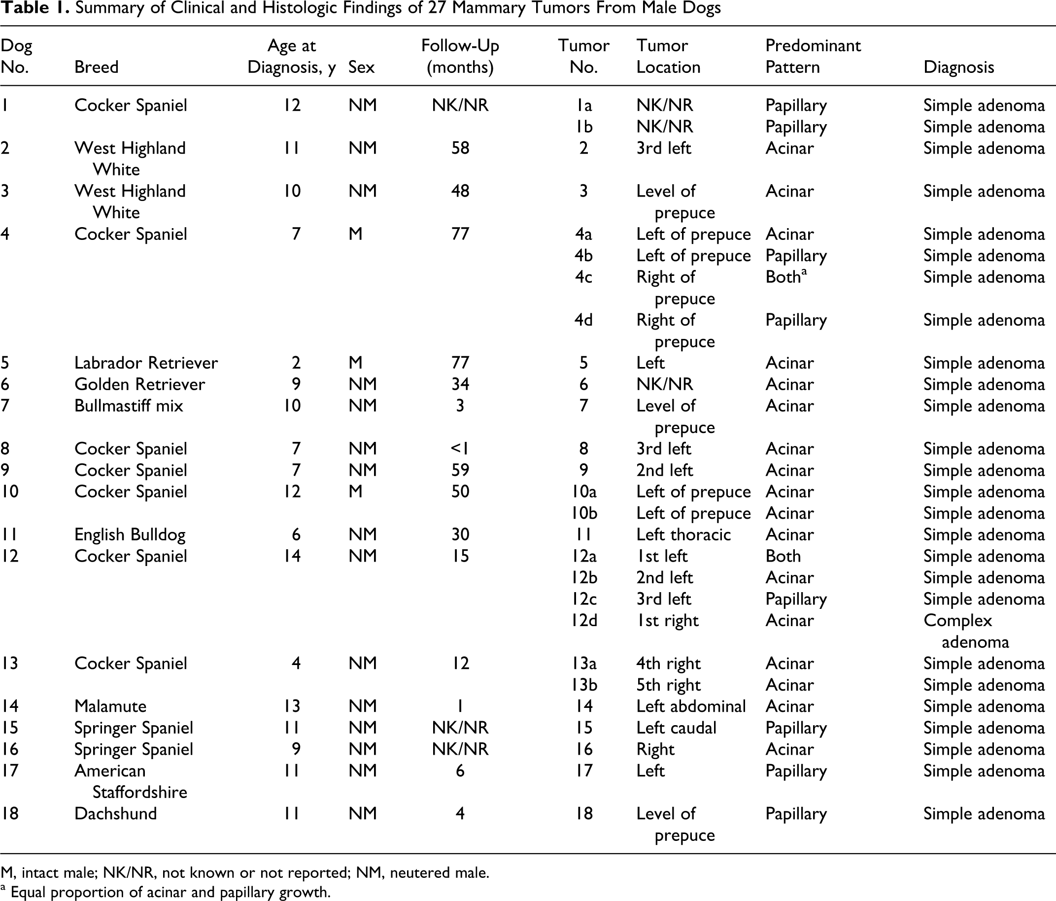
M, intact male; NK/NR, not known or not reported; NM, neutered male.
a Equal proportion of acinar and papillary growth.
Three of the dogs were intact; 15 were neutered. The age at castration could not be accurately determined from the medical records in most cases but ranged from 7 months to 10 years in the cases for which the age was documented. Nine dogs were spaniels (7 Cocker Spaniels, 2 Springer Spaniels), 3 were terriers (2 West Highland White Terriers, 1 American Staffordshire Terrier), and there was 1 each of the following breeds: Labrador Retriever, Golden Retriever, Bullmastiff cross, English Bulldog, Malamute, and Dachshund. All 5 of the dogs that had multiple mammary tumors were Cocker Spaniels. The average age of the dogs at diagnosis was 9.2 years (range, 2–14 years).
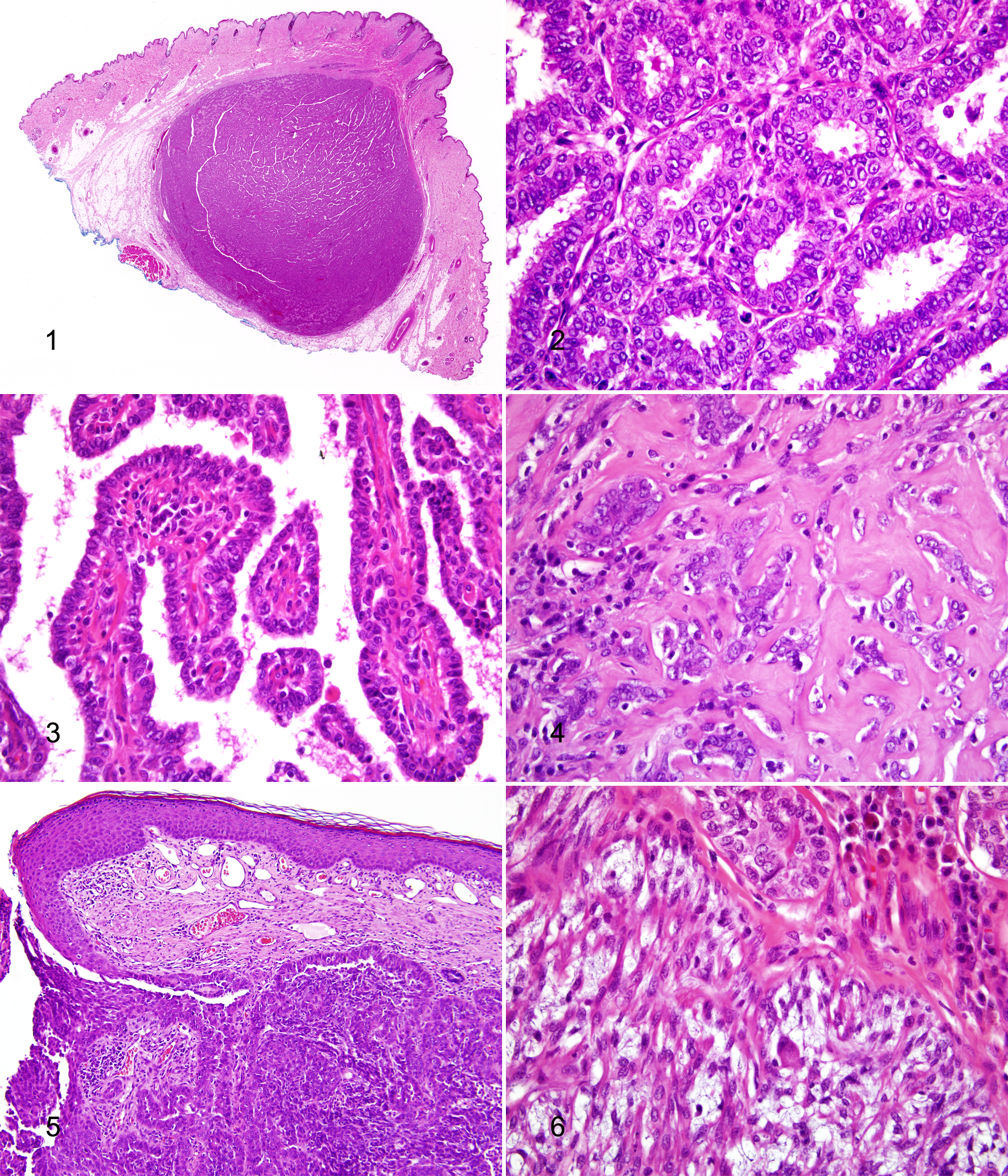
Histologically, all but 1 of the 27 neoplasms were diagnosed as simple adenomas. A single complex mammary adenoma (also known in human medicine as an adenomyoepithelioma) was removed from dog No. 12, which had multiple mammary tumors. Simple adenomas were characterized by single- or multiple well-circumscribed nodules, composed of numerous acinar structures and papillary projections lined by a single or multiple layers of cuboidal to columnar epithelium (Figs. 1–3). Neoplastic cells were arranged in a moderately thick fibrovascular stroma. Mitotic figures averaged one per ten 400× fields. Most (17/26) simple adenomas were arranged predominantly in an acinar pattern (Fig. 2), with small areas of papillary growth, whereas 9 of the 26 tumors had predominantly a papillary pattern (Fig. 3) or roughly equal proportions of acinar and papillary growth. One simple adenoma had a central aggregate of thick, keloidal collagen surrounded by adenomatous growth, consistent with a sclerotic papilloma (Fig. 4). Another simple adenoma had direct transition from the stratified squamous epithelium of the nipple to the cuboidal epithelium of the adenoma, consistent with a nipple adenoma (Fig. 5). Both of these entities are recognized subtypes of simple adenomas in human breast pathology but do not carry any additional prognostic or behavioral significance. The single case of complex adenoma was characterized by acinar adenomatous growth as seen in the simple adenomas, punctuated by several small islands of spindled myoepithelial cells in a myxomatous matrix (Fig. 6).
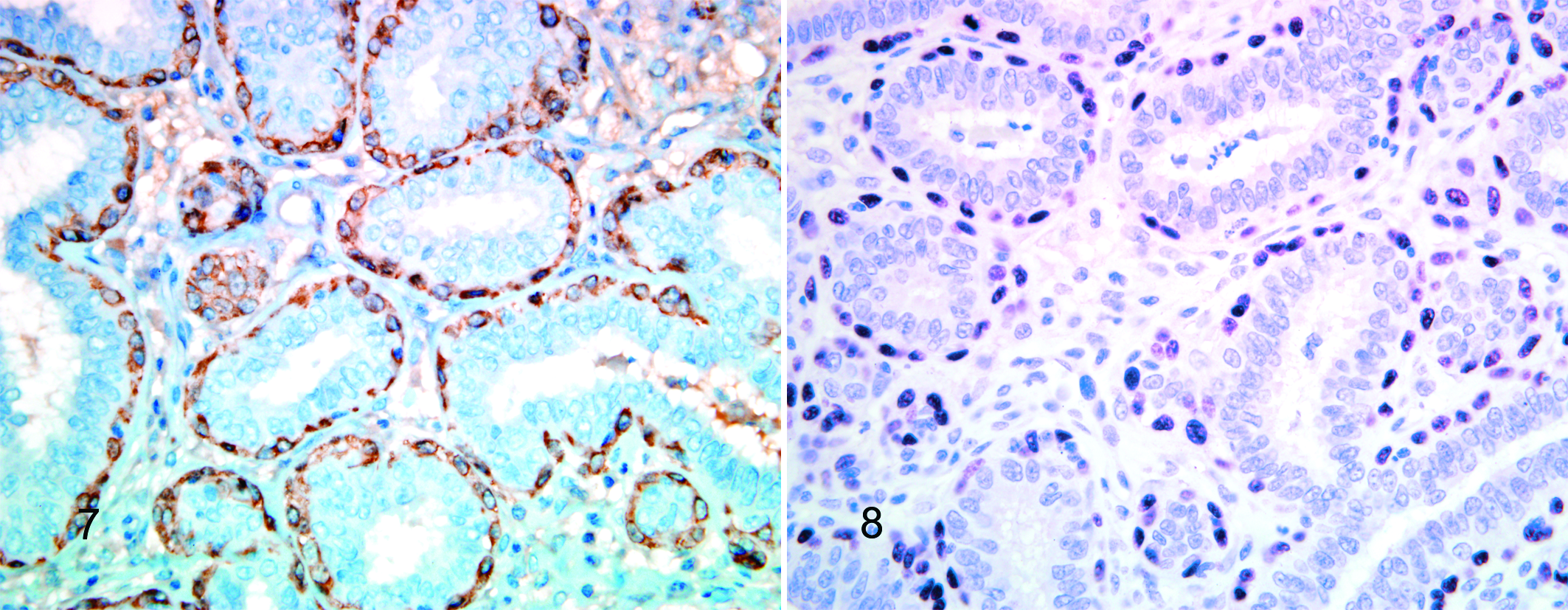
All neoplasms in this study had a continuous layer of spindle-shaped myoepithelial cells immediately subjacent to the epithelial layer lining acini and papillary projections. These myoepithelial cells had strong cytoplasmic immunopositivity for calponin and strong nuclear immunopositivity for p63 (Figs. 7, 8) in 25 of the 25 tumors that had tissue available for immunohistochemical testing. Additionally, the islands of spindle-shaped myoepithelial cells identified in the single complex adenoma were immunopositive for both calponin and p63.
At least some follow-up information was available for 15 of the dogs. The follow-up period ranged from 2 weeks to 77 months (average, 31.9 months; median, 32 months). None of the 14 dogs for which body condition was reported was considered obese. None (0/14) of the dogs had a history or evidence of a testicular tumor. Five dogs were alive at the end of the follow-up period; the other 10 had died or were euthanatized for causes unrelated to the mammary tumors. Necropsy was not performed on any dog. Of the 14 cases with relevant information available, none had evidence of recurrence or metastasis, although 1 dog reportedly had developed “abdominal” masses, 1 of which involved a nipple, 1 year after removal of the initial mammary tumors; these lesions were not biopsied. Surgery was the only reported treatment for the mammary tumors in 15 of the 15 cases. Two dogs were being treated for hypothyroidism; a third was suspected of being hypothyroid; 2 were suspected to have Cushing disease; 2 were treated with cyclosporine for keratoconjunctivitis sicca; 2 were being treated with prednisone for allergies; and 1 was treated with topical steroids for otitis externa.
Discussion
Mammary tumors are common in female dogs, accounting for up to 42% of all tumors in the bitch in one study. 5 Mammary tumors in male dogs are far less common, accounting for between 0 and 2.7% of all mammary tumors in dogs.3,5,9–11,15 Previous reports of mammary neoplasms in male dogs included the following diagnoses: ductal papillary adenoma, carcinoma, adenocarcinoma perithelioma, papillary cystadenocarcinoma, chondroadenocarcinoma, adenocarcinoma, cystic carcinoma, mixed tumor, osteosarcoma, sarcocarcinoma, cystadenochondroma, complex adenoma, simple adenoma, papillary cystadenoma with squamous cell carcinoma, benign mixed tumor, malignant mixed tumor, fibroadenoma, and spindle cell sarcoma.5,7–10,12,13,17
Mammary cancer in men is similarly rare, accounting for less than 1% of all breast carcinomas. 14 Reported mammary neoplasms in men include papilloma, nipple duct adenoma, myofibroblastoma, adenomyoepithelioma, mesenchymoma, and mammary carcinoma. 14 Many mammary neoplasms in men appear be related to a hormonal abnormality, and one study found that approximately 50% of mammary cancer in men were associated with 1 or more of the following risk factors: undescended testes, orchiectomy, testicular injury or inflammation, hormonal therapy, high blood cholesterol, rapid weight gain, obesity, amphetamine use, diabetes, and cigar smoking. 16 Some previous reports of mammary neoplasia in male dogs implicated hormonal abnormalities, particularly in association with testicular neoplasms, as the cause of the tumors.9,12,17 None of the dogs in this study for which detailed history was available had a history of testicular neoplasia or other testicular abnormalities. Additionally, there was no history of obesity, diabetes, or sex hormone therapy.
The prevalence of mammary neoplasia in female dogs has been associated with age and breed.1,7,8,10,13 Breed and age appear to be significant factors in the development of mammary tumors in the male dogs of this study. Nine of 18 were spaniels, and 3 were terrier breeds. This is similar to previous reports of mammary neoplasia in dogs, both male and female, in which Cocker Spaniels, terrier breeds, and poodles appear to be overrepresented.1,7,8,10,13 This is not surprising, given that heredity has been found to play a pivotal role in mammary cancer in humans, where a family history of breast cancer in first-degree relatives is commonly reported. 16 The average age of onset for the tumors in our study was 9.2 years, which is within the range of age of onset reported in most female dogs (6–10 years).1,7,10
Like mammary tumors in female dogs, most of the tumors for which location was specified occurred in the third through the fifth sets of glands. This predilection for the caudal mammary glands has been attributed to the greater mass of mammary tissue in these glands as well as greater potential for injury attributable to the more pendulous nature of these glands.7,10,13 The higher prevalence of mammary tumors in the caudal glands in male dogs may also be attributable to increased mass of mammary tissue in which a tumor might arise, although male mammary tissue is composed of only ductal elements and is devoid of fully developed terminal duct lobular units. 14
All tumors in this study were well circumscribed and had benign histologic features. Demonstration of a continuous layer of myoepithelium between the neoplastic mammary epithelium and the stroma indicates that the neoplastic epithelium has not invaded the stroma and, thus, correlates with benignancy.2,14 Myoepithelial cells can be difficult to identify in HE-stained sections; therefore, immunohistochemical markers for calponin and p63 were used to demonstrate their presence. Calponin is a smooth muscle-specific protein that has been shown to be a sensitive marker of normal and neoplastic myoepithelium in the canine mammary gland; however, other cell types (stromal myofibroblasts and chondroblasts) can also stain positively. 4 P63 is a recently characterized p53 homologue that is consistently expressed in canine myoepithelial cells and the basal layer of multilayered epithelium and does not stain other cell types in the canine mammary stroma. 6 Despite the size and dense cellularity of some of the tumors in the current series, a continuous layer of myoepithelium around acinar and papillary structures in all neoplasms was demonstrated with calponin and p63 immunohistochemistry, supporting the classification of benignancy in all cases. The benign nature of these tumors was corroborated by the lack of recurrence, metastasis, or tumor-related deaths.
This is in contrast with previously reported mammary gland neoplasms in male dogs, for which 28 of 51 were classified as malignant.5,7–10,13,17 This discrepancy may be attributed to differences in criteria for malignancy. With the exception of a sarcocarcinoma with metastasis to regional lymph nodes and lung 7 and a cystic carcinoma with metastasis to axillary lymph nodes, 8 the clinical and/or histological characteristics that were used to differentiate the reported malignancies from their benign counterparts were not defined. Another potential reason for this discrepancy may be a difference in the reproductive, and thereby hormonal, status of the reported population. Most of the dogs in our study (15/18) were neutered; however, the reproductive status of most dogs in the previous reports was not indicated, so comparison of this factor is not possible. Whether the relatively large number of cases in this report represents an increase in incidence of mammary neoplasia in male dogs or is simply a function of increased recognition and reporting cannot be determined.
The differential diagnosis for benign glandular proliferations of the mammary gland includes adenomas and hyperplasia. The distinction between adenoma and hyperplasia can be subjective. For the purpose of this study, the following criteria were used based on one of the authors’ (D. C.) experience in human breast pathology: adenomas are clinically evident, discrete, grossly visible (>4 mm diameter), and often contained in a larger lactiferous duct, whereas hyperplasia is not grossly or clinically evident (usually <1 mm diameter) and is present along existing smaller ducts.
In summary, mammary neoplasia in male dogs is rare. In the current series, middle-aged, intact, and neutered male Cocker Spaniels appear to be predisposed. Almost all tumors were simple adenomas, with variation in predominant growth pattern. Only 1 tumor was classified as a complex adenoma. All tumors were histologically benign without evidence of recurrence or metastasis. Despite the absence of complete histories in some cases, there does not appear to be a strong association between the development of mammary neoplasms in male dogs and testicular disease, obesity, or other previously implicated hormonal influences. Future studies on mammary neoplasia in male dogs could examine the role of hormone receptors, specifically progesterone and estrogen receptors, in the development of these tumors, as well as possible correlations between castration and the incidence and types of mammary neoplasia in male dogs.
Footnotes
Acknowledgements
We thank all the submitting veterinarians and owners for their willingness to contribute to the study. We thank Dr. T. P. Lipscomb for providing cases and reviewing the manuscript. Ms. L. Zimmerman of Marshfield Laboratories, Marshfield, Wisconsin, provided invaluable logistic support. J. Bearss is a Major in the US Army. The opinions and assertions contained herein are the private views of the authors and are not to be construed as official or as reflecting the views of the Department of the Army or Department of Defense.
The authors declared that they had no conflicts of interest with respect to their authorship or the publication of this article.
This article was supported in part by the American Registry of Pathology.