
Abstract
Canine liposarcoma is classified as well differentiated (WDL), dedifferentiated (DDL), myxoid (ML), and pleomorphic (PL). Overexpression of the protooncogene MDM2 has been reported in WDL and DDL, but little is known regarding the role of p53 in their tumorigenesis. The aim of this study was to assess p53 expression in canine liposarcoma and compare it with subtype, grade, mitotic count (MC), Ki67 labeling index (LI), and MDM2 expression. Forty-seven cases were included (13 WDL, 3 DDL, 7 ML, and 24 PL); 17 were MDM2-positive (13 WDL, 3DDL, and 1ML). Five were p53-positive (4 ML and 1 WDL) but DDL and PL were consistently negative. p53 expression correlated with higher Ki67-LI, higher MC, and myxoid histotype. No correlation was found with grade and MDM2 expression. Based on these results canine liposarcoma seems to embody a group of neoplasms whose subtypes, especially ML, may represent distinct diseases rather than morphological variants of the same entity.
Canine liposarcoma is an uncommon soft tissue sarcoma (STS) arising most frequently in the subcutis. 3,4,8 Four histological subtypes are recognized: well-differentiated liposarcoma (WDL), dedifferentiated liposarcoma (DDL), myxoid liposarcoma (ML), and pleomorphic liposarcoma (PL), 3,8 paralleling the classification used in human medicine. 6 In veterinary medicine, the diagnosis of liposarcoma is based mainly on histomorphologic features, and specific immunohistochemical markers are lacking. 3 On the contrary, in human medicine, the diagnosis relies on the combination of histologic aspects and recognition of specific cytogenetic anomalies, which allow grouping of the 4 histological variants into 3 distinct biological entities. The first includes WDL and DDL, representing the 2 ends of the morphologic and biological spectrum of a single entity characterized by the amplification of genes encoding MDM2 and CDK4. The second is ML, a type of liposarcoma consistently displaying a reciprocal translocation between chromosomes 12 and 16: t(12;16)(q13;11). The third is PL, the least common type that exhibits complex structural genetic rearrangements. In PL, dysregulation of several tumor suppressor pathways (such as p53 and Rb1) typically occurs. 6 While some similarities have been reported between canine and human liposarcoma, such as MDM2 protein overexpression in WDL and DDL, 3 as well as some differences, such as the lack of association between CDK4 expression and histotype in canine liposarcoma, 3 little is known regarding the role of p53 in canine liposarcoma and STS tumorigenesis in general. 9
The aim of this study was to assess by immunohistochemistry the expression of p53 in canine liposarcomas for which MDM2 expression and Ki67 labeling index were previously evaluated. 3
Of the original 53 tumors of the previous caseload, 47 cases for which sufficient material was available were included in this study. Histotype, grade, 5 mitotic count, MDM2 expression, and Ki67 labeling index were retrieved. 2,3 Histotype was determined following previously published criteria (Supplemental Materials). 2,3
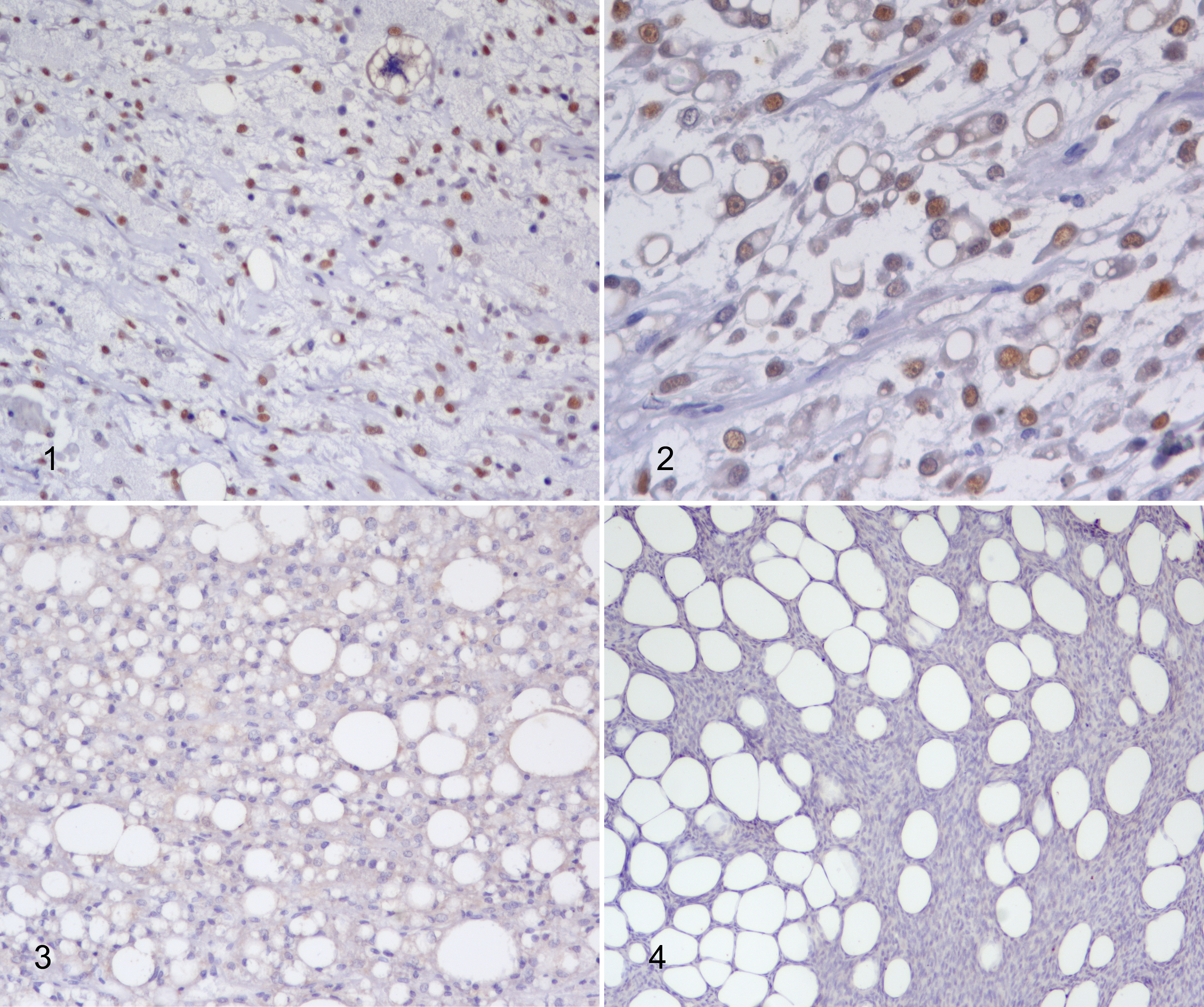
p53 expression was evaluated by immunohistochemistry on 3-μm-thick sections that were dewaxed and rehydrated. Endogenous peroxidase was blocked by immersion in H2O2 3% in methanol for 30 minutes. For antigen retrieval, sections were immersed in citrate buffer (pH 6.0), heated in a microwave oven at 750 W for two 5-minute cycles, and cooled at room temperature for 20 minutes. Incubation with primary antibody (mouse monoclonal, clone PAb 240, dilution 1:100, BD Bioscience) was performed overnight. The clone PAb 240 utilized in this report was raised against human p53 protein to detect wild type and mutated p53. This and several other PAb antibody clones have been characterized and demonstrated to cross-react with normal and mutated canine p53 protein by means of cell vector expression, tissue arrays, western blot, and immunocytochemistry of UVB-irradiated cultured canine keratinocytes for assessment of endogenous p53 overexpression. 1,7 The reaction was amplified by the avidin-biotin method (ABC kit elite, Vector, Burlingame, CA) and visualized with 3,3′-diaminobenzidine (0.04% for 4 minutes). Sections were counterstained with Papanicolaou’s hematoxylin, rinsed in tap water, dehydrated, and coverslipped. Sections of canine mammary carcinoma in which the expression of p53 was known were used as positive controls. Negative controls comprised slides incubated with omission of the primary antibody. Expression of p53 was scored semiquantitatively and considered positive if more than 5% of neoplastic cells showed nuclear staining, following the criteria reported for human liposarcoma. 10 Ki67 expression was evaluated as the labeling index and defined as the percentage of Ki67 positive cells. 3
Data were analyzed with the Shapiro and Wilk’s W test for normality. Because data were not normally distributed, Mann-Whitney test was used for continuous data and χ2 test for categorical variables using GraphPad 8.3.1 software. A conventional 5% level was used to define statistical significance.
Thirteen cases were WDL, 3 were DDL, 7 were ML, and 24 were PL; 15 were grade I, 26 were grade II, and 6 were grade III. Seventeen cases were MDM2-positive (13/13 WDL, 3/3 DDL, 1/7 ML, and 0/24 PL). Median Ki67 labeling index was 15.3% (range 1.1% to 75.1%). Median mitotic count was 5 (range 1–56). p53 expression was present in 5 cases (11%), including 4 ML (57% of the ML), and 1 WDL (8% of WDL; Figs. 1-3). DDL and PL were consistently p53-negative (Fig. 4). Expression of p53 was correlated with a higher Ki67 labeling index (Mann-Whitney test; P = .003), a higher mitotic count (Mann-Whitney test; P = .002), and myxoid histotype (χ2 test; P = .0002). No significant correlation was found with histologic grade (χ2 test; P = .1544) and MDM2 expression (χ2 test; P = .85).
Canine liposarcoma is a rare STS for which few studies are available. Recently, knowledge of this neoplasm has been expanded in dogs by the description of DDL, demonstration of MDM2 expression in a subset of cases, 3 identification of the expression of markers of myoid differentiation, 8 demonstration of a higher proliferative activity of ML and DDL, and identification of cases of liposarcomas expressing PDGFRβ. 2,3
Liposarcoma is the most common STS in humans, and the genetic anomalies correlating to the different LPS variant have been well characterized: they include MDM2 and CDK4 gene amplifications in WDL/DDL, chromosome translocations in ML, and varying genetic anomalies often involving p53 and Rb1 in PL. 6 Little is known about p53 anomalies in canine liposarcoma.
This report describes the expression of p53 assessed by immunohistochemistry with a monoclonal antibody recognizing both wild type and several mutated forms of p53 known to cross-react with canine tissue. 1,7 Based on the principle that mutations lead to a greater stability of p53, thus to an easier detection by immunohistochemistry, a cutoff of 5% of positive neoplastic cells was applied, a cutoff established for human liposarcoma. 10
In our caseload, only 5 out of 47 cases were p53 positive, and unexpectedly, all PL were negative. The majority of positive cases (4/5) were ML and represented more than 50% of ML included in the study. The correlation with the histotype was statistically significant even though p53 expression was not present in all ML tested. This result, taken together with the previously documented differential expression of MDM2 in canine liposarcoma subtypes, 3 leads to a scenario where different phenotypes exist among canine liposarcomas including MDM2+/p53− (WDL/DDL), MDM2-/p53+ (ML), and a last group of MDM2−/p53− (PL). This phenotypic variability parallels, on one side, what is reported in humans, where liposarcomas are divided into 4 histological variants but 3 distinct entities. 6 On the other side, the expression of p53 by canine ML differs from human ML, suggesting that despite being histologically similar, they may bear different genetic alterations. Further studies evaluating the mutational status of p53 in canine liposarcomas may clarify if any specific mutations are present in this subtype and if immunohistochemically p53-negative ML carries abnormalities in the gene sequence.
Furthermore, p53 expression was associated with higher proliferative activity, assessed both by mitotic count and Ki67 labeling index. This result may be a secondary effect of the correlation between p53 expression and histotype, as ML is a variant with high proliferative activity. 3
In summary, based on these results and previously published studies, 3 canine liposarcoma includes adipocytic tumors whose subtypes, especially ML, may represent distinct diseases rather than mere morphological variants of the same neoplasm. Studies evaluating genetic anomalies in canine liposarcomas are needed to confirm this hypothesis.
Supplemental Material
Supplemental Material, Combined_supplemental_materials-Avallone_et_al - p53 Expression in Canine Liposarcoma Correlates With Myxoid Variant and Higher Proliferative Activity
Supplemental Material, Combined_supplemental_materials-Avallone_et_al for p53 Expression in Canine Liposarcoma Correlates With Myxoid Variant and Higher Proliferative Activity by Giancarlo Avallone, Luisa V. Muscatello, Alessandro Leoni, Paola Roccabianca, Elvio Lepri, Luca Crippa and Barbara Bacci in Veterinary Pathology
Footnotes
Declaration of Conflicting Interests
The author(s) declared no potential conflicts of interest with respect to the research, authorship, and/or publication of this article.
Funding
The author(s) received no financial support for the research, authorship, and/or publication of this article.
Supplemental Material
Supplemental material for this article is available online.
References
Supplementary Material
Please find the following supplemental material available below.
For Open Access articles published under a Creative Commons License, all supplemental material carries the same license as the article it is associated with.
For non-Open Access articles published, all supplemental material carries a non-exclusive license, and permission requests for re-use of supplemental material or any part of supplemental material shall be sent directly to the copyright owner as specified in the copyright notice associated with the article.