
Abstract
Canine liposarcoma is an uncommon soft tissue sarcoma usually arising in the subcutis. While liposarcoma classification in dogs is based solely on histology, in humans it depends on the detection of genetic abnormalities that can lead to specific protein overexpression. This study is an immunohistochemical evaluation of MDM2 and CDK4 expression in canine liposarcoma designed to assess the correlation of these proteins with histologic type, grade, mitotic index and Ki67 labeling index and evaluate their utility in improving tumor classification. Fifty-three liposarcomas were retrospectively collected: 24 were well differentiated liposarcomas (WDL), 16 of which expressed MDM2 and 21 CDK4; 7 were myxoid liposarcomas (ML), 1 of which expressed MDM2 and 5 expressed CDK4; 18 were pleomorphic liposarcomas (PL), all were MDM2 negative and 12 expressed CDK4. Four tumors were morphologically consistent with dedifferentiated liposarcoma (DDL) a subtype described only in humans: 3 expressed MDM2 and 4 expressed CDK4. MDM2 expression correlated with histotype (highly expressed in WDL and DDL) and grade (highly expressed in grade 1 tumors). Histotype correlated with the Ki67 labeling index (lowest in WDL and highest in DDL). A revised classification, considering MDM2 expression, allowed 8 WDL to be reclassified as PL and correlated significantly with mitotic and Ki67 labeling index (both significantly lower in WDL and progressively higher in ML and DDL). These results partially parallel data reported for human liposarcomas, suggesting that WDL and DDL are distinct neoplastic entities characterized by MDM2 expression, which may represent a useful diagnostic and potentially prognostic marker for canine liposarcoma.
Canine liposarcoma is an uncommon soft tissue sarcoma (STS) that most commonly arises in the subcutis with a 28% recurrence rate and low metastatic potential. 2,6,9 –11 The World Health Organization (WHO) classifies canine liposarcomas into 3 histological types: well differentiated (WDL), pleomorphic (PL), and myxoid (ML). 11 WDL is composed of round to polygonal cells arranged in solid sheets with little or no stromal collagen production, 9,10 in which the majority of cells contain easily recognizable intracytoplasmic lipid droplets. 10,11 The percentage of mature neoplastic cells containing a single large lipid vacuole and resembling normal adipocytes varies from a moderate number 9 to the majority of cells. 11 ML is characterized by abundant background mucin resembling myxosarcoma, from which it is differentiated by the presence of cytoplasmic lipid-laden vacuoles within the cytoplasm of the neoplastic cells. 9 –11 ML frequently present a delicate anastomosing capillary vasculature. 10 PL is characterized by severe cellular pleomorphism, variable numbers of multinucleated neoplastic cells, and scarce to absent intracellular lipids. 2,9 –11 In addition, atypical lipoma has been included in the list of canine liposarcoma types, and represents a lipocytic neoplasm characterized by low malignancy, and histological features resembling lipoma containing scattered atypical lipoblasts. 10
Human liposarcoma is the most common STS, usually developing in the extremities and in the retroperitoneum, and its biological behavior varies among the different histotypes. 7,8 Its classification is based on histological appearance and immunohistochemical and cytogenetic evaluation including: atypical lipomatous tumor/well differentiated liposarcoma (ALT/WDL), dedifferentiated liposarcoma (DDL), ML liposarcoma, and PL liposarcoma. 7,8 ALT/WDL and DDL are characterized by the same specific genetic abnormalities, consisting of a giant ring chromosome and amplification of the genes encoding for MDM2 and CDK4. 5,7,8 Bearing the same genetic abnormalities, ALT/WDL and DDL are regarded as the 2 extremities of a morphological spectrum characterizing the same biological entity. 7,8 While ALT/WDL are histologically similar to canine atypical lipoma and WDL, DDL is defined as ALT/WDL juxtaposed to areas of nonlipogenic sarcoma usually resembling a fibrosarcoma or undifferentiated pleomorphic sarcoma. 7,8 Fluorescence in situ hybridization (FISH) for the detection of MDM2 and CDK4 gene amplifications or immunohistochemical assessment of MDM2 and CDK4 expression are highly diagnostic for ALT/WDL and DDL since these anomalies are not present in the other subtypes of liposarcoma. 3,8,20,21 Likewise, ML in humans, which is morphologically similar to canine ML, is characterized by a specific genetic anomaly consisting in a reciprocal translocation, t(12;16)(q13.3;p11.2), with fusion of the DDIT3 (CHOP) and FUS gene. 7,8
The aims of this study were to evaluate the immunohistochemical expression of MDM2 and CDK4 in canine liposarcoma; to assess their association with the histotype, grade, mitotic index (MI) and Ki67 labeling index and how the expression of these proteins could improve histological classification of liposarcoma as in man.
Materials and Methods
The histological archives of the diagnostic services of 3 academic institutions and 1 private laboratory were searched for cases diagnosed as liposarcoma in dogs between 2001 and 2012. Hematoxylin and eosin stained sections were prepared from the original blocks and cases were classified histologically.
Cases with intermediate morphology between WDL and PL were classified as PL based on the presence of atypical and/or multinucleated neoplastic cells regardless of the number of lipid vacuoles.
A revised classification scheme was also applied, and included the expression of MDM2 and CDK4 as diagnostic criteria indicative of WDL and DDL, paralleling the features reported in human liposarcoma. 7,8 For each case, the grade was calculated according to the guidelines reported in the literature, 6 and based on the degree of differentiation, mitotic index and percentage of necrosis within the tumor. 6 Mitotic index was calculated as absolute number of mitoses counted in 10 continuous high power fields (40×) from the most cellular part of the tumor as previously reported. 6
Immunohistochemistry
Three micrometer thick sections were dewaxed and rehydrated. Endogenous peroxidase was blocked by immersion in 3% H2O2 in methanol for 30 minutes. Sections were then rinsed in Tris buffered saline (pH 7.0). For antigen retrieval, sections were immersed in citrate buffer (pH 6.0), heated in a microwave oven at 750 W for 2 cycles of 5 minutes each (for MDM2 and CDK4) and 4 cycles of 5 minutes (for Ki67), and cooled at room temperature for approximately 20 minutes. CDK4 (Rabbit polyclonal, dilution 1:200, Santa Cruz Biotech, Santa Cruz, CA, USA), MDM2 (mouse monoclonal, clone 2A10, dilution 1:100, Abcam, Cambridge, UK) and Ki67 (Mouse monoclonal, clone MIB-1, dilution 1:600, Dako, Glostrup, Denmark) specific antibodies were applied overnight at 4°C. Sections were incubated for 30 minutes at room temperature with the appropriate biotin-conjugated secondary antibody (dilution 1:200, Dako, Glostrup, Denmark). The reaction was amplified with an avidin-biotin method (ABC kit elite, Vector, Burlingame, CA, USA) and visualized with 3,3′-diaminobenzidine (0.04% for 4 minutes). 1 Sections were counterstained with Papanicolaou’s stain, rinsed in tap water, dehydrated and coverslipped. Sections of normal canine testis were used as positive controls for all the antibodies, and hyperplastic hepatoid gland specimens were also utilized for MDM2 expression. Negative controls comprised slides incubated with a nonspecific antibody or by omission of the primary antibody. Cases were considered positive for MDM2 and CDK4 when at least 1 nucleus was positive per each high power field (40×), as reported in the literature. 3 The count of Ki67 positive cells was performed in 10 high power fields (40×) counting at least 1000 cells for each case, using the manual count tool of ImageJ 1.48 analysis software. Ki67 expression was evaluated as the labeling index and was defined as the percentage of Ki67 positive cells.
Statistical Analysis
Data were analyzed with the Shapiro and Wilk’s W test for normality. Because data did not
fit a normal distribution, the Spearman test was used to compare MDM2 and CDK4 expression among tumor subtypes, comparing the group of WDL plus DDL
versus PL or ML (based on the histological classification) MI, grade, or Ki67 labeling index versus MDM2 expression, CDK4 expression, and
tumor subtype (based on both the histological classification and the revised
classification)
A conventional 5% level was used to define statistical significance.
Results
A total of 53 canine liposarcomas in 53 dogs were collected. Liposarcoma was diagnosed mainly in crossbreed dogs (15 cases) and retrievers (14 cases). Breed was not reported in 4 cases. The remaining 20 dogs were purebred and included German Shepherd (3 cases), Rottweiler (3 cases), Dachshund (2 cases), Beagle (2 cases), Fox Terrier (2 cases), Maremma Sheepdog (2cases), Pointer (2 cases), Great Dane (1 case), Poodle (1 case), Belgian Shepherd (1 case), French Bulldog (1 case). Fifteen cases were female and 37 male, female to male ratio was 0.41. Sex was not reported in 1 case. Age was collected in 51/53 cases and ranged between 4 and 16 years (mean 10.7, median 10). The majority of liposarcomas were located in the subcutis (46/53 cases), 39 of which were located in the trunk and proximal limbs, 5 in the distal limbs, 1 on the tail and 1 on the face. Three tumors were primary splenic, 1 was located in the spinal canal (intraspinal and extradural), and 3 were intramuscular (1 in the thorax and 2 in perianal region).
Histology identified 24 WDL (Figs. 1 and 2), 7 ML (Figs. 3 and 4), and 18 PL (Figs. 5 and 6). Four cases did not fit any category reported by the WHO classification of domestic animal tumors. These cases were densely cellular and were composed of interlacing bundles of spindle cells closely resembling fibrosarcoma (Fig. 7). Neoplastic cells had a small amount of eosinophilic cytoplasm and indistinct cell borders. Nuclei were central, oval to elongated, with finely granular to dense chromatin and absent or poorly evident nucleolus. Cells were intermixed with a minimal amount of thin collagen fibers. In all cases, adipocytic differentiation was disclosed in small multifocal areas accounting for 5 to 25% of the tumors (Fig 7). These areas were characterized by cells containing a single, sharply demarcated, optically empty vacuole displacing the nucleus at the periphery of the cell, and resembling well differentiated adipocytes. A small number of cells, with multiple optically empty vacuoles scalloping the hyperchromatic nucleus (lipoblasts) (Fig. 8) were consistently evidenced and supported the adipocytic origin of the neoplasms. 7 These features were consistent with DDL morphology as described in human medicine 7 and the cases were therefore classified as DDL.
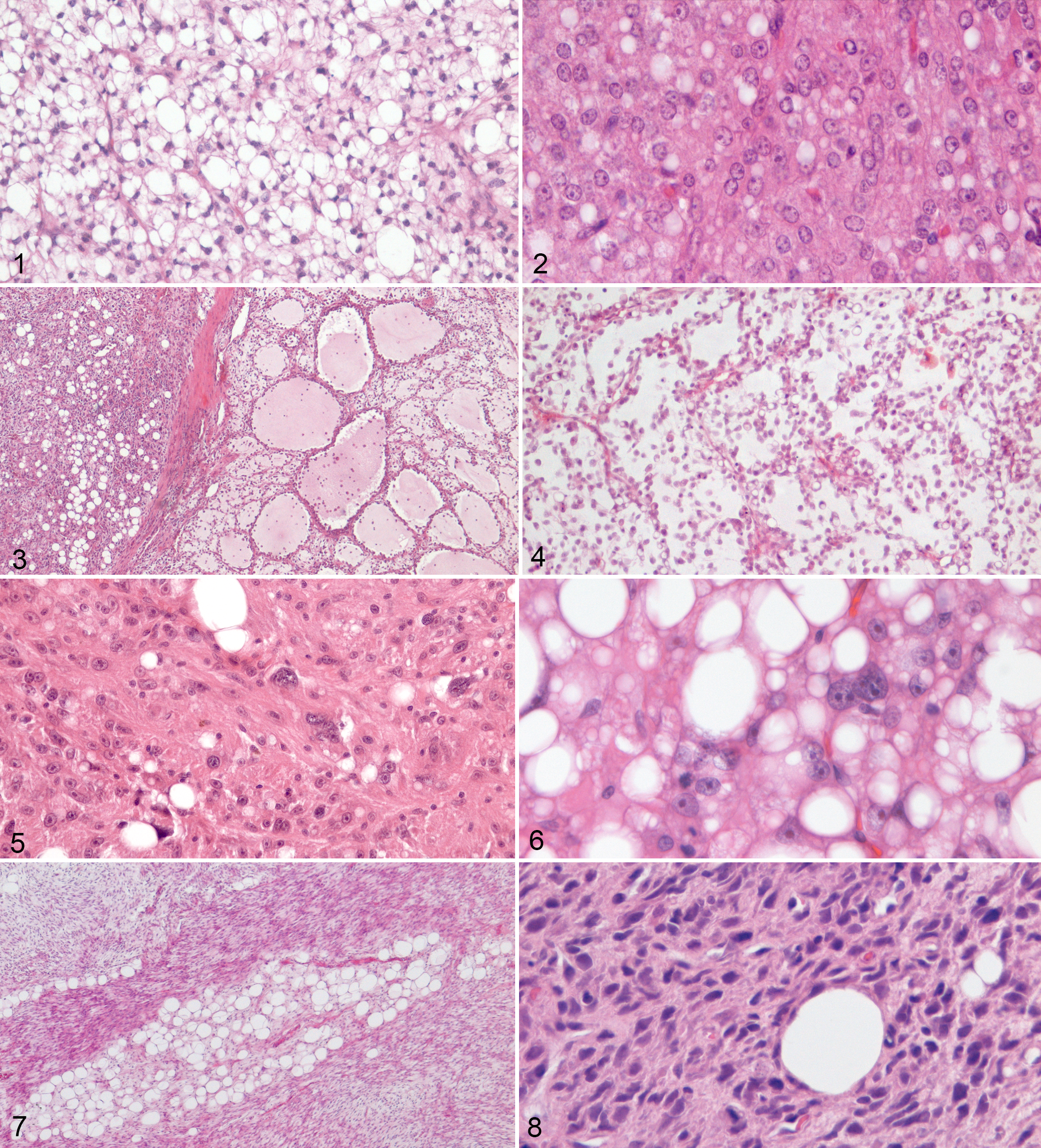
Liposarcomas, dog. Histological features of canine liposarcoma subtypes. Hematoxylin
and eosin stain.
Nineteen cases were grade 1, 27 were grade 2 and 7 cases were grade 3. Mitotic index ranged from 1 to 56 (mean 7.7, median 5). Assessment of Ki67 was not possible in 3/53 cases because of insufficient material in the original blocks and it was therefore evaluated in 50 out of 53 cases. Ki67 labeling index ranged from 1.1% to 76.8% (mean 20.6%, median 14.9).
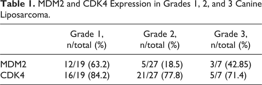
Cross-reactivity of the anti-MDM2 antibody with the canine molecule was assessed by western blot and immunohistochemistry on canine testis and canine hyperplastic hepatoid glands (see supplemental material). MDM2 was expressed in 20 cases, of which 16/24 (66.7%) WDL (Fig. 9 and 10), 3/4 (75%) DDL (Fig. 11 and 12), 1/7 (14.3%) ML, and 0/18 (0.0%) PL. CDK4 was expressed in 42 cases of which 21/24 (87.5%) WDL (Fig. 13), 4/4 (100%) DDL (Fig. 14), 5/7 (71.4%) ML (Fig. 15), and 12/18 (66.7%) PL (Fig. 16). Concurrent expression of MDM2 and CDK4 was identified in 17 cases, 14 of which were WDL and 3 DDL. MDM2 expression was significantly correlated to histological subtypes (R = .66, P < .01), being expressed more commonly in WDL and DDL than in ML and PL (Fig. 17), and with histological grade (R = –.29, P = .03), being expressed more commonly in grade 1 tumors. A statistically significant correlation between CDK4 expression and histotype or grade was not observed (Fig. 18) (Table 1).
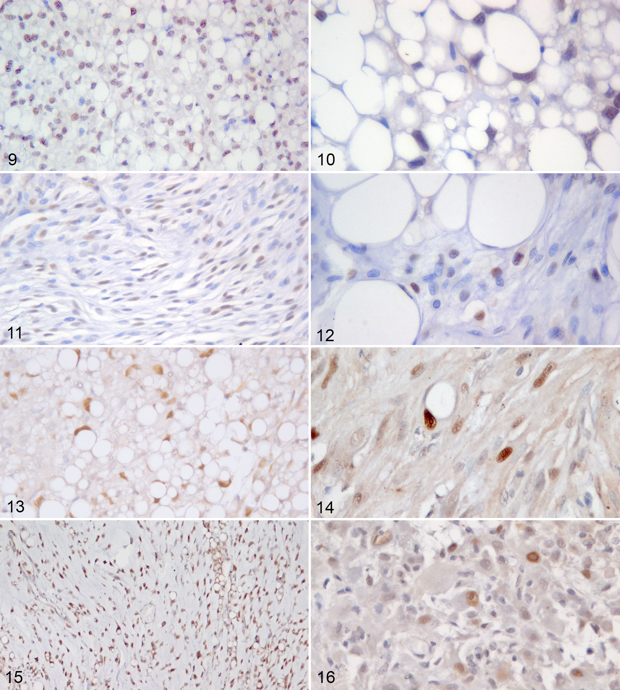
Liposarcomas, dog, subcutis. Immunohistochemistry for MDM2 and CDK4.
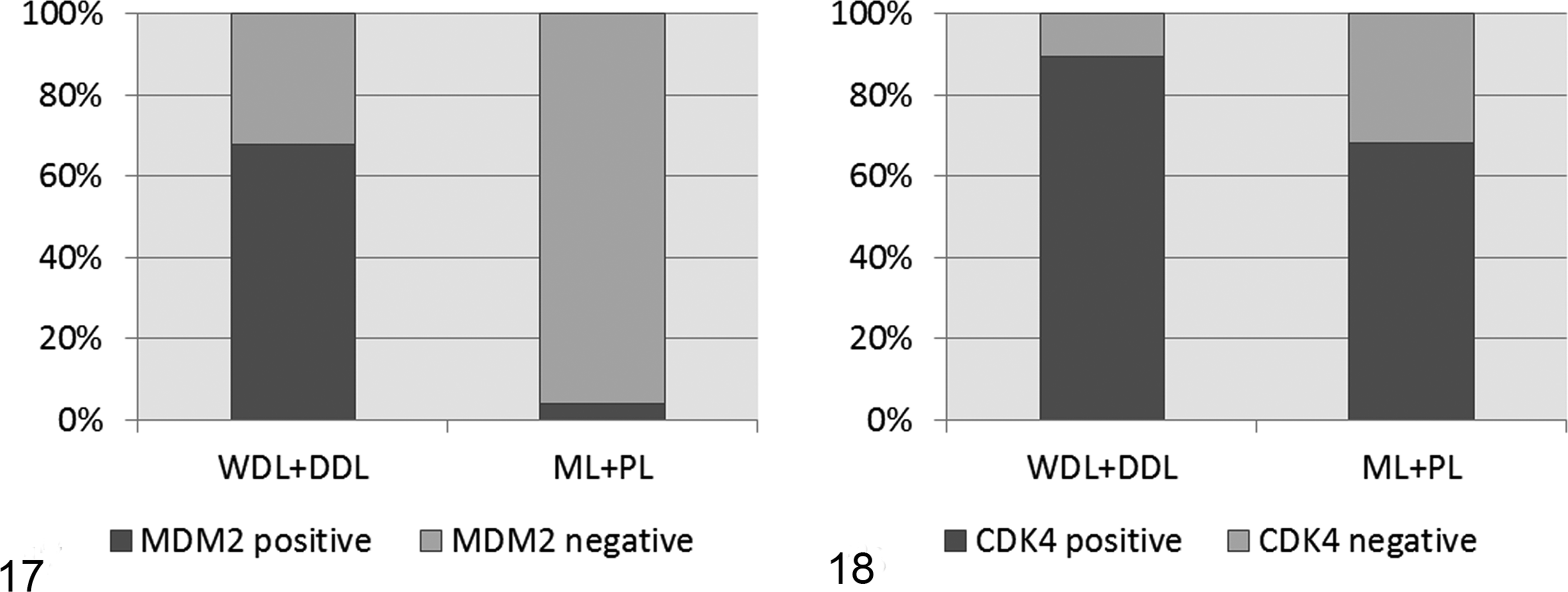
Histograms showing the expression of MDM2 (Fig. 17) and CDK4 (Fig. 18) in well differentiated (WDL) and dedifferentiated (DDL) liposarcoma compared with myxoid (ML) and pleomorphic liposarcoma (PL), diagnosed by histological classification. MDM2 is mainly expressed in the group of WDL and DDL. By contrast, CDK4 expression did not differ significantly between the 2 groups of tumors.
MDM2 and CDK4 Expression in Grades 1, 2, and 3 Canine Liposarcoma.
Therefore, only MDM2 expression was included in a proposed revised classification as an additional diagnostic criterion to differentiate WDL from PL in cases with ambiguous histological appearance. Based on this revised classification, 8 cases, previously diagnosed as WDL and for which cytological atypia was considered insufficient for the diagnosis of PL, were reclassified as PL because of the lack of MDM2 expression, while ML and DDL were not reclassified on these grounds. A total of 16 WDL, 26 PL, 7 ML, and 4 DDL were diagnosed with the revised classification (Table 2).
Percentage of Grades 1, 2, and 3 Cases in Each Subtype of Canine Liposarcoma.
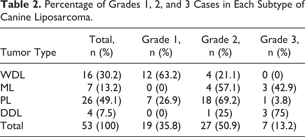
The majority of WDL were grade 1 (67% and 75% based on histological classification and revised classification, respectively). Grade 3 WDL were not identified. The majority of PL were grade 2 (78% and 69% based on histological and revised classification, respectively). All the grades were represented in the group of PL both by the histological and the revised classification. ML were grades 2 and 3 only (57% and 43%, respectively). DDL were grades 2 and 3 only (25% and 75%, respectively). Grade distribution in the different subtypes of canine liposarcoma is summarized in Table 2. No correlation between grade and subtype was found by either histology or the revised classification.
Tumor subtype obtained with the histological classification correlated with Ki67 labeling index (R = .43, P < .01). Tumor subtype obtained with the revised classification correlated with both MI (R = .30, P = .03) (Fig. 19) and Ki67 labeling index (R = .44, P < .01) (Fig. 20). In all the correlation tests, WDL showed the lowest proliferation rate, while an increase was apparent in the DDL (mitotic index, Fig. 19) or in the ML (Ki67 index, Fig. 20).
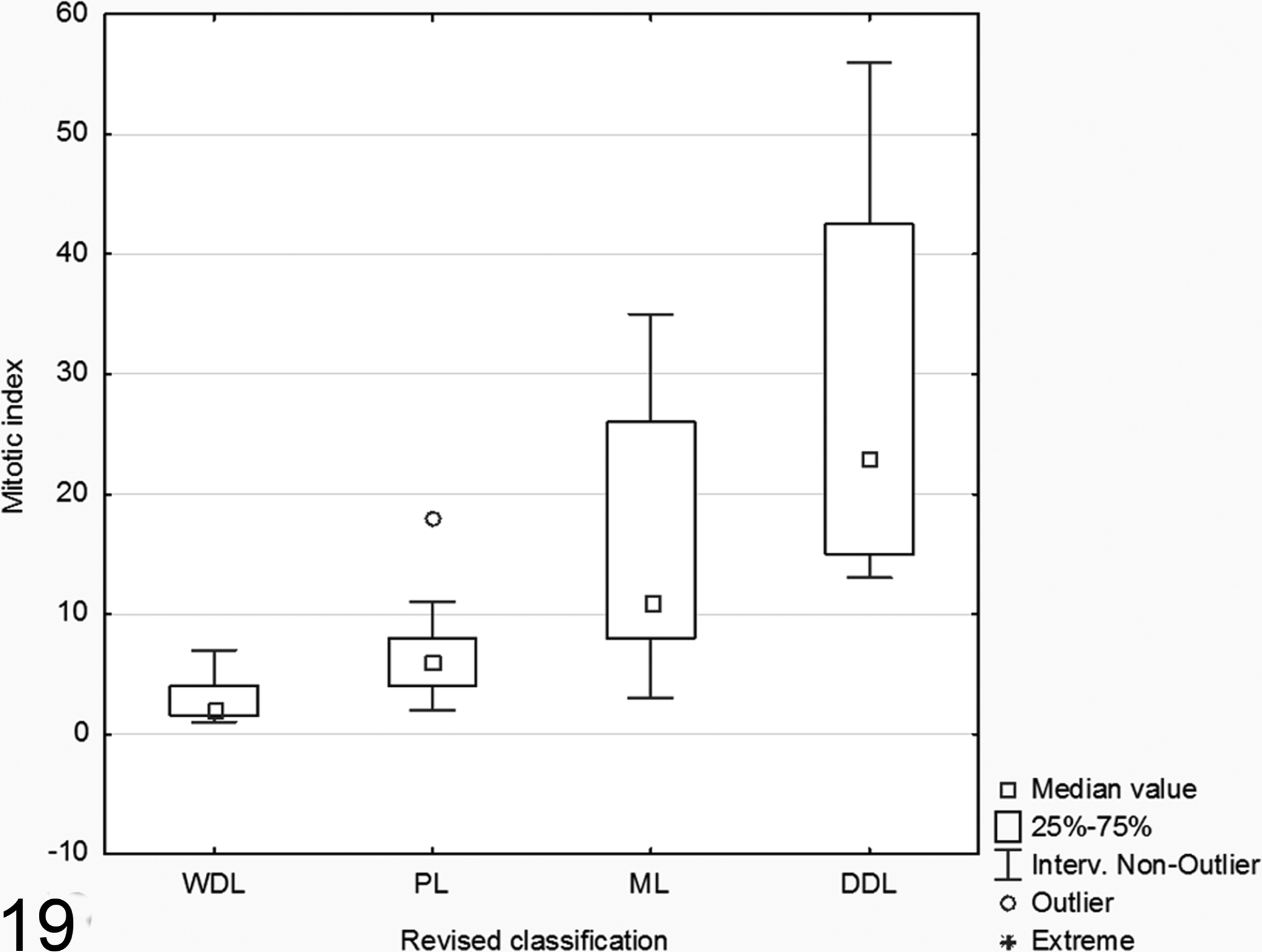
Boxplot showing the mitotic index in the different subtypes of liposarcoma evaluated with the revised classification. The mitotic index progressively increases from well differentiated to dedifferentiated liposarcoma. DDL, dedifferentiated liposarcoma; PL, pleomorphic liposarcoma; WDL, well differentiated liposarcoma; ML, myxoid liposarcoma.
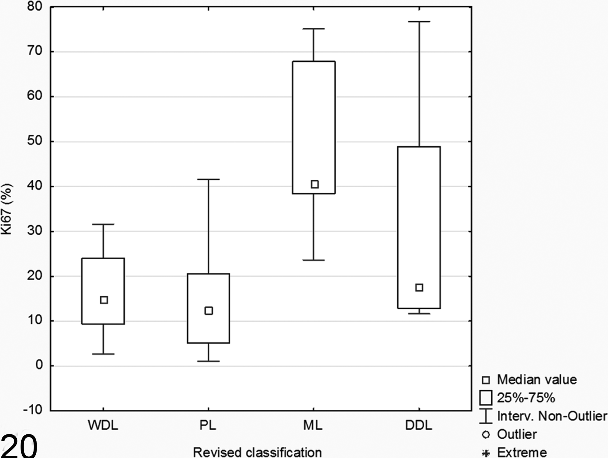
Boxplot showing the Ki67 labeling index (Ki67%) in the different subtypes of liposarcoma evaluated with the revised classification. Myxoid liposarcoma (ML) is the subtype with the highest Ki67 labeling index. DDL, dedifferentiated liposarcoma; PL, pleomorphic liposarcoma; WDL, well differentiated liposarcoma.
Discussion
The majority of canine liposarcomas included in this study were located in the subcutis of the axial region and proximal legs, consistently with literature reports in the dog 2,9 –11 and differently from humans, in which liposarcoma is mainly retroperitoneal and deep-seated. 7,8 Overrepresented breeds in our caseload included crossbreeds and retrievers (Labrador and Golden Retrievers). This is in contrast to the literature that reports a predisposition of Shetland Sheepdogs and Beagles. 2,9 –11 This discrepancy may be related to the popularity of retrievers in Italy.
Based on the histological classification, the majority of liposarcomas were diagnosed as WDL and PL, while a minority of cases was ML. Hibernoma was ruled out because of the absence of the typical round or polygonal cells with moderate amounts of granular or multivacuolated, pale to deeply eosinophilic cytoplasm. 17 The low percentage of ML identified in our study is consistent with previous reports. 9 –11 By contrast, there are discrepancies regarding the occurrence of WDL and PL that are alternately indicated as the most common subtypes by different authors. 2,10
These discrepancies may stem from a partial overlap of the morphology of some cases that does not completely fit the definitions reported in the WHO classification, such as cases with a large amount of lipids (typical of WDL) associated with severe atypia (typical of PL), or cases with a small amount of lipids and an absence of striking cellular atypia. In such cases, we considered the severity of atypia the main histological criterion indicative of PL regardless of the number of lipid vacuoles. The application of this criterion followed the description, for human liposarcoma, of lipid-poor, epithelioid areas in WDL and of lipid-rich areas in PL. 7,8
Interestingly, 4 cases in this caseload fitted the morphological criteria of DDL. 7,8 To the best of our knowledge, this entity has not been previously described in dogs. The spindle and fusiform appearance of neoplastic cells with scarce adipocytic differentiation makes the diagnosis of DDL challenging, especially if only a small sample is examined (eg, core needle biopsies) since DDL can resemble and be misinterpreted as an infiltrative STS containing entrapped adipocytes. Nevertheless, the presence of lipoblasts confirms the adipocytic differentiation of DDL and is one of the major diagnostic morphologic criteria for DDL in accordance with the human literature. 7,8 Interestingly, DDL identified in our caseload had a higher mitotic index than other liposarcoma subtypes. This is consistent with human DDL, that is an aggressive and usually high grade sarcoma. 7,8
Furthermore, the histological classification applied in this caseload demonstrated a statistical correlation with the Ki67 labeling index, that was higher in ML. The discrepancy identified between mitotic index, higher in DDL, and Ki67 labeling index, higher in ML, can be related to the relatively low cellularity of ML, that could lead to an underestimation of the mitotic index compared with the highly cellular DDL. Nevertheless, both the mitotic index and the Ki67 labeling index seems to indicate that ML and DDL are the 2 subtypes of liposarcoma with the highest proliferative activity. This finding suggested that the morphological subtypes of liposarcoma in the dog may represent neoplastic entities with distinct biological behavior as in humans. 7,8 The distribution of grade among the different histotypes, obtained with either the old or the revised classification further supported this hypothesis. Despite the lack of a statistically significant correlation between histotype and grade, it is noteworthy that WDL were mainly low grade, PL mainly intermediate grade, and DDL and ML exclusively intermediate to high grade.
MDM2 and CDK4 expression were observed in a subset of canine liposarcomas. MDM2 expression was restricted to WDL and DDL, with the sole exception of 1 ML. By contrast, CDK4 was variably expressed in all groups of liposarcoma.
MDM2 is an ubiquitine-ligase that binds to the N-terminal transcription activation domain of p53, promoting its degradation. 4,13,22 MDM2 expression has been reported in several types of canine neoplasms, including hepatoid gland tumors, 12,14 cutaneous mast cell tumors, 23 and mammary gland carcinoma. 18 Furthermore, MDM2 gene amplification in a small percentage of canine STS was demonstrated in 1 study in which the few cases of canine liposarcoma tested did not show MDM2 gene amplification. 15 MDM2 expression is considered highly specific and sensitive for WDL and DDL in humans, 7,8 and represents the consequence of a specific genetic disorder leading to amplification of the gene encoding for MDM2. 7,8
The present study identified MDM2 expression in the majority of WDL and DDL tested, suggesting that this protein bears a specific role in canine WDL and DDL development as in humans. Nevertheless, 36% of WDL in our caseload were MDM2 negative. This result can be explained by several hypotheses. First, MDM2 expression may not be a specific feature of WDL/DDL in dog. However, this seems unlikely since, with the exception of 1 ML, MDM2 expression was significantly correlated with the group of WDL/DDL, suggesting that these 2 types of liposarcoma are linked, as in human. 8 A second hypothesis is that the histological classification traditionally used for canine liposarcoma inadequately discriminates the different subtypes, especially WDL from PL. For this reason, MDM2 expression was included in the revised classification as a possible additional parameter to discriminate WDLP from PL in cases with equivocal histological appearance. Interestingly, this approach allowed a group of WDL to be reclassified, and supports a correlation between the mitotic index and the histotype. This finding indicates that MDM2 expression can be useful for a more accurate classification of canine liposarcoma.
Last, MDM2 expression is correlated with histological grade, being more common in low grade liposarcoma. This finding parallels what is reported in human medicine, where MDM2 expression is typical of ALT/WDL, that are more commonly low grade. 7,8
On the contrary, CDK4 less specifically discriminates between the different liposarcoma subtypes. CDK4 is a cyclin-dependent kinase that, in association with cyclins, phosphorylates the retinoblastoma protein, reducing its ability to suppress RNA polymerase I and III activity and gene transcription. 16,19
In our study, CDK4 was expressed in the majority of WDL and DDL, but also in a large proportion of ML and PL. This differs from liposarcoma in humans, in which the coexpression of MDM2 and CDK4 and the coamplification of the corresponding genes is specific for WDL and DDL and is reported in 90% of cases, with the net result of reduced apoptosis and increased cell proliferation. 8 By contrast, CDK4 expression in the dog could reflect an alteration of cell cycle progression (eg, functional impairment of proteins inhibiting CDK4) characterizing the group of canine liposarcoma at large.
A further hypothesis is that canine liposarcoma could bear several independent genetic or cell cycle abnormalities leading to overexpression of MDM2 and/or CDK4, respectively. This option could explain why the expression of MDM2 and CDK4 is not associated as occurs in humans. 8
Summarizing, a small subgroup of canine liposarcomas is histologically consistent with DDL that, to the best of our knowledge, has never been described in the dog. Classification based on histological appearance is not always precise, especially because of the partial overlap of histological features of WDL and PL, that may be better differentiated based on MDM2 expression. Finally, the expression of MDM2 in canine liposarcoma may bear prognostic significance since it correlates with histological grade, and is limited to WDL and DDL. In addition, the findings of this work suggest that these 2 types of canine liposarcoma may represent a specific biological entity rather than a mere morphological variant, as occurs in humans. 7,8
Supplemental Material
Supplemental Material, DS1_VET_10.1177_0300985815626573 - Histological Classification and Immunohistochemical Evaluation of MDM2 and CDK4 Expression in Canine Liposarcoma
Supplemental Material, DS1_VET_10.1177_0300985815626573 for Histological Classification and Immunohistochemical Evaluation of MDM2 and CDK4 Expression in Canine Liposarcoma by G. Avallone, P. Roccabianca, L. Crippa, E. Lepri, B. Brunetti, C. Bernardini, M. Forni, A. Olandese, and G. Sarli in Veterinary Pathology
Supplemental Material
Supplemental Material, DS2_VET_10.1177_0300985815626573 - Histological Classification and Immunohistochemical Evaluation of MDM2 and CDK4 Expression in Canine Liposarcoma
Supplemental Material, DS2_VET_10.1177_0300985815626573 for Histological Classification and Immunohistochemical Evaluation of MDM2 and CDK4 Expression in Canine Liposarcoma by G. Avallone, P. Roccabianca, L. Crippa, E. Lepri, B. Brunetti, C. Bernardini, M. Forni, A. Olandese, and G. Sarli in Veterinary Pathology
Footnotes
Declaration of Conflicting Interests
The author(s) declared no potential conflicts of interest with respect to the research, authorship, and/or publication of this article.
Funding
The author(s) received no financial support for the research, authorship, and/or publication of this article.
References
Supplementary Material
Please find the following supplemental material available below.
For Open Access articles published under a Creative Commons License, all supplemental material carries the same license as the article it is associated with.
For non-Open Access articles published, all supplemental material carries a non-exclusive license, and permission requests for re-use of supplemental material or any part of supplemental material shall be sent directly to the copyright owner as specified in the copyright notice associated with the article.