
Abstract
Human melanoma is one of the deadliest forms of cancer, with poor prognosis and high resistance to chemotherapy and radiotherapy. The discovery of immunosuppressive mechanisms in the human melanoma microenvironment led to the use of new prognostic markers and to the development of immunotherapies targeting immune checkpoint molecules. Immunoescape mechanisms in canine melanoma have not yet been investigated, and no such immunotherapy has been tested. The aim of this study was to provide preliminary data on the expression of transcription factor forkhead box protein P3 (FoxP3) and indoleamine 2,3-dioxygenase (IDO) in primary canine melanocytic tumors and to investigate their prognostic role. Formalin-fixed, paraffin-embedded samples from 74 canine melanocytic tumors (26 oral melanomas, 23 cutaneous melanomas, and 25 cutaneous melanocytomas) were retrospectively evaluated by immunohistochemistry to explore the expression of FoxP3 and IDO. An increased risk of death due to melanoma was associated with a higher number of FoxP3+ cells per high-power field (FoxP3+/HPF), a higher percentage of CD3+ cells that were also FoxP3+ infiltrating and surrounding the tumor (%FoxP3), and a higher number of IDO+ cells/HPF (IDO+/HPF). A prognostic value for FoxP3 and IDO is suggested by our study, with optimal cutoffs of 14.7 FoxP3+ cells/HPF, 6.1 IDO+ cells/HPF, and 12.5% FoxP3+ cells. Both markers were also associated with tumor type. Multivariable analysis identified IDO+/HPF (P < .001) as an independent prognostic marker. Even though stratification by diagnosis caused a loss of significance, results from the present study suggest a prognostic role for IDO and FoxP3, possibly related to the establishment of an immunosuppressive microenvironment.
Keywords
Although melanoma accounts for only 5% of human cutaneous tumors, it represents the deadliest form of skin cancer, being difficult to treat and resistant to common therapies such as chemotherapy and radiotherapy. 27,49,53 Oral human melanoma occurs rarely and also has a poor prognosis. 14 Melanocytic tumors in dogs are common, since melanoma represents 7% of all malignancies diagnosed; 38,63 canine oral melanoma is particularly aggressive, scarcely responsive to conventional therapies and associated with a poor prognosis. 1,4 On the other hand, cutaneous melanocytic neoplasms display a less aggressive behavior, and surgical excision is usually curative. 50 In the past few years, the characterization of the immune microenvironment has been considered critical for assessing the prognosis and predicting the response to therapy in human melanoma. 2,13,20,25,26,30,37,56 In contrast, in canine melanoma, 62,64 the role, the characteristics, and the potential prognostic value of immune microenvironment profiling are still largely unexplored. 1
The capability of tumor cells to escape from immune destruction has been classified as one of the emerging hallmarks of cancer, confirming the pivotal role of the immune system in the control of tumor formation and progression. 22 The interaction between the immune system and tumor progression has been best described by the process of “immunoediting,” which consists of 3 phases: elimination, as cancer cells are destroyed by the immune system; equilibrium, when the tumor enters a dynamic balance with the immune system; and escape, with the establishment of peripheral tolerance and of an immunosuppressive microenvironment. 12,29,42,69
Current knowledge on the immunology of human melanoma postulates that in the melanoma microenvironment, different systems of immunoescape can be activated to reduce the capability of cytotoxic (CD8+) T cells to destroy tumor cells. 29 Among these systems is the production of immunosuppressive factors, including transcription factor forkhead box protein P3 (FoxP3) and indoleamine 2,3-dioxygenase (IDO).
FoxP3 is a pivotal intracellular molecule for the development of Tregs, an immunoregulatory subset of CD4+ T cells. 70 Tregs are divided into different subgroups: the 2 major subsets are naive Tregs (nTregs), which develop in the thymus and contribute to maintaining peripheral tolerance, and inducible Tregs (iTregs), which are induced at the periphery and mediate suppression by producing transforming growth factor (TGF)–β and interleukin (IL)–10. 18,70 FoxP3 is the most reliable Treg marker so far, even though this molecule can be transiently induced in non-Treg cells, including cancer cells. 16,33,34,51,72 Tregs suppress the activation and function of other leukocytes, including CD4+ and CD8+ T cells, B cells, and antigen-presenting cells (APCs) such as dendritic cells (DCs). 18 In the past few years, growing evidence sheds some light on antitumor immune response, where Tregs seem to act to maintain an immunological tolerance in host tissues. 33,34,61 Recently, different studies focused on the role of FoxP3+ Tregs in cancer and in melanoma, suggesting their role in immunosuppression and in cancer resistance to immunotherapy. 46 The usefulness of FoxP3 as a prognostic factor in cancer is still controversial, but different studies highlighted a negative prognostic role of FoxP3+ Tregs in cancer, including melanoma. 17,31,55,61 In human medicine, a link between melanoma malignant behavior and the presence of FoxP3+ Tregs has been observed, 16,19,40,44,51 whereas data in dogs are still limited. 24,59,68
IDO (also called IDO1) is a cytosolic enzyme involved in the acquisition of peripheral tolerance. 46 It is constitutively expressed in different mammalian tissues, including the epididymis, placenta, spleen, thymus, lung, and digestive tract. 11 IDO can be expressed by APCs (macrophages and DCs), epithelial cells, vascular endothelium, and cancer cells. This molecule is involved in the catabolism of tryptophan (TRP) to kynurenine (KYN) and KYN-pathway metabolites. The depletion of TRP is recognized as a mechanism of immunoregulation, because its metabolites suppress T-cell function and antitumor immune response by decreasing the immunogenicity of DCs and promoting differentiation of FoxP3+ Tregs. FoxP3+ Tregs can, in turn, promote IDO activation. 23,46 –48 The expression of the enzyme IDO has been reported in different human cancers and has usually been associated with a worse prognosis. Different studies aimed at the evaluation of IDO as a prognostic marker in human melanoma have pointed out an association between IDO expression and a poorer prognosis (clinical recurrence and/or shorter overall survival) in primary melanoma, lymph nodes, and metastases. 8,9,32,52,56,57,65 IDO activity has been hypothesized to play a role in different stages of tumor development, being expressed very early in melanoma development, as well as in blood and metastatic tissue of patients years after surgery. It has been recently shown that the use of IDO inhibitors in melanoma patients can enhance tumor immunity and improve chemotherapy response, although recent clinical trials have shown that better results can be obtained when IDO inhibition is combined with either chemotherapy or other immunotherapeutic strategies. 7
The aim of this study was to characterize FoxP3 and IDO expression in primary canine melanocytic tumors and to investigate their potential use as prognostic markers.
Materials and Methods
Sample Selection and Histological Evaluation
Seventy-four formalin-fixed, paraffin-embedded (FFPE) samples of canine primary melanocytic tumors were selected from the archives of the Department of Veterinary Medicine at the University of Perugia and of the Department of Veterinary Science at the University of Torino (Italy). All samples were collected by clinicians and submitted for histological diagnosis between 2009 and 2016.
The amelanotic tumors were included in the study only if positivity for either Melan-A or PNL-2 was demonstrated. Since there was only 1 tumor classified as oral melanocytoma, it was excluded from the statistics but submitted for immunohistochemistry for descriptive purposes.
The final study population included 26 oral melanomas, 23 cutaneous melanomas, and 25 cutaneous melanocytomas. Samples were reexamined by a boarded pathologist (C.B.) to confirm the diagnosis. Mitotic count was recorded. 39 The presence of metastases was histologically assessed if original submission included regional lymph nodes and/or distant organs. Referring veterinarians were asked about the tumor staging (TNM), follow-up, local recurrence, and the cause of death. Presence or absence of metastases at time of diagnosis was recorded based on feedback from referring veterinarians.
Immunohistochemistry
Samples were cut into slices of 5 μm and mounted on poly-L-lysine–coated slides, dewaxed, and rehydrated. Heavily pigmented tumors were bleached overnight at room temperature with 30% H2O2, following a modified protocol. 36 In a preliminary experiment, we performed bleaching before and after the application of the primary antibodies and found no effect on the immunohistochemical results. Therefore, for practical reasons, we decided to use the preimmunohistochemistry bleaching protocol. Immunohistochemistry was performed on serial sections following standard protocols 6 with antibodies against CD3 (rabbit polyclonal, 1:200, A 0452; Dako, Glostrup, Denmark), FoxP3 (rat monoclonal, 1:100, clone FJK-16s; Thermo Fisher, Waltham, MA), and IDO (rabbit polyclonal; 1:50; Biorbyt, Cambridge, UK). Heat-induced epitope retrieval was performed in a microwave for 20 minutes in TRIS-EDTA buffer (pH 9.0). After incubation with primary antibodies (2 hours for IDO and CD3 and overnight for FoxP3), slides for IDO and CD3 immunolabeling were treated with an ABC ready-to-use kit (Abcam, Cambridge, UK) following the manufacturer’s instructions. For FoxP3 immunolabeling, a rat biotinylated secondary antibody was incubated after the primary antibody, and then streptavidin was applied. Positive reaction was revealed with 3-amino-9-ethilcarbazole (Dako), and Mayer’s hematoxylin was applied as a counterstain. Reactive canine lymph node was used as a positive control for all antibodies of this study. Negative controls were incubated with Tris-buffered saline (TBS), omitting the primary antibody.
Evaluation of FoxP3 and IDO Expression
The number of FoxP3+ nuclei of cells interpreted as lymphocytes was counted in 5 high-power fields (HPF) (field number 20, FN20), selecting “hot spots” and avoiding areas near necrosis or ulceration; the mean value was then calculated. The amount of infiltrating and peripheral CD3+ lymphocytes was given a semiquantitative evaluation considering the whole section (occasional lymphocytes scattered in the mass = mild; moderate number of lymphocytes admixed with neoplastic cells = moderate; numerous lymphocytes, sometimes arranged in aggregates = severe). Two operators independently quantified the percentage of FoxP3+ cells in the total amount of infiltrating and peripheral CD3+ lymphocytes: in serial sections of the same case stained with the 2 antibodies, 10 high-magnification and nonoverlapping fields were respectively considered for each marker, paying attention that the observed field of the section was the same for the 2 antibodies (anatomical structures or vessels were used as references). In each magnification field, the FoxP3-immunolabeled cells and the total of CD3-immunolabeled cells were counted and a percentage was calculated; the mean value of the 2 operators was finally considered. Furthermore, the presence or absence of FoxP3+ neoplastic cells with nuclear positivity was also considered. IDO+ inflammatory cells were also counted by 2 operators in 5 HPF (FN20), choosing “hot spots” and avoiding areas near necrosis or ulceration; the mean value was then calculated. In this case, the expected labeling was granular and cytoplasmic. The presence of IDO+ cells with neoplastic morphology was recorded.
Statistical Analysis
Diagnostic graphics and Shapiro-Wilk and Levene tests were used to test assumptions and outliers. Mean value of FoxP3+ lymphocytes/HPF (FoxP3+/HPF) and the percentage of FoxP3+ lymphocytes were log and the log(x + 1) transformed, respectively. Mitotic count was included in the statistical evaluation, being one of the most reliable prognostic factors in canine melanocytic tumors. 3,38,62,64
We used univariate analysis of variance by general linear model (GLM) procedure or independent t test to evaluate associations between IDO+/HPF, FoxP3+/HPF, and %FoxP3+ with diagnosis, quantity of tumor-infiltrating and peripheral CD3+ T cells (classified in 3 categories: mild, moderate, and severe), or presence of metastasis. Sidak correction was used for multiple comparisons. The χ2 and z test (with Bonferroni method) was used for IDO+ and FoxP3+ neoplastic cells, while association between quantitative variables was evaluated by using Spearman’s correlation coefficient (ρ). The GLM was also used to evaluate the association between mitotic count and neoplastic IDO+ or FoxP3+ cells (classified in 2 categories: yes or no). We used the life table and Kaplan-Meier methods to calculate disease-free and survival times. The differences in survival rate according to diagnosis were evaluated by the log-rank test. We used the Cox proportional hazards model (both on all cases and stratifying for diagnosis) to evaluate the influence of all the parameters characterizing IDO and FoxP3 on survival. All variables with P < .05 on the univariate analysis were entered into the multivariable model using the stepwise backward selection method, and P < .1 was set for inclusion in the multivariable model. The prognostic significance of each variable was expressed as a hazard ratio (HR) with corresponding 95% confidence intervals (CIs) and P values. The receiver operating characteristic (ROC) analysis method was used to assess the diagnostic accuracy of the parameters (continuous variables) to detect death due to melanoma. On the basis of the area under the curve (AUC) statistic, the diagnostic test can be noninformative (AUC = 0.5), less accurate (0.5 < AUC ≤ 0.7), moderately accurate (0.7 < AUC ≤ 0.9), highly accurate (0.9 < AUC < 1), or perfect (AUC = 1). 21,66 ROC curves were compared by a critical ratio z. 21 Optimal cutoff values were determined as points on the curve closest to (0, 1) and by the Youden index. Values are expressed as median with interquartile range (IQR) or number and percentage. Statistical analysis was performed using SPSS 23.0 (SPSS, an IBM Company, Chicago, IL), and P ≤ .05 was considered statistically significant.
Results
Sample Population
The signalment of the dogs with melanocytic tumors of this study is summarized in Supplemental Table S1. No marked breed predilection was observed, but a higher number of Dachshund dogs was present in our study group, compared to the other breeds (8%). Among the total population, 43 of 75 were males (57.3%), of which 5 of 43 (11.6%) were neutered; 28 of 75 were females (37.3%), of which 8 of 28 (28.6%) were spayed. In 4 cases, there was no information about the sex of the dog. No significant difference was found between these groups. Median age was 10.4 years. A higher median age was observed for oral melanomas (12 years) compared to cutaneous melanomas and melanocytomas (11 years and 9 years, respectively) (P < .001). Twelve cases had evidence of local or distant metastasis, which was determined in 5 of 12 (42%) cases via histology, in 3 of 12 (20%) cases via necropsy, in 1 of 12 (8%) cases via fine-needle aspiration of the draining lymph node, in 2 of 12 (17%) cases via clinical assessment, and in 1 case (8%) via radiography (lung metastasis). Metastasis was identified in only the draining lymph node in 7 of 12 cases (58.3%), in both a local lymph node and the lung in 1 case (8.3%), and in abdominal organs (liver and spleen) in 2 of 12 (16.7%) cases. Two cases (16.7%), both submitted to necropsy, showed systemic neoplastic dissemination. Therapy was limited to surgical excision and pain management therapy, except in 2 cases where chemotherapy was attempted.
FoxP3 Immunohistochemistry
In our samples, positive FoxP3 immunolabeling was observed both in the nuclei of small mature lymphocytes and, occasionally, also in irregular and large nuclei, often with a prominent nucleolus, of neoplastic cells (Table 1).
FoxP3 and IDO Expression in Melanocytic Tumors of Dogs, According to the Diagnosis, Amount of T Lymphocytes, and Presence of Metastasis.
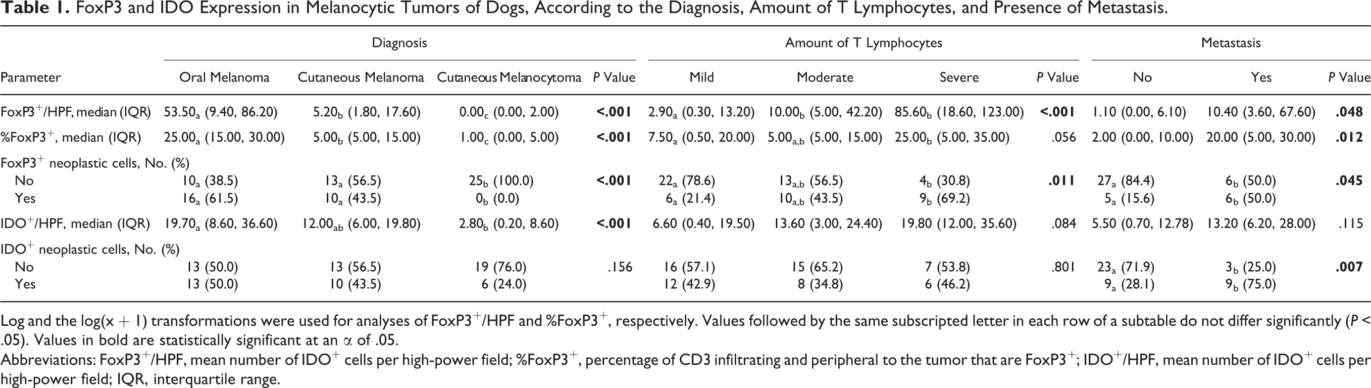
Log and the log(x + 1) transformations were used for analyses of FoxP3+/HPF and %FoxP3+, respectively. Values followed by the same subscripted letter in each row of a subtable do not differ significantly (P < .05). Values in bold are statistically significant at an α of .05.
Abbreviations: FoxP3+/HPF, mean number of IDO+ cells per high-power field; %FoxP3+, percentage of CD3 infiltrating and peripheral to the tumor that are FoxP3+; IDO+/HPF, mean number of IDO+ cells per high-power field; IQR, interquartile range.
FoxP3+ lymphocytes were observed in 24 of 26 (92%) oral melanomas (Fig. 1), 20 of 23 (87%) cutaneous melanomas (Fig. 2), and 12 of 25 (48%) melanocytomas. The number of FoxP3+ lymphocytes was positively associated with a worse histological diagnosis (P < .001), with a decreasing quantity from oral melanomas to cutaneous melanomas to cutaneous melanocytomas. CD3+ lymphocytes were absent (mucosal melanomas: 2/26, 8%; cutaneous melanomas: 1/23, 4%; cutaneous melanocytomas: 7/25, 28%), mild (mucosal melanomas: 8/26, 31%; cutaneous melanomas: 10/23, 44%; cutaneous melanocytomas: 10/25, 40%), moderate (mucosal melanomas: 9/26, 35%; cutaneous melanomas: 8/23, 35%; cutaneous melanocytomas: 6/25, 24%), or severe (mucosal melanomas: 7/26, 27%; cutaneous melanomas: 4/23, 17%; cutaneous melanocytomas: 2/25, 8%).
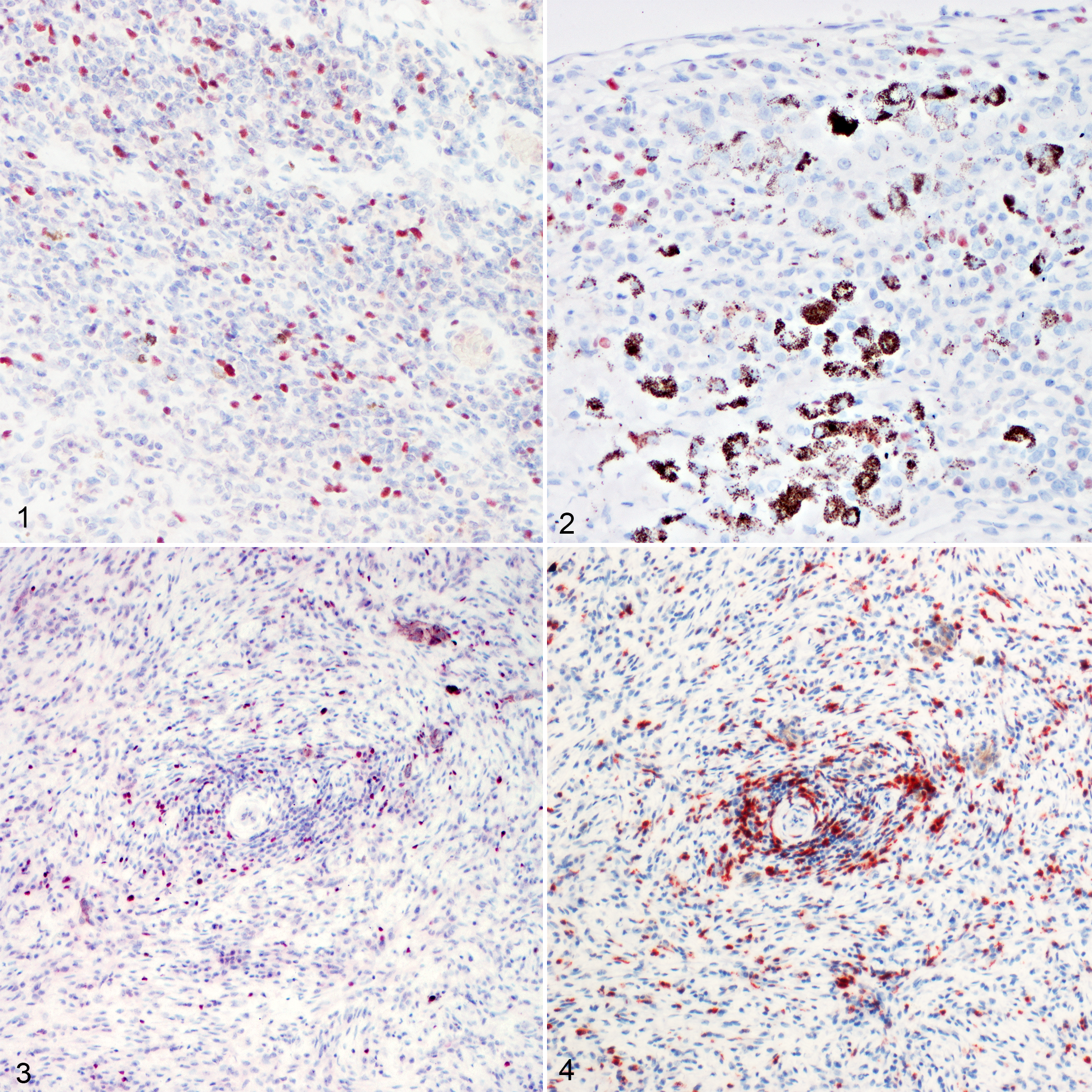
An association with the histological diagnosis was also observed when the percentage of CD3+ cells that were positive for FoxP3 (%FoxP3+) was assessed (P < .001; Figs. 3, 4). As additional information, the only oral melanocytoma that was excluded from the statistical analysis showed no FoxP3+ nuclei.
FoxP3+/HPF value correlated to %FoxP3+ (P ≤ .05) and to the mitotic count (ρ = 0.634; P < .001) and was associated with the presence of metastases (P < .05). The percentage of FoxP3+ cells showed a correlation with the mitotic count (ρ = 0.551; P < .001) and an association with the presence of metastasis (P = .012).
An association with the number of CD3+ lymphocytes was observed for FoxP3+/HPF (P < .001). However, an association with the number of CD3+ lymphocytes was not present for %FoxP3+ lymphocytes, even though the P value was bordering on statistical significance (P = .056).
The presence of FoxP3+ neoplastic cells was observed in 16 of 26 oral melanomas (61%), in 10 of 23 cutaneous melanomas (43%), and in none of the cutaneous melanocytomas (0.0%), thus being associated with histological diagnosis (P < .001). In the samples with FoxP3+ neoplastic cells, the CD3+ lymphocytic infiltrate was more often severe than mild, while in the samples without FoxP3+ neoplastic cells, it was usually mild. Mitotic count was higher in samples with FoxP3+ neoplastic cells (P < .001). Also, the absence of FoxP3+ neoplastic cells was associated with the lack of detected metastasis (P < .05). No FoxP3+ neoplastic cells were seen in the oral melanocytoma excluded from the study.
IDO Immunohistochemistry
IDO positivity was seen as a cytoplasmic granular labeling in cells interpreted as DCs or macrophages, neoplastic cells, and rarely endothelial cells (Figs. 5–8). IDO+ cells, morphologically interpreted as DCs or macrophages, were observed in 26 of 26 (100%) oral melanomas, 21 of 23 (91%) cutaneous melanomas, and 19 of 25 (76%) melanocytomas (Table 1).
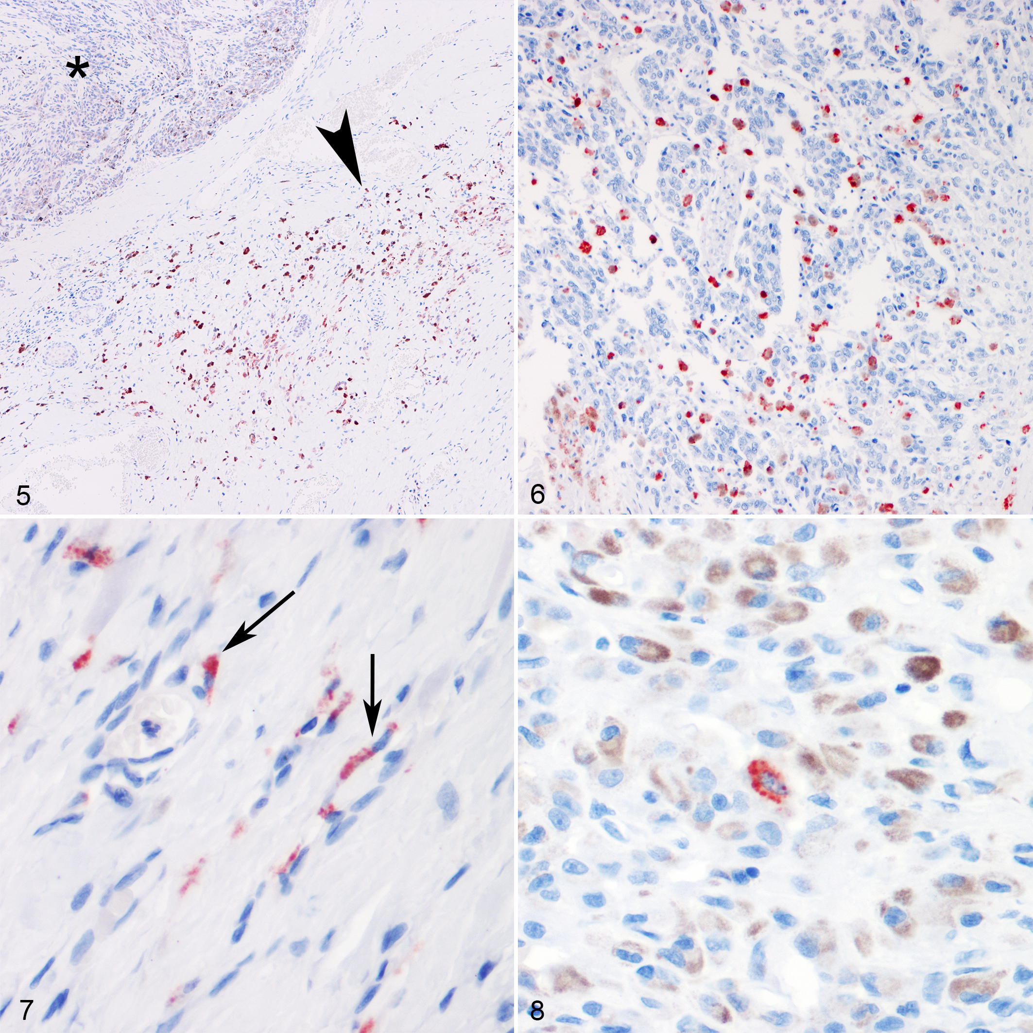
Immunohistochemistry revealed an association between the number of IDO+ inflammatory cells/HPF and the histological diagnosis (P < .001); specifically, in oral melanomas, their number was higher than in cutaneous melanocytomas (P < .05), while no differences were seen between cutaneous melanomas and melanocytomas. The only oral melanocytoma showed the presence of 7.2 IDO+/HPF. IDO+ inflammatory cells/HPF showed a positive correlation with FoxP3+ lymphocytes/HPF (ρ = 0.421; P < .001) and with %FoxP3+ (ρ = 0.294; P < .05); moreover, IDO+ inflammatory cells/HPF were correlated with the mitotic count (ρ = 0.447; P < .001), whereas they did not show any association with the total number of CD3+ cells or to the presence of metastasis.
The presence of IDO+ neoplastic cells was observed in 13 of 26 (50%) oral melanomas, 10 of 23 (43%) cutaneous melanomas, and 6 of 25 (24%) cutaneous melanocytomas. It was positively associated with the presence of metastasis (P < .01), whereas it did not show any association with the quantity of CD3+ cells. There were no IDO+ neoplastic cells in the oral melanocytoma.
Survival Analysis
Follow-up at the end of the study ranged from 127 to 2013 days, with a median follow-up of 821 days. Only 2 cases had a follow-up <1 year (the dogs died of causes unrelated to melanocytic tumors), with 55% of cases being followed for >2 years.
Considering disease-specific survival (death due to melanoma) as an end point, the 6-month and 1-year estimated survival probabilities were 74% ± 10% and 25% ± 11% for dogs with oral melanoma and 87% ± 9% and 78% ± 11% for those with cutaneous melanoma, respectively. The survival probability of dogs with melanocytoma was 100% ± 0% at 1 year.
Dogs with oral melanoma (median = 249 days, IQR = 165–355 days) had a lower survival probability than those with cutaneous melanoma (median not reached, P = .002) and melanocytoma (median not calculated because all cases were censored, P < .001). The difference was also significant between cutaneous melanoma and melanocytoma (P = .015).
When considering all tumors together, all the parameters related to IDO and FoxP3 were significantly associated with an increased risk of death (Table 2). The stratification according to the diagnosis resulted in a loss of statistical significance except for the IDO+/HPF in oral melanomas (HR, 1.038; 95% CI, 1.006–1.070; P = .020).
Results of Univariate and Multivariable Cox Proportional Regression Analyses of Overall Survival in the Entire Cohort of Dogs With Melanoma.
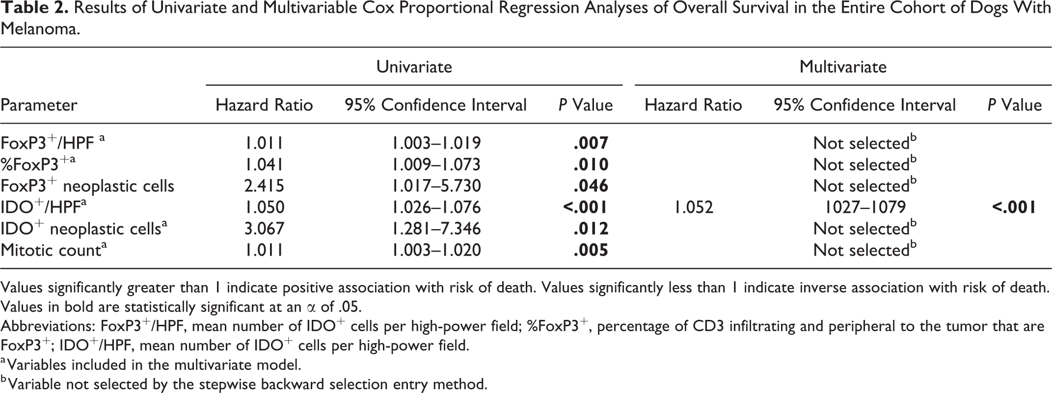
Values significantly greater than 1 indicate positive association with risk of death. Values significantly less than 1 indicate inverse association with risk of death. Values in bold are statistically significant at an α of .05.
Abbreviations: FoxP3+/HPF, mean number of IDO+ cells per high-power field; %FoxP3+, percentage of CD3 infiltrating and peripheral to the tumor that are FoxP3+; IDO+/HPF, mean number of IDO+ cells per high-power field.
a Variables included in the multivariate model.
b Variable not selected by the stepwise backward selection entry method.
FoxP3+/HPF, FoxP3%, FoxP3+ neoplastic cells, IDO+/HPF, IDO+ neoplastic cells, and mitotic count were included in the multivariable model. Multivariable Cox hazard regression analysis identified IDO mean (P < .001) as an independent variable positively associated with the risk of death (Table 2).
ROC Analysis
Variables with a P value of <.05 from the univariate analyses were considered candidate variables for predicting tumor-related death and submitted to ROC analysis (ie, FoxP3+/HPF, FoxP3%, and IDO+/HPF). Of the 75 animals included in the ROC analysis, 21 were dead due to melanoma and 54 were alive or dead due to other causes. Results showed that FoxP3+/HPF and IDO+/HPF were moderately accurate parameters for predicting death while FoxP3% was less accurate (Table 3). However, there were no significant differences with the AUC of mitotic counts (z = 0.88, z = 0.56, and z = 1.79 for FoxP3+/HPF, IDO+/HPF, and FoxP3% respectively; P > .05). The optimal cutoffs for predicting death were 6.1 for FoxP3+/HPF (Fig. 9), 12.5 for FoxP3% (Fig. 10), and 14.7 for IDO+/HPF (Fig. 11 and Table 3).
Results of Receiver Operator Characteristic Analysis Including the AUC With Corresponding 95% Confidence Intervals and P Values, Optimal Cutoff With Associated Sensitivity and Specificity Values to Predict Mortality Due to Melanoma.
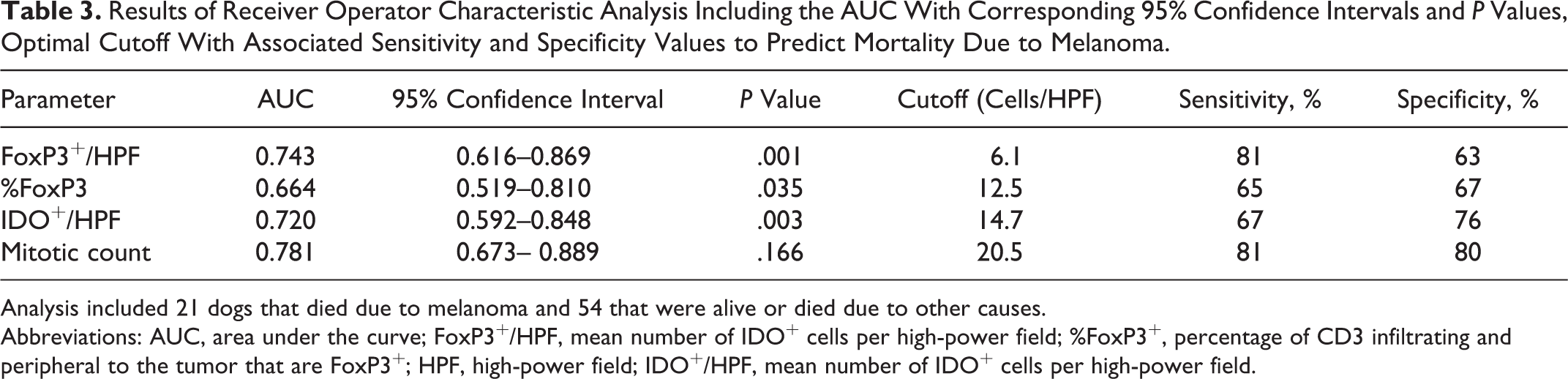
Analysis included 21 dogs that died due to melanoma and 54 that were alive or died due to other causes.
Abbreviations: AUC, area under the curve; FoxP3+/HPF, mean number of IDO+ cells per high-power field; %FoxP3+, percentage of CD3 infiltrating and peripheral to the tumor that are FoxP3+; HPF, high-power field; IDO+/HPF, mean number of IDO+ cells per high-power field.
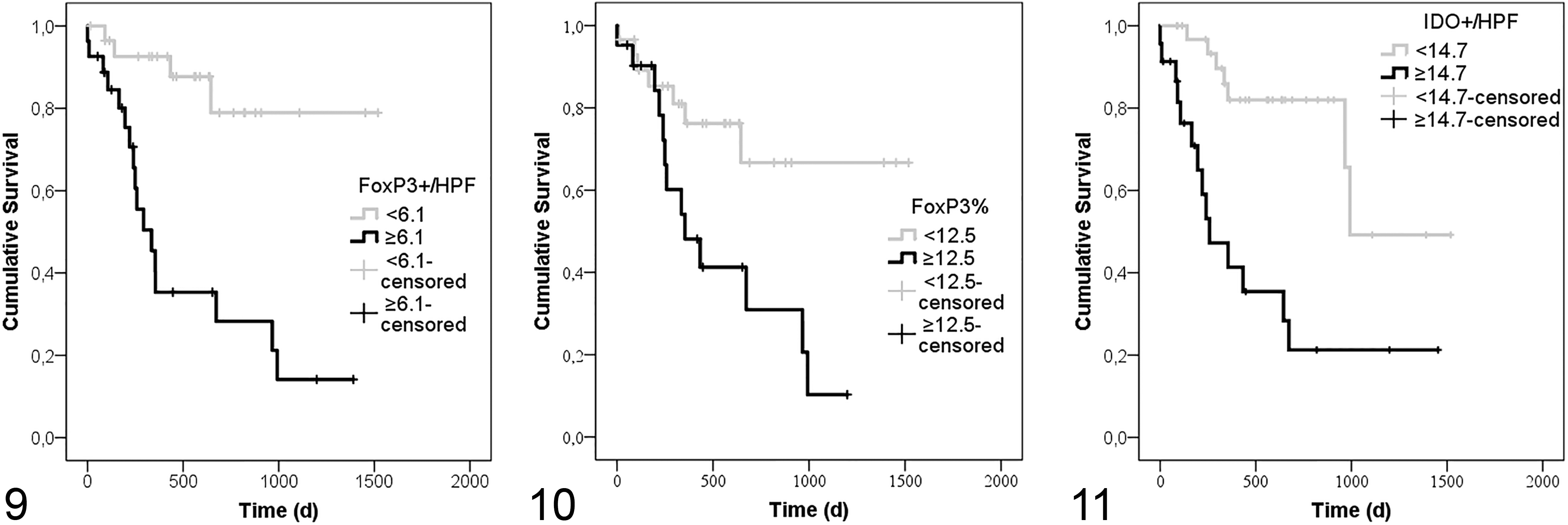
The risk of death was higher for dogs with FoxP3+/HPF ≥6.1 (HR, 6.774; 95% CI, 2.259–20.312; P = .001; Fig. 9) and FoxP3% ≥12.5 (HR, 3.016; 95% CI, 1.200–7.583; P = .019; Fig. 10). Similarly, IDO+/HPF ≥14.7 (HR, 4.707; 95% CI, 1.890–11.719; P = .001; Fig. 11) increased the risk of death by 4.7 times.
Discussion
The role and the profiling of the immune environment is gaining attention in oncology and in melanoma research because of the growing evidence of its impact on both prognosis and response to immunotherapy. 28,29 To our knowledge, studies on canine melanoma immunology are few, and the use of immunotherapy targeting immune checkpoint molecules is still limited to in vitro data. 1,5,35,67 This study was intended to provide a first overview on the expression of FoxP3 and IDO in primary melanocytic tumors of the dog; moreover, it investigates the possible value of these molecules as additional prognostic immunohistochemical markers in canine melanoma.
Results from this study showed that FoxP3+/HPF and FoxP3% were associated with both the diagnosis (decreasing quantity from oral melanomas > cutaneous melanomas > cutaneous melanocytomas) and the presence of metastasis, suggesting that Tregs might play an important role in the establishment of immune tolerance mechanisms, as elsewhere suggested, for dogs and humans. 40,44,59 In addition, in univariate analysis, both FoxP3+/HPF and FoxP3% were shown to be prognostic factors in canine melanocytic tumors, independently of the diagnosis. FoxP3+/HPF was associated with the quantity of CD3+ lymphocytes, but the percentage of FoxP3+ lymphocytes was not. This last result points out an inconstant fraction of FoxP3+ cells among the total number of T lymphocytes. Thus, the prominent Treg infiltration observed in canine oral melanoma could be one of the factors contributing to a marked immunosuppressive microenvironment and to the activation of immunoescape pathways, thereby contributing to the aggressive clinical behavior of these tumors, similar to what is described in humans. 18,39,40,51 However, other studies suggest that Tregs can comprise a “bad” subset that contributes to antitumor immunity and a “good” one, with an anti-inflammatory role, which are pivotal in maintaining self-tolerance and of benefit to the host. 71 Therefore, further investigations are needed to better understand the different subsets and the biologic activity of Tregs in canine oncology. Another point of concern is that Tregs do not have a specific marker to confirm their identity: FoxP3 is a reliable marker in mice but is less reliable in humans. Little information is available for dogs, but some recent studies seem to validate FoxP3 as a specific marker. 15,59,68,70
Our results showed the expression of FoxP3 by neoplastic cells, as demonstrated by the presence of FoxP3+ nuclei in cells with phenotypic features of neoplastic cells. They were present only in oral and cutaneous melanoma, and their presence was associated with the diagnosis, with the quantity of lymphocytes, and also with the presence of metastasis; moreover, in univariate analysis, the results suggested a prognostic value in canine melanocytic tumors. This finding could support the hypothesis proposed by different authors that tumor cells could exert a FoxP3-dependent, Treg-like suppressive effect on T cells, suggesting that this mimicking of Treg functions could act as an immunosuppressive process, further contributing to melanoma cells’ immunoescape and survival. 41,51,67 Evaluation of a larger caseload for the coexpression of melanocytic markers and FoxP3 to better identify the population of neoplastic cells expressing the transcription factor and their role in immunoescape is needed.
The role of IDO in canine immune regulation is still under examination, and few data are available in the scientific literature; only 1 study has investigated the potential use of blocking IDO to revert tumor immune suppression and potentiate the effects of radiation therapy. 10,43,60 To our knowledge, this is the first study that has investigated the expression and the potential biological and prognostic role of this molecule in canine melanocytic tumors. Our results showed that the mean number of IDO+ inflammatory cells/HPF was associated with the histological diagnosis (decreasing from oral melanomas > cutaneous melanomas > cutaneous melanocytomas), but no significant association with the presence of T lymphocytes or with metastasis was found. The results of univariate analysis identified IDO+ inflammatory cells/HPF as a prognostic marker that was associated with a higher hazard of death. Multivariable analysis also selected it as an independent factor. Although the low number of cases resulted in the loss of significance when stratifying according to tumor type, in oral melanomas, IDO+ cells were still associated with an increased risk of death. Interestingly, the presence of IDO+ neoplastic cells was associated with the presence of metastasis, and dogs with IDO+ neoplastic cells had a 3 times higher hazard of death compared to those without. In human medicine, the expression of IDO in melanoma cells has been observed, 8,54 and beyond its role in immunosuppression, it has been suggested to be a part of the genetic changes associated with malignant transformation. 45 From the ROC curve, we can infer that optimal cutoffs for predicting death were 14.7 for IDO+/HPF, 6.1 for FoxP3+/HPF, and 12.5 for FoxP3%, with IDO+/HPF being the most specific (76%) and FoxP3+/HPF the most sensitive (81%). However, since we performed the analysis on all cases, the values could have been affected by the differences of expression in the different tumor types (oral/cutaneous); thus, the values provided have to be considered indicative and should be confirmed in a wider sample size and analyzing cases stratified by diagnosis. Furthermore, due to the presence of many dogs lost to follow-up or dead for other causes, these results and the results of the survival curves must be interpreted with caution and further assessed.
Our results also indicated a positive correlation between IDO+/HPF and both FoxP3+/HPF and FoxP3%. These data seem to confirm the strong interconnection between the 2 molecules; in the past few years, it has been demonstrated that IDO can both induce Tregs and activate Treg-mediated suppression with different mechanisms, and in turn, Tregs can induce IDO. 46,73 Our results suggest that the immune profile of the tumor should be carefully taken into consideration together with the tumor cell biology and, if substantiated by further studies, could help to define the prognosis in canine melanocytic tumors.
Cancer immune microenvironment is gaining more and more attention in oncology, since evidence of its role in prognosis and response to therapy is accumulating. Immunotherapy in the past few years has emerged as a new approach for different types of malignancies, including melanoma. Particularly, anti–CTLA-4 therapy demonstrated effectiveness in depletion of intratumoral Tregs, and its combination with pharmacological IDO inhibition even resulted in superior responses. 7,58
The current study suggests, for the first time, a potential role of both FoxP3 and IDO in influencing the biological behavior of canine melanocytic tumors and thus the importance that an immunosuppressive tumor microenvironment may play in melanoma progression. Our results, including the identification of cutoff values, suggest that FoxP3 and IDO immunohistochemistry could help to estimate the prognosis of patients with melanocytic tumors. These parameters should be evaluated in association with more commonly used prognostic indicators (eg, mitotic index, nuclear atypia), providing useful information about the immune microenvironment of the canine melanocytic tumors. However, this is a preliminary retrospective study, and the small sample size may be responsible for the loss of significance of the IDO and FoxP3 parameters after stratification according to the diagnosis. Further studies, with a larger sample population, are advised to confirm the potential role of IDO and FoxP3 as prognostic indicators in canine melanocytic tumors and to evaluate if these parameters can be useful in predicting canine melanoma response to immunotherapeutic approaches.
Supplemental Material
Supplemental Material, DS1_VET_10.1177_0300985818808530 - FoxP3 and IDO in Canine Melanocytic Tumors
Supplemental Material, DS1_VET_10.1177_0300985818808530 for FoxP3 and IDO in Canine Melanocytic Tumors by Ilaria Porcellato, Chiara Brachelente, Livia De Paolis, Laura Menchetti, Serenella Silvestri, Monica Sforna, Gaia Vichi, Selina Iussich, and Luca Mechelli in Veterinary Pathology
Footnotes
Acknowledgements
We thank Sara Leto, Valeria Migni, and Luca Stefanelli for their valuable technical assistance.
Declaration of Conflicting Interests
The author(s) declared no potential conflicts of interest with respect to the research, authorship, and/or publication of this article.
Funding
The author(s) received no financial support for the research, authorship, and/or publication of this article.
Supplemental material for this article is available online.
References
Supplementary Material
Please find the following supplemental material available below.
For Open Access articles published under a Creative Commons License, all supplemental material carries the same license as the article it is associated with.
For non-Open Access articles published, all supplemental material carries a non-exclusive license, and permission requests for re-use of supplemental material or any part of supplemental material shall be sent directly to the copyright owner as specified in the copyright notice associated with the article.