
Abstract
Bovine abortion is a worldwide problem, but despite extensive histopathologic and molecular investigations, the cause of abortion remains unclear in about 70% of cases. Cellular debris is a commonly observed histopathologic finding in the fetal placenta and is often interpreted as necrosis. In this study, the nature of this cellular debris was characterized, and histologic changes in the normal fetal placenta during pregnancy and after delivery were assessed. In addition, the presence of the most common abortifacient pathogens in Switzerland (Chlamydiaceae, Coxiella burnetii, Neospora caninum) was tested by polymerase chain reaction. We collected 51 placentomes and 235 cotyledons from 41 and from 50 cows, respectively. In total, cellular debris was present in 48 of 51 (94%) placentomes and in 225 of 235 (96%) cotyledons, inflammation occurred in 1 of 51 (2%) placentomes and in 46 of 235 (20%) cotyledons, vasculitis was seen in 1 of 51 (2%) placentomes and 46 of 235 (20%) cotyledons, and 18 of 51 (35%) placentomes and 181 of 235 (77%) cotyledons had mineralization. The amount of cellular debris correlated with areas of positive signals for cleaved caspase 3 and lamin A. Therefore, this finding was interpreted as an apoptotic process. In total, 10 of 50 cotyledons (20%) were positive for C. burnetii DNA, most likely representing subclinical infections. The results of our study indicate that histologic features in the fetal placenta such as cellular debris, inflammation, vasculitis, and mineralization must be considered physiological processes during pregnancy and after delivery. Therefore, their presence in placentae of aborted fetuses must be interpreted with caution and might not be necessarily linked to an infectious cause of abortion.
Keywords
Abortion in cattle, with an estimated abortion rate of 2% to 5%, 1,35 is a major cause of economic losses worldwide. 6,10,41 In Switzerland, 14 000 to 28 000 bovine abortions are reported annually, accounting for losses of 22 to 45 million Swiss Francs per year. 21 Knowledge of the cause of abortion is important when deciding on control measures or improved management. Despite extensive histopathologic, microbiologic, and molecular investigations, most bovine abortion cases remain without an etiologic diagnosis 4,8,16 : in a retrospective Swiss study on 347 bovine abortions, an infectious cause was detected in 28.5% and a noninfectious cause in 3.8% of the cases, but the etiology was unknown in 67.7%. 36
During histologic examination of placental membranes from bovine abortions, cellular debris in the chorionic epithelium and adjacent cotyledonary stroma, often termed necrosis, is a very common finding. 8,11 However, the pathogenesis and cause of such lesions often remain undetermined. Moreover, it has not been determined if these areas represent necrotic or apoptotic changes or if they are part of the normal fetal membrane separation process during delivery. Literature describing this detachment process in the fetal part of the placentomes (cotyledons) is limited, whereas most studies focus on the maternal component (caruncle) and/or the placentomes as a whole.
The microscopic components of the placentomes are the trophoblasts and the cryptal epithelium. 7 Fetal villi arising from the chorionic plate of the cotyledon interdigitate with a widely ramified corresponding system of maternal caruncular crypts. Initially, the bovine trophoblast was described to be composed of uninucleated and larger binucleated cells. Later, it was realized that, throughout gestation, terminally differentiated trophoblast giant cells (TGC) derive permanently from uninucleated trophoblast cells by a series of acytokinetic mitoses. 31 TGC invade the caruncular epithelium, where they soon undergo apoptotic cell death after degranulation, or they may fuse with caruncular epithelial cells, giving rise to short-lived trinucleate feto-maternal hybrid cells. The function of this weakly invasive activity is obviously the transfer of fetal-derived substances into the maternal compartment. 31,43
In our study, we assessed the normal histology of placental membranes during pregnancy and after delivery. Thereby, we investigated the presence of cellular debris, inflammation, as well as vasculitis and placental mineralization. The placental membranes were collected from uneventful parturition case;, however, we examined them for the most common abortifacient pathogens prevalent in Switzerland, namely, Chlamydiaceae, Coxiella burnetii, and Neospora caninum, using polymerase chain reaction (PCR). Testing for the aforementioned pathogens was deemed necessary for the correct interpretation of the results of this study, in view of the fact that necrotizing and/or purulent placentitis and purulent to necrotizing vasculitis are the key histologic lesions induced by these agents. 10
Materials and Methods
Samples, Questionnaire, and Macroscopic Examination
In total, 51 placentomes were collected from 41 cows. These included 26 hematoxylin and eosin– (HE) stained slides of placentomal tissues that were available from 16 Swiss cows euthanized at different pregnancy stages (range, 69–270 days) because of unrelated diseases such as abomasal ulcers or metabolic problems. Placentomes were collected by a veterinary pathologist only from fresh fetuses, in the absence of pathologic changes in the uterus. In addition, 25 formalin-fixed and paraffin-embedded (FFPE) placentomal tissue samples and corresponding HE-stained slides from 25 cows covering the period between day 100 of pregnancy until normal at-term delivery originated from a previous study. 38 Sample collection from living animals was approved by the Regierungspräsidium Giessen (permission No. II25.3-19c20/15cGI18/14).
In collaboration with the herd health service (Section for Herd Health, Department for Farm Animals, Vetsuisse Faculty, University of Zurich), 235 cotyledonary tissue specimens were collected from 50 Swiss healthy cows from 12 different farms after birth and expulsion of the fetal membranes. Permission by the State Veterinary Office of Zurich was not required since all specimens were collected during spontaneous birth. Figure 1 summarizes the samples collected.
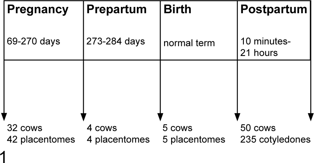
Normal placentome and cotyledon sample collection at different gestation periods. In total, 42 placentomes from 32 cows were collected during pregnancy (69 to 270 days of pregnancy), 4 placentomes from 4 cows were collected in the prepartal period (12 to 48 hours before birth), 5 placentomes from 5 cows were collected at birth, and 235 cotyledons from 50 cows were collected in the postpartum period (10 minutes to 21 hours after birth).
Information about the 50 Swiss cows including the name of the farmer, name of the cow, unique animal identification number, date of fertilization, date of birth, time of birth, any assistance during birth, date, time point and place of placenta collection, and pretreatment was collected using a questionnaire. Of those, macroscopic inspection was performed on 107 cotyledonary samples derived from 30 cows.
Histologic Examination
In total, 235 FFPE blocks containing cotyledonary tissue taken postpartum from 50 cows were prepared (45 cases with 3 blocks, 5 cases with 20 blocks). After fixation in a 10% buffered formalin solution, cotyledonary tissue from random locations was trimmed and embedded in paraffin blocks (Sakura, Horgen, Switzerland). Then, 2-μm sections were prepared and stained with HE according to standard procedure.
A total of 286 slides were examined (at a magnification of 400×) using a light microscope (Olympus BH 2, Tokyo, Japan). Four main microscopic findings were identified and assessed qualitatively: cellular debris, inflammation, vasculitis, and mineralization. This was followed by a semiquantitative analysis of these parameters by evaluation of the whole section. Cellular debris, inflammation, and mineralization were classified as mild (+, n = 1–10 foci per section), moderate (++, n = 11–30), and severe (+++, n = >30). Vasculitis was classified as mild (+, n = 1–6), moderate (++, n = 7–12), and severe (+++, n = > 12). Depending on the distribution of the microscopic findings, the presence of cells debris and inflammation was classified as focal (F), multifocal (MF), and multifocal to coalescing (MF-C). The presence of hemorrhage or hyperemia was also noted.
High-resolution digital scans of 186 HE-stained slides were acquired using the Hamamatsu Photonics’s NanoZoomer HT2.0 (Hamamatsu, Japan). The virtual slides were imported into the image analysis software Visiopharm (Visiopharm Integrator System [VIS], version 6.7.0.2590, Visiopharm, Hoersholm, Denmark).
Total section area was used for the semiquantitative analysis of the microscopic parameters described above (cellular debris, inflammation, vasculitis, and mineralization), which were expressed as number of foci per 100 mm2.
Immunohistochemistry for Cleaved Caspase 3 and Lamin A
Immunohistochemical labeling to detect the cleaved caspase 3 and lamin A antigen was performed on 186 FFPE sections derived from 91 cases. The sections were mounted on positively charged slides and dried overnight at 37°C. After deparaffinization, antigen retrieval was performed as previously described. 13 Incubation with a 1:100 diluted primary antibody for cleaved caspase 3 (cleaved caspase 3 rabbit monoclonal antibody, nr. 9664; Cell Signaling Technology, Leiden, the Netherlands) or with a 1:50 diluted primary antibody for cleaved lamin A (cleaved lamin A rabbit polyclonal antibody, nr. 2035; Cell Signaling Technology) was performed for 1 hour at room temperature (RT). Then, slides were rinsed with Tris-buffered saline–Tween (TBS, nr. 3006; Dako, Jena, Germany) before adding peroxidase-blocking buffer (nr. S2023, Dako) as previously described. 28 Slides were rinsed again with TBS followed by addition of a peroxidase-labeled polymer conjugated goat anti-rabbit antibody (HRP Rabbit, nr. K4003; Dako) for 30 minutes at RT. Finally, the substrate aminoethylcarbazol (AEC; nr. K3464; Dako) was added for 10 minutes at RT before the sections were counterstained with hematoxylin and coverslipped.
High-resolution digital scans of 372 immunohistochemical slides derived from 91 cases (186 slides labeled with cleaved caspase 3 and cleaved lamin A, respectively) were acquired using the Hamamatsu Photonics’s NanoZoomer HT2.0. The virtual slides were imported into the image analysis software VIS. Quantitative analysis using a threshold classification method allowed recognition of positive (red, AEC) and negative (blue, hematoxylin counterstain) placentomal and cotyledonary tissue, and the results were expressed as positive area versus total area in the whole tissue section. Data are presented as average (±SEM) percentage of tissue in the section undergoing cellular debris.
Testing for Infectious Agents
In total, 91 cases were tested for the presence of N. caninum, C. burnetii, and Chlamydiaceae DNA by PCR (N. caninum) or by real-time PCR (C. burnetii, Chlamydiaceae). DNA was extracted as described previously 12 and examined with the Nanodrop 1000 version 3.7.1 (Thermo Fisher Scientific, Waltham, MA) to determine DNA quantity and quality.
The N. caninum PCR was carried out using the specific primers Np6+ and Np21+ and a previously described protocol amplifying a 328-bp fragment of the Nc5 gene of N. caninum. 33 N. caninum tachyzoites and DNA extracted from FFPE tissues from a naturally infected bovine were used as positive controls, whereas RNAsefree water (QIAGEN, Hilden, Germany) was used as negative control.
For the 23S rRNA gene Chlamydiaceae family-specific real-time PCR, 15 all DNA samples (n = 91) were analyzed on an Applied Biosystems 7500 Real-Time PCR System (Thermo Fisher Scientific, Waltham, MA) using a previously described protocol 8 with primers Ch23SF (5′CTGAAACCAGTAGCTTATAAGCGGT3′), Ch23SR (5′ACCTCGCCGTTTAACTTAACTCC3′), and probe Ch23Sp (5′FAMCTCATCATGCAAAAGGCACGCCGTAMRA3′), including an internal positive control (enhanced green fluorescent protein [eGFP] 24 ) with primers EGFP1F (5′GACCACTACCAGCAGAACAC3′), EGFP10 R (3′CTTGTACAGCTCGTCCATGC5′), and probe EGFP1HEX (5′HEXAGCACCCAGTCCGCCCTGAGCABHQ13′). All samples were tested in duplicate. Duplicate mean Ct values of <38 were considered positive, while values >38 were classified as questionable positive.
For the C. burnetii PCR, the extracted DNA (n = 91) was investigated on a real-time PCR system using the kit LSI VetMAX Triplex C. burnetii and Chlamydia spp. (catalog No. TFQQCHP; Thermo Fisher Scientific) according to the manufacturer’s instructions. Interpretation of the results was performed as recommended by the manufacturer.
Statistical Analysis
Initially, a qualitative check of the data was performed using the program Stata (Stata Statistical Software Release 12; StataCorp, College Station, TX) with <codebook var> (<STATA-command Variable>). Continuous data were tested for normal distribution by the Wilk-Shapiro test <swilk var>. Values not normally distributed were transformed to normal distribution by STATA command <ladder var>. The data were statistically analyzed, and a P value of ≤0.05 was considered significant. The differences in the continuous data were tested using analysis of variance (ANOVA) <anova vary varx> (vary = y variable, dependent variable; varx = x variable, independent variable), linear regression <regress vary varx>, fractional polynomial regression <fracpoly, degree(1): glm vary varx>, and 1-way ANOVA using the Bonferroni post hoc test <oneway vary varx, bonferroni>.
Results
Survey Results
All 50 cows, from which cotyledonary samples were obtained, lived on Swiss farms. Information about sample collection was incomplete in 8 cases: missing date of breeding and/or place of placenta collection (n = 2), missing time of birth and the time of placenta collection (n = 1), missing date and time of placenta collection (n = 2), missing time of placenta collection (n = 2), and missing identification number (n = 1).
The gestation period in all 50 cases was calculated based on each month comprising 30 days. The gestation period in all 50 cases ranged from 259 to 297 days. The time between birth and placenta collection ranged from 10 minutes to 21 hours (mean duration, 7.25 hours). In total, 44 placentas were collected from the floor, 3 from the vagina, 1 from the uterus, and 2 from an unknown location. Assistance during birth was recorded in 8 cases, and 2 cows received pretreatment with vitamin D3 suspension.
Macroscopic Findings
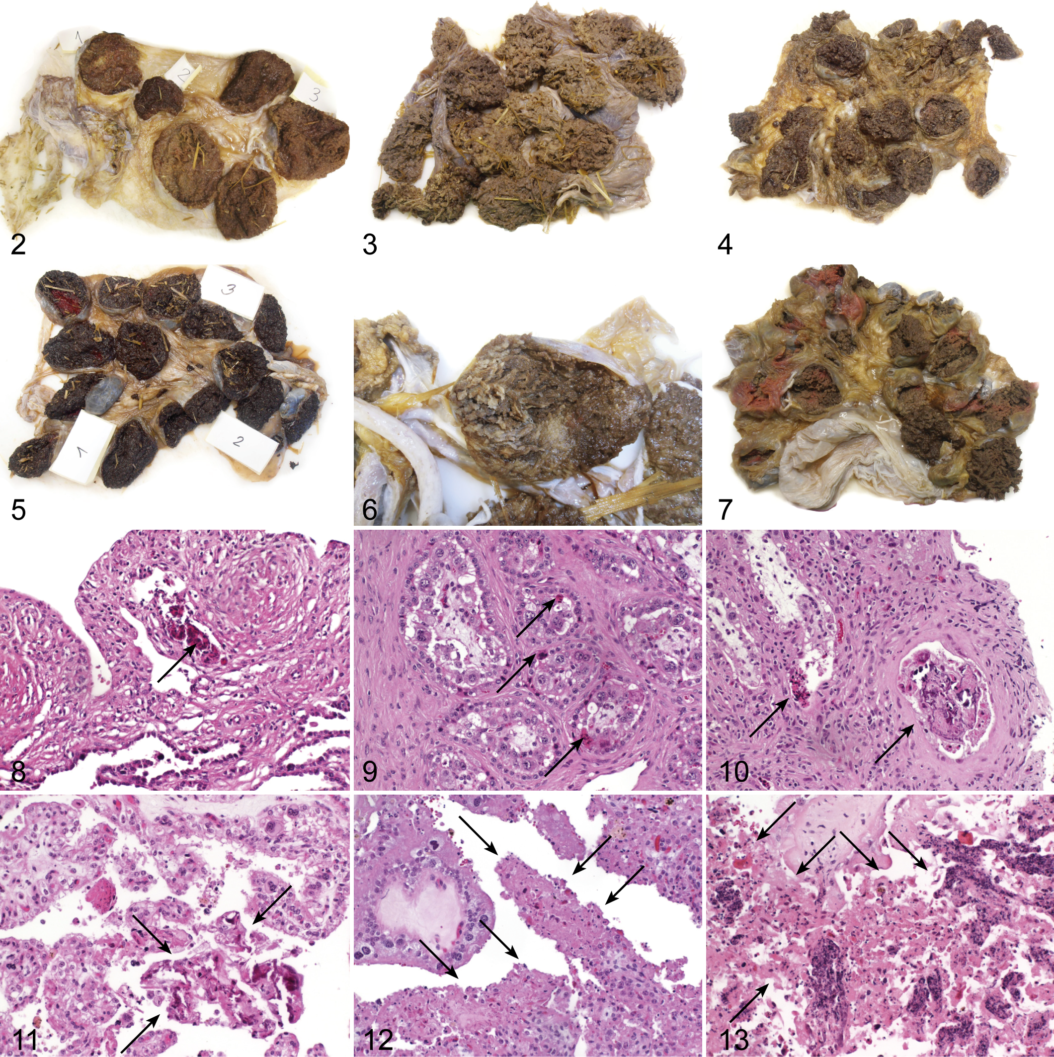
From each case, 3 to 19 cotyledons were available. The macroscopic colors of 107 cotyledonary samples derived from 30 cases ranged from light brown (Fig. 2), brown (Fig. 3), dark brown (Fig. 4), dark red (Fig. 5), to marbled (Fig. 6). A few cotyledons were insufficiently fixed in formalin (Fig. 7). The intercotyledonary stroma was transparent and mildly edematous in all cases. There was no correlation between the color of the cotyledons and the histologic findings (P > .05).
Histologic Findings
Histologic findings of placentomes (n = 51) are summarized in Table 1. The most commonly observed histologic finding was cell debris (94%; Figs. 8–10). Inflammation and vasculitis were present in only 1 of 51 placentomes (2%), and mineralization was observed in 18 of 51 placentomes (35%). Hemorrhage or hyperemia was found in 2 of 51 placentomes (4%). Only 3 of 51 placentomes (6%) showed no histologic findings. From the first histologic section of each case (n = 41), the total area of tissue of the 41 placentomes ranged from 25.4 mm2 to 368.3 mm2.
Histologic Findings in Placentomes From Cows (n = 51).
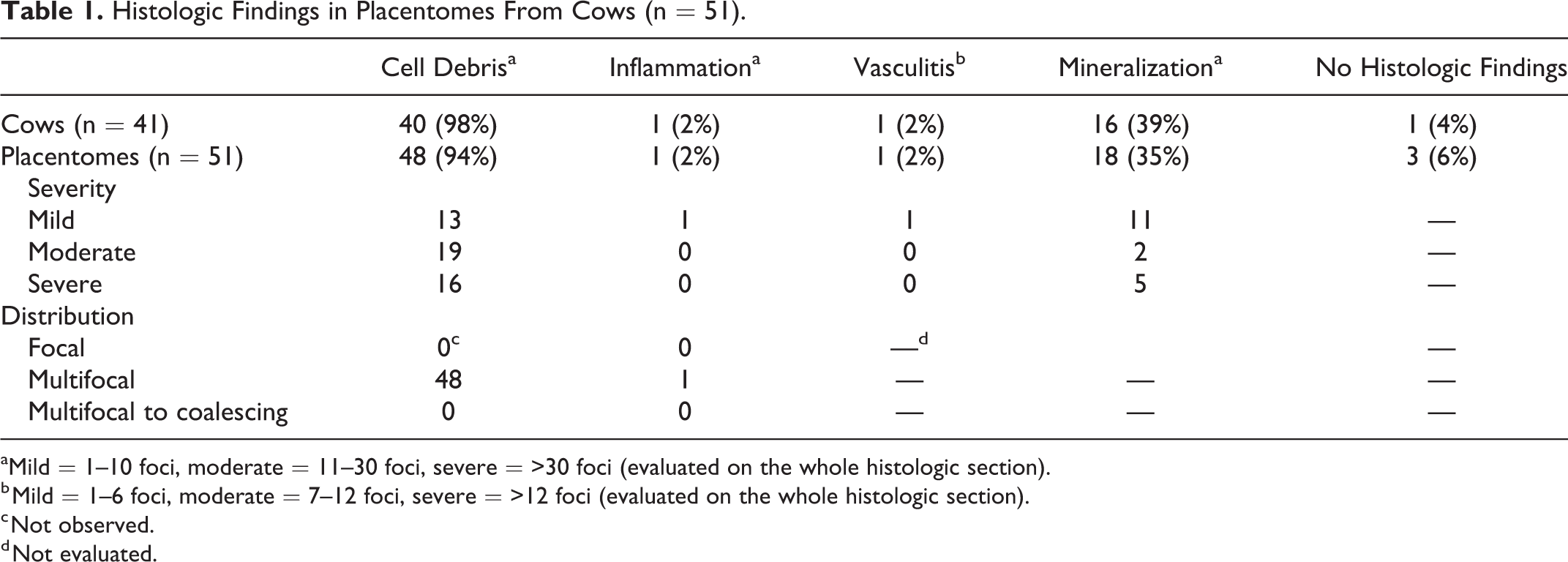
aMild = 1–10 foci, moderate = 11–30 foci, severe = >30 foci (evaluated on the whole histologic section).
b Mild = 1–6 foci, moderate = 7–12 foci, severe = >12 foci (evaluated on the whole histologic section).
c Not observed.
d Not evaluated.
Histologic findings of cotyledons (n = 235) are summarized in Table 2. First, we collected 20 cotyledons from each cow (n = 5) to determine whether the distribution of the histologic findings was equal across the 20 cotyledons. As our pilot study showed an equal distribution, we collected 3 cotyledons from the remaining 45 cases.
Histologic Findings in Cotyledons From Cows With Uneventful Parturition (n = 235).
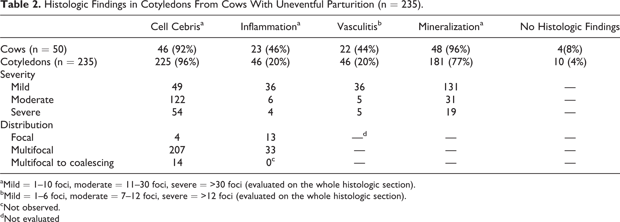
aMild = 1–10 foci, moderate = 11–30 foci, severe = >30 foci (evaluated on the whole histologic section).
bMild = 1–6 foci, moderate = 7–12 foci, severe = >12 foci (evaluated on the whole histologic section).
cNot observed.
dNot evaluated
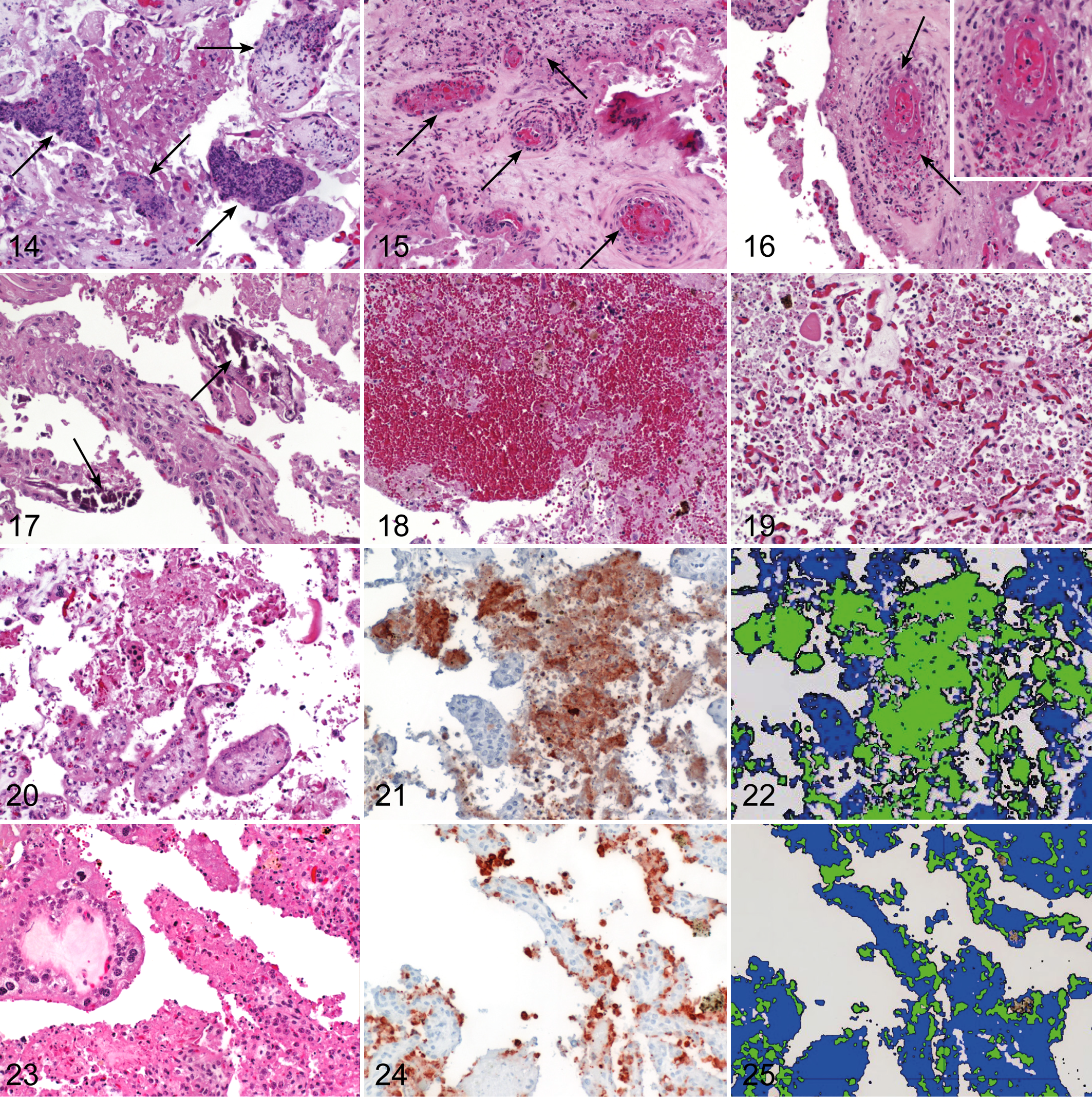
The most frequent histologic finding in the cotyledons was cellular debris (96%; Figs. 11–13). Most cell debris was moderately severe and had a multifocal distribution. Purulent inflammation and vasculitis were detected in 46 of 235 cotyledons (20%; Figs. 14–16), and mineralization was observed in 181 of 235 cotyledons (77%; Fig. 17). Hemorrhage or hyperemia was found in 130 cotyledons of 235 (55%; Fig. 18). Only 10 cotyledons of 235 (4%) showed no histologic findings. One slide exhibited severe autolytic changes (Fig. 19). From the histologic sections of cotyledons in 145 slides, the total area of tissue of the 145 cotyledons ranged from 12.8 mm2 to 535.2 mm2.
The amount of cellular debris positively correlated with the time between birth and collection of placenta (P = .004) but did not correlate with the gestation period (P = .8). There was no significant increase in the amount of cellular debris in the postpartum period (P = .45). Purulent inflammation and purulent to necrotizing vasculitis was most common in the postpartum period, but statistical analysis was not possible because of low case numbers. Furthermore, purulent inflammation was not correlated with purulent to necrotizing vasculitis (P = .08). The histologic finding of mineralization was not correlated with the presence of cellular debris (P = .17). Semiquantitative and qualitative presence of cellular debris was not correlated with the progress of pregnancy until birth (P = .4486, P = .290).
Immunohistochemical Findings
Immunohistolabeling of placentomes for cleaved caspase 3 and lamin A was quantified on one histologic slide of each case (n = 41). The area of positive immunolabeling ranged from 0.04 mm2 to 5.2 mm2 for cleaved caspase 3 and from 0.03 mm2 to 1.8 mm2 for cleaved lamin A. The proportion of tissue with positive immunolabeling ranged from 0.02% to 3.54% for cleaved caspase 3 and from 0.01% to 2.7% for cleaved lamin A.
Immunohistolabeling of cotyledons for cleaved caspase 3 and lamin A was quantified on 145 histologic sections. The area of positive immunolabeling ranged from 0.002 to 58.8 mm2 for cleaved caspase 3 and from 0.002 mm2 to 43.1 mm2 for cleaved lamin A. The proportion of tissue with positive immunolabeling ranged from 0.01% to 27.43% for cleaved caspase 3 (Figs. 20–22) and from 0.33% to 12.03% for cleaved lamin A (Figs. 23–25). Figure 26 shows the range of caspase 3 and lamin A immunolabeling for placentomes and cotyledons.
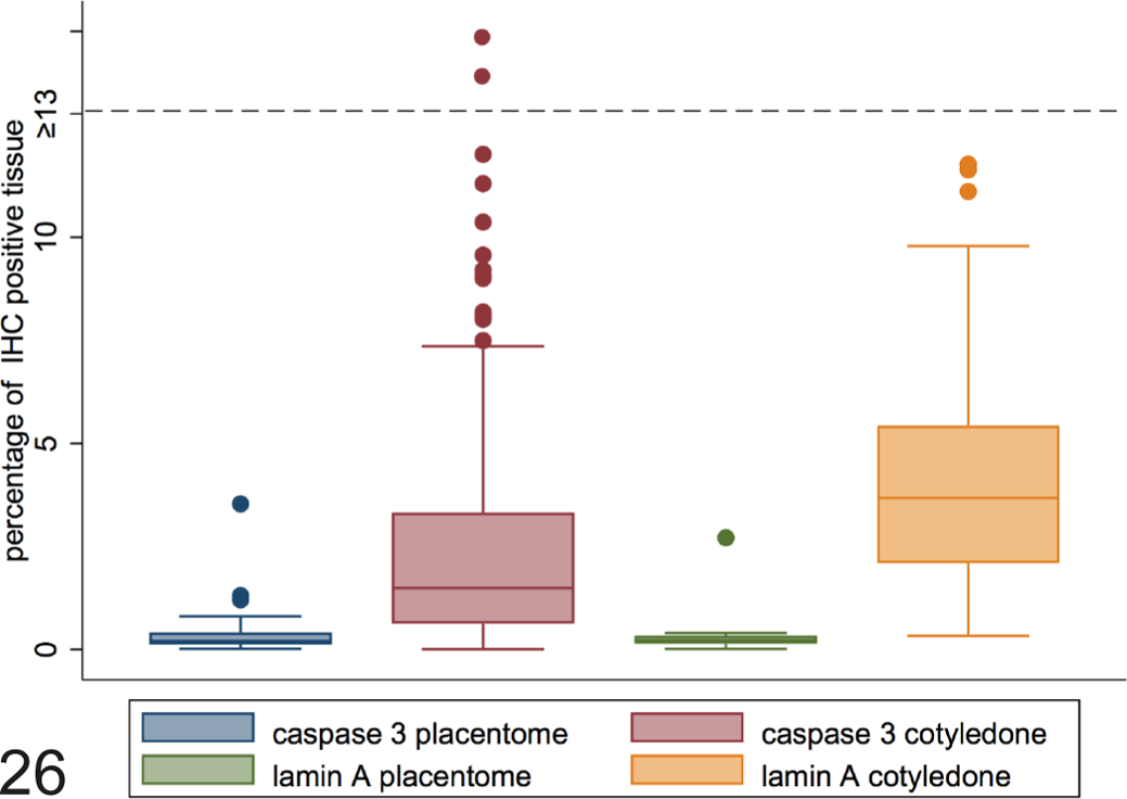
Percentage of immunolabeling for cleaved caspase 3 and cleaved lamin A in normal bovine placentomes and cotyledons. The horizontal line within the box represents the median, and the boxes show the 25th to 75th percentiles. The whiskers show the range from the 10th to 25th percentiles (bottom whisker) and 75th to the 90th percentiles (top whisker). Single dots represent outliers (below 10th or above 90th percentile).
The amount of qualitatively and semiquantitatively assessed cellular debris correlated with an increased expression of cleaved caspase 3 (P = .003, P = .016) and cleaved lamin A (P = .01, P = .004). Positive signals for cleaved caspase 3 (P = .001) and cleaved lamin A (P = .001) increased significantly during pregnancy until the postpartum period, and positive signals for cleaved caspase 3 correlated with positive signals for cleaved lamin A (P = .001). The histologic finding of mineralization was not correlated with the increased expression of cleaved caspase 3 (P = .3) but did correlate with positive signals for cleaved lamin A (P = .040).
Molecular Investigations for Infectious Agents
All 91 cases investigated for N. caninum and Chlamydiaceae by PCR or real-time PCR, respectively, were negative. C. burnetii was detected by real-time PCR (threshold cycle-value [Ct-value] less than 45) in 10 of 91 (11%) cotyledon samples of the postpartum period (Table 3). Multifocal or multifocal to coalescing accumulation of cell debris was severe in 4, moderate in 2, and minimal in 3 of these 10 cases. Purulent inflammation of the stroma with purulent to necrotizing vasculitis was present in 3 cases.
Histologic Details of Cases Positive by Real-Time Polymerase Chain Reaction for Coxiella burnetii.a
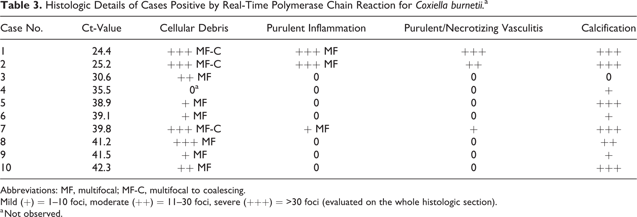
Abbreviations: MF, multifocal; MF-C, multifocal to coalescing.
Mild (+) = 1–10 foci, moderate (++) = 11–30 foci, severe (+++) = >30 foci (evaluated on the whole histologic section).
a Not observed.
Discussion
Studies centered on the fetal part of the placenta are limited. Therefore, we focused on the physiological processes in the fetal placenta during and after pregnancy and tried to differentiate between physiological and pathologic processes. We investigated 51 placentomes of 41 cows during pregnancy and at birth, as well as 235 placental cotyledons collected shortly after delivery from 50 cows.
In total, 107 cotyledons from 30 cows were subjected to macroscopic examination describing color of the cotyledonary and the intercotyledonary tissue. Macroscopic findings could not be correlated to any specific histologic findings, indicating that the macroscopic color is not a predictive parameter for the histologic examination. From each case, 3 to 19 cotyledons were present, and their color varied from light brown to dark brown, marbled, and dark red. These color variations can be interpreted as normal physiological findings. Other authors 32,37 described the gross anatomy of the placentomes during pregnancy, but they did not investigate the color variations. The total number of cotyledons in our study was lower compared with the number of placentomes in the study by Laven et al, 32 as we collected only parts of the placental tissue.
In total, 51 placentome and 235 cotyledonary tissue samples were subjected to a qualitative and semiquantitative histologic examination. In a recent study, 20 the authors also applied a semiquantitative histologic scoring of cotyledonary changes, ranging from grade 0 to ++++, but the distribution of the different histologic changes in the cotyledons was not mentioned. Other authors assessed neutrophilic infiltration in placentomes using a qualitative grading system ranging from moderate to severe, which was not further clarified and did not include the distribution of the inflammatory process. 42
The microscopic examination of placentomes and cotyledonary tissue collected during pregnancy and in the postpartum period revealed the presence of histologic changes in most samples. The major histologic feature in both was the presence of cellular debris. In total, 48 of 51 (94%) placentome samples and 225 of 235 (96%) cotyledonary samples showed cellular debris ranging from a mild (+) to severe (+++) degree and from a focal (F) to multifocal to coalescing (MF-C) distribution according to our histologic score.
The amount of qualitatively and semiquantitatively assessed cellular debris correlated with an increased expression of cleaved caspase 3 and cleaved lamin A. Therefore, the cellular debris was consistent with an apoptotic process. Furthermore, the area of positive tissue labeling of both markers increased progressively from early to late pregnancy state until the postpartum period. These results are in agreement with another study describing a decline in the proliferation and an increase in cell death in maternal crypt epithelium, maternal stroma, and fetal chorionic epithelium during pregnancy and near parturition as physiological processes. 9 The same processes were observed by other authors 23 : they described the regulation of cell proliferation and regression as key for placental maturation during late pregnancy and for detachment of the fetal membrane in the early postpartum period. It was mentioned in another study that increased apoptosis is involved in placental maturation, 19 and similar results were found in the placentome of yaks. 27 Other authors 38 described the degeneration of caruncular epithelium as an important process for the remodeling of the placenta. Consequently, there is formation of cell debris that plays an important role for the histiotrophic nutrition. 7,39 Another theory for the formation of cellular debris was described elsewhere: after migration of chorionic binucleate cells into the maternal epithelium and release of granules by exocytosis, there is a loss of cytoplasmic volume together with some nuclear shrinkage. These cellular changes could produce the binucleate cell residues. 43 Other authors studied the morphologic changes in the interplacentomal wall of the gravid uterine horn of cattle during pregnancy and observed that the structural elements of the interplacentomal uterine wall exhibited hypertrophic changes, but they did not describe apoptotic processes. 3
Necrotic trophoblast changes were found in 69 of 110 (62.7%) bovine placental specimens after birth and necrotizing placentitis in 13 of 110 samples (11.8%). 20 However, these authors 20 did not further characterize the necrotic processes with immunohistochemical markers and did not mention apoptotic processes. Moreover, they studied the correlation between infection with C. burnetii and histologic findings. Most of the placental samples with necrotic trophoblasts and necrotizing placentitis were not associated with C. burnetii infection. 20
In our study, the amount of cellular debris correlated statistically with the time period between birth and fixation of the cotyledonary sample in formalin. This correlation suggests that the histologically detected cellular debris in the cotyledonary tissue represents in part an autolytic process, which might be due to delayed time to fixation. This limitation was inevitable, as the samples were collected by farmers and not all samples could be fixed in formalin immediately after birth, a situation that can be similar for diagnostic samples, which may often be fixed in formalin long after the placenta is delivered. In contrast, the placentomal samples collected during pregnancy at the slaughterhouse and in the peripartal period were freshly taken from the cow and were immediately fixed, thus limiting the autolytic processes. Therefore, caution is warranted when assessing cellular debris in samples that have not been fixed promptly, as they might represent autolytic processes. For these reasons, markers for apoptosis as used in our study were helpful to distinguish real apoptotic processes from autolytic changes.
In contrast to the relationship between positive signals for cleaved caspase 3 and cleaved lamin A and duration of pregnancy/birth, semiquantitative and qualitative presence of cellular debris was not correlated with the progress of pregnancy until birth. This might further confirm previous observations that cellular debris cannot be clearly distinguished from autolytic processes on HE-stained sections.
In our study, 1 of 51 placentome samples (2%) and 46 of 235 cotyledonary samples (20%) showed purulent inflammation ranging from mild (+) to severe (+++) with focal (F) to multifocal (MF) distribution according to our histologic scores. This finding suggests that placental purulent inflammation is more common in the postpartum period. However, because of low numbers of cases showing inflammation in placentomal samples, statistical analysis to confirm this hypothesis was not possible. The pathogenesis of purulent inflammation was previously investigated in another study 42 by examining placentomes collected immediately after normal parturition. Neutrophilic infiltration in the placenta was considered a consequence of physiological changes in blood coagulation of the mother, resulting in an increase of plasma fibrinogen and prothrombin and a decrease of fibrinolytic activity in the placentome tissue. Consequently, it is suggested that there is an increase in thrombus formation in the uterus, which is aimed at preventing possible bleeding at the placental interface during parturition. However, a consequence of these processes is slowing of the blood flow, which was suggested to facilitate the extravasation of neutrophils into the placentome tissue. 42
In contrast, another theory 18 was proposed: during birth, the blood supply to the placenta ceases, and the placenta is recognized as a foreign tissue. It is suggested that the maternal immune system recognizes and attacks the fetal placenta as a foreign tissue to expel it. 18 This theory was supported by other authors, who focused on the innate immunity mediated by neutrophils, as the fetal placenta is usually expelled within 6 hours after parturition, and this time interval is too short for lymphocytes to recognize the fetal placenta as foreign. 29 In contrast, another study did not observe significant infiltration with leucocytes in placentomes of cows with and without retained placenta. 40
Stromal infiltration with inflammatory cell infiltrates in the cotyledonary tissue of 17 of 110 (15.5%) cotyledons has been reported, 20 but in only 1 case was the cellular infiltrate dominated by neutrophils. This study found a correlation between cellular stromal infiltration and positive real-time PCR results for C. burnetii. As such, the authors did not interpret the inflammatory cell infiltration as a physiological process but rather a consequence of the bacterial infection. 20
In our study, purulent inflammation was detected in 1 of 51 slides of placentomal tissue during pregnancy. This slide originated from a cow at day 270 of gestation, thus near term, and, in the absence of associated postmortem findings, the inflammatory reaction most likely represents a physiological process in line with the studies mentioned before. In total, 38 of 46 cotyledonary tissue slides (83%) with purulent inflammation revealed concurrent purulent to necrotizing vasculitis; however, a positive correlation between these 2 findings could not be confirmed statistically. This suggests that vasculitis can occur in normal bovine placental tissue. However, vasculitis as a physiological feature in the bovine placenta has not been described before. Vascular changes such as edema, thrombosis, and vasculitis in placentomes of cows with retained placenta after normal birth has been reported by other authors, but they did not describe the histologic findings in cows without retained fetal placenta. 2
Thrombosis in placental vessels has been reported as a physiological process in normal expelled bovine placenta 42 ; however, this was not a common histologic feature in our study. One study 20 described thrombosis in 7 of 110 cases (6.4%), but the authors did not interpret this histologic finding as a physiological process, in contrast to Woicke et al. 42 Other authors studied the retained placenta after normal birth, and they also found thrombosis in retained placenta but did not describe this feature in normal expelled placenta. 2 Further studies are needed to clarify the significance of thrombosis and vasculitis in normal expelled bovine placentae.
Finally, we also examined mineralizations in the placentomes and the placental samples. In total, the foci of mineralization were detected in 18 of 51 placentomes (35%) and in 181 of 235 cotyledons (77%), ranging from mild (+) to severe (+++) according to our histologic score. In all 18 placentomes (100%) and in 176 of 181 cotyledonary samples (97%), mineralizations were detected alongside the presence of cellular debris, but a statistical correlation between mineralization and cell debris was not detected. The presence of mineralization was correlated with positive signals for cleaved lamin A but not with positive signals for cleaved caspase 3. It is known from the literature that mineralization can occur as a result of apoptotic processes. 14,30 A similar mechanism is thought to take place during the mineralization of cellular debris in the placentome or the cotyledons.
In our study, immunohistochemical labeling for cleaved caspase 3 and cleaved lamin A antigen was performed to detect apoptotic processes. Positive signals were detected with both markers, and the amount of positive labeling correlated between the 2 markers. Caspase 3 is an important endoprotease involved in apoptotic cellular processes and is commonly used for the detection of apoptosis. 17,25,26,34 In contrast, literature about the apoptotic marker cleaved lamin A is limited.
Abortion in cattle occurs worldwide and causes significant economic losses. 6,10,41 The most important bovine abortifacient pathogens prevalent in Europe have been reviewed recently 10 and include bovine herpesvirus type 1, Pestiviruses, Schmallenberg virus, Brucella spp., C. burnetii, and N. caninum. Occasional pathogens are bluetongue viruses, Chlamydia abortus, miscellaneous bacteria, Tritrichomonas foetus, and Aspergillus fumigatus. 10 In Switzerland, the bovine abortifacient pathogens Brucella abortus, Campylobacter foetus, bovine herpesvirus, bovine viral diarrhea virus, and Tritrichomonas foetus are eradicated, 5 and the abortigenic agents C. burnetii, N. caninum, Listeria monocytogenes, and Chlamydiaceae are monitored as notifiable diseases. 5
In our study, we examined the placental membranes for the most common abortifacient pathogens prevalent in Switzerland: Chlamydiaceae, N. caninum, and C. burnetii. They are known to cause purulent and/or necrotizing placentitis and purulent to necrotizing vasculitis and might be also present in placental membranes taken during normal birth. By PCR, all 91 investigated cases were negative for Chlamydiaceae. In another study, the prevalence of Chlamydiaceae in bovine late-term abortion cases in the canton of Grison (Switzerland) was examined, and placentitis and chlamydial antigen and/or Chlamydia (C.). abortus/C. psittaci DNA within the placental lesions were found in 9 of 235 cases (3.8%) of late-term cattle abortion cases. 11 In a follow-up study, cases of bovine abortion from 3 representative regions of Switzerland were investigated. In total, 35 of 343 (10.2%) cases showed purulent and/or necrotizing lesions and were positive or questionable for Chlamydiaceae PCR. 8 Most recent data from Switzerland showed that 41 of 242 placental samples (16.9%) and 2 of 57 abomasal content samples of bovine abortion cases (3.5%) were positive by using a pan-Chlamydiales real-time PCR. In total, 14 of 43 positive samples for Chlamydiales real-time PCR showed placentitis and necrosis, 12 of 14 samples mixed inflammation, and 7 of 14 samples vasculitis. 41
By PCR, all 91 investigated cases were negative for N. caninum. Another study examined the relevance of N. caninum as a bovine abortifacient pathogen in Swiss farms with abortion problems (n = 113) compared with control farms (n = 113). In total, the authors examined 242 abortions out of 226 dams and detected N. caninum in fetal brain by PCR and by serology (dam serum sample) in 42 cases (21%); however, they did not differentiate between problem and control farms. 16
In another Swiss study, 36 PCR-positive results for N. caninum in fetal brains in 36 of 223 bovine abortion cases (16.1%) were reported. Other authors 22 investigated the epidemiology of abortions due to N. caninum on Swiss dairy farms and detected an antibody prevalence of 31% in the positive and 25.2% in the control herds.
In our study, 10 of 91 cases (11%) were positive for C. burnetii by PCR, and placental inflammation seemed to be more common in cases with lower Ct values (higher DNA copy load), but case numbers were too low to draw any final conclusions. Thus, these 10 positive cases might represent subclinical infections of C. burnetii, whereas the histologic features seen in these cases could be secondary to physiological processes of placental detachment during normal delivery. Furthermore, these farms had no abortion problems in the past, and the cows calved normally as reported by the farmer. Moreover, similar histologic features as in C. burnetii–positive cases were also detected in the remaining negative cases.
Other authors 20 observed that significant C. burnetii–associated lesions of the bovine placenta at parturition are rare. The absence of severe lesions at parturition indicates that placental dysfunction is not considered a typical feature of C. burnetii infection in cattle, and this might explain why bovine Q fever is mostly subclinical. 20 In a recent study from Switzerland, positive results for C. burnetii were detected in 28 of 242 (11.6%) placental samples and 7 of 57 (12.3%) abomasal content samples of bovine abortion cases by real-time PCR. Of these, 35 positive samples of placentitis were present in 12 of 13 cases, necrosis was present 11 of 13 cases, mixed inflammatory infiltrates were found in 9 of 13 cases, and vasculitis was present in 5 of 13 cases. 41
The diagnostic investigation of abortion is a stepwise process, which includes collection of appropriate samples, recording of a complete case history, macroscopic examination, microbiologic investigations, as well as histopathology and molecular analyses. Linking molecular results to macroscopic and/or microscopic findings is crucial to confirm the cause of abortion and to prevent false-positive results. 10 Such an extended workflow, including molecular and histologic analysis, is also important to avoid misinterpretation of molecular results. 41 In our study, we observed histologic changes such as the presence of cellular debris, stromal inflammation, and vasculitis in normal placental specimens. This would mean that these histologic features occur in physiological and pathologic processes during pregnancy and birth.
Conclusions
Histologic interpretation of placental lesions occurring in bovine abortion cases can be difficult and might be limited due to the presence of autolysis. The objective of the present study was to investigate the histology of the normal fetal bovine placenta to provide essential information to pathologists assessing placental membranes of bovine abortion cases. Moreover, we linked findings of HE-stained placentomes and cotyledons with the clinical history, macroscopic and molecular investigations, as well as labeling with markers for apoptosis. The results of our study indicate that histologic features such as cellular debris, inflammation, vasculitis, and mineralization must be considered as part of physiological processes during pregnancy and after birth. As such, features frequently occur in placental samples after normal delivery, and their presence in the placentae of bovine abortions must be interpreted with caution and might not be necessarily linked to an infectious abortion cause. Moreover, autolytic changes might be misinterpreted as cellular debris, and the latter can be identified as part of the apoptotic processes by using antibodies against cleaved caspase 3 and cleaved lamin A.
Footnotes
Acknowledgement
The authors wish to thank the farmers and the herd health service (Section for Herd Health, Department for Farm Animals, Vetsuisse Faculty, University of Zurich) for their helpful work by collecting of cotyledonary samples and by the filling of the questionnaire. The authors wish to thank Professor Dr Alois Boos and Dr med vet Karl Klisch of the Institute of Veterinary Anatomy, Vetsuisse Faculty, University of Zurich, for their helpful input and discussions.
Declaration of Conflicting Interests
The author(s) declared no potential conflicts of interest with respect to the research, authorship, and/or publication of this article.
Funding
The author(s) received no financial support for the research, authorship, and/or publication of this article.