
Abstract
A muskox neonate (Ovibos moschatus) that died of starvation was diagnosed with congenital lenticular anomalies that included spherophakia and hypermature cataract associated with probable lens-induced lymphocytic uveitis and neutrophilic keratitis. Impaired sight as a result of cataract and associated inflammation likely contributed to abandonment and starvation, although maternal death cannot be excluded definitively. Ocular lesions, such as congenital cataracts and spherophakia in neonates, may be important factors affecting survival in free-ranging animals.
On May 7, 2019, a male muskox neonate (Ovibos moschatus) was observed alone on the tundra in the North Slave Region of the Northwest Territories. This region consists of boreal forest and barrenlands, is located on the traditional land of the people of Treaties 8 and 11, the Yellowknives Dene First Nation that includes Tłı̨chǫ Ndè, Dënéndeh, Akaitcho, and Denendeh (Dënësųłinë Nëné), and is also home to the North Slave Métis Alliance and the Northwest Territories Métis Nation. Local lay personnel captured, transported, and released the calf near the presumed source herd with permission from the Government of the Northwest Territories. They noted that the herd seemed to accept the calf but saw no nursing events. Photographs of the calf taken during the reunification attempt showed that it had a normal hair coat; however, gross assessment of the eyes was not possible given image composition (animal was positioned too far away, and angles were inadequate). The individuals involved, who were neither wildlife experts nor veterinarians, did not describe ocular abnormalities. It is probable that people inexperienced with animal health could readily miss ocular abnormalities such as cataracts, given that even experienced farmers have failed to notice them.2,12 The calf was found dead on May 22, 2019, and was submitted frozen to the Canadian Wildlife Health Cooperative Alberta Region, Faculty of Veterinary Medicine, University of Calgary, for diagnostic investigation. The University of Calgary Animal Care Committee approved this work. The University of Calgary, where this work was completed, is located on the traditional territories of the people of the Treaty 7 region in southern Alberta and is also home to the Métis Nation of Alberta (Region 3).
The calf weighed 11 kg, was emaciated with no visible fat stores, and had poor muscle mass, severe dehydration, and no milk in the forestomachs. Locally extensive alopecia extended over ~60% of the dorsum, and the thymus was diffusely atrophic. The right eye had been scavenged. We fixed the left globe and samples of major organs in 10% neutral-buffered formalin for 48 h, then trimmed the globe in the sagittal plane (roundness of the lens went unnoticed at the time of trimming); however, histologic examination of multiple sections of the globe confirmed the identity of the anterior and posterior lens capsules. No other significant gross intraocular abnormalities were detected at the time of trimming. Tissues were processed by routine methods. Using light microscopy, an anatomic veterinary pathologist (J.L. Rothenburger), certified by the American College of Veterinary Pathologists (ACVP), examined 4-μm thick sections of paraffin-embedded tissues stained with hematoxylin and eosin, including 6 sections of the globe. A veterinary ophthalmologist certified by the American College of Veterinary Ophthalmologists (B.H. Grahn) and an ACVP-certified anatomic pathologist (E.G. Clark) also examined sections of the globe and skin, respectively.
Histologically, the eye had moderate autolysis and freeze–thaw artifact. Sub-grossly, the lens was round (spherophakia; Fig. 1A). It was assumed to be of approximately normal size, despite being completely round. The lens cortex was liquified, and the lens capsule was wrinkled. The lens epithelium was multi-layered and consisted predominantly of bladder cells, which were present subjacent to both the anterior and posterior lens capsule (Fig. 1B). A diffuse lymphocytic infiltrate was present throughout the iris, choroid, and ciliary body, consistent with secondary lens-induced (phacolytic) uveitis (Fig. 1C). There was mild mixed-cell keratitis with neutrophils scattered throughout the stroma, and stromal vascularization at the limbus (Fig. 1D). Most of the retina was autolytic and lay in fragments within autolytic vitreous humor in the posterior segment of the globe. The autolysis and fragmentation precluded meaningful histologic examination. Although artifactual changes caused by freezing were present, we interpreted the lenticular changes as significant and tissue preservation adequate to conclude that they were consistent with spherophakia and accompanying congenital cataract.
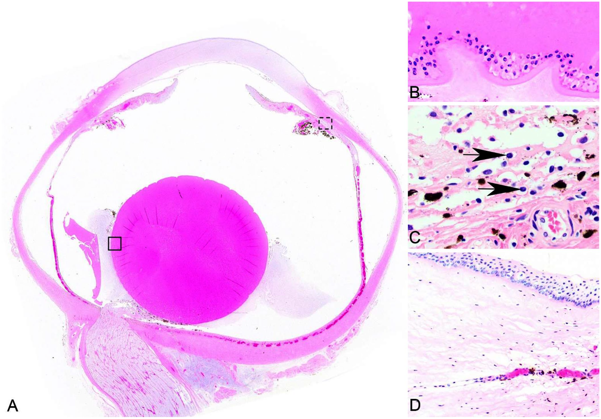
Spherophakia and hypermature cataract of the left eye of a wild muskox (Ovibos moschatus) neonate from the Northwest Territories, Canada. H&E.
Compared to the grossly normal haired skin, samples from alopecic areas had more superficial hair bulbs and increased prominence of dermal papillae, indicative of the telogen phase of the hair cycle. Additional lesions included moderate lymphoid depletion of the thymus, spleen, and mesenteric lymph node. Paraffin-embedded skin tissue, submitted to the Animal Health Laboratory, University of Guelph (Guelph, Ontario, Canada) for bovine viral diarrhea virus (BVDV) immunohistochemistry (a test that may cross-react with other pestiviruses), was negative. Trace mineral and vitamin E analysis and routine bacterial culture at Prairie Diagnostic Services (Saskatoon, Saskatchewan, Canada) were within normal limits (compared to domestic species), and significant bacterial pathogens were not identified. Vitamin A levels were not assessed in this analysis.
Spherophakia is a congenital lesion wherein the lens is round instead of oval and is accompanied by cataract. 9 Congenital cataracts develop in utero, whereas developmental cataracts are the result of heritable genetic defects that cause bilateral lesions any time after birth. 9 Causes of congenital cataracts include sporadic defects in organogenesis caused by in utero viral infections, toxin exposure, nutritional deficiencies, and genetic mutations; they are often associated with additional developmental abnormalities. 9 No other congenital abnormalities were identified in this calf. Based on its age, the absence of systemic disease, and no identifiable trace mineral or vitamin E deficiencies, the cataract was assumed to be congenital, with uveitis and keratitis developing secondarily. Alopecia was attributed to starvation, stress, and/or potentially excessive grooming activities by adult conspecifics given that it had an apparently normal hair coat at the time of capture (~2 wk prior to death).
Muskoxen are susceptible to gammaherpesvirus (malignant catarrhal fever viruses; Rhadinovirus), pestivirus (such as Bovine viral diarrhea virus, BVDV), and orbivirus (Bluetongue virus) infections, all of which could induce panuveitis. 1 However, we considered infectious causes for the cataract in this calf as unlikely. Immunohistochemical labeling of tissues for pestivirus was negative, and no other lesions were consistent with gammaherpesvirus or orbivirus infection.
Congenital or neonatal cataracts are described only rarely in free-ranging wildlife. Reports include a harbor seal (Phoca vitulina richardsi), a coyote (Canis latrans), and white-tailed deer (Odocoileus virginianus), with no identified cause.4,7,10 Among domestic species related to muskoxen, congenital cataracts are best described in cattle and have been associated with multiparous dams, birth in warmer months, hypovitaminosis A, BVDV infection, inbreeding, and genetic mutations.2,9,12–14 In an investigation of congenital cataracts in Ayrshire dairy cattle, the highest prevalence occurred in bull calves, which suggests the potential for X-linked inheritance. 12 Although the calf in our case was a male, it is uncertain what role sex may have in the development of cataracts in muskox.
In domestic and captive animals, congenital cataracts have also been attributed to genetic abnormalities, particularly in species with low genetic diversity. For example, “puppy eye syndrome” in Portuguese Water Dogs features bilateral congenital cataracts that are linked to a genetic anterior segment malformation. 16 Hereditary cataracts have been identified in closely related captive-bred vervet monkeys (syn. grivet; Chlorocebus aethiops). 6 Muskoxen have experienced multiple population bottlenecks, resulting in extremely low genetic diversity, 15 which could contribute to congenital abnormalities including cataracts, as seen in this calf.
In an interesting parallel, human studies have identified that a 5°C increase in environmental temperature significantly increases the risk of congenital cataracts in infants if the temperature is high during the critical lens development period (during organogenesis that occurs 3–8 wk following conception). 17 Abnormally warm temperatures are occurring across Canada and are particularly high in northern areas, where the annual average temperature has increased by 2.3°C from 1948–2016, which is more than 3 times the global average. 18 Temperature increases range from 4–6°C during winter for northern areas including the Northwest Territories, where this case originated. 18 This creates the potential for abnormally high heat exposure during gestation for muskoxen, which typically breed in August and September followed by an ~8-mo gestational period. 11 Lens organogenesis in domestic cattle occurs at 30–60 d of gestation. 3 Assuming lens development in the muskox is similar, this may be a highly susceptible time for muskox fetuses to develop lens abnormalities, which corresponds to late fall and early winter, when temperatures are abnormally high. During the presumed critical window of this muskox’s lens development (Sept–Nov 2018), the daily average temperature was 0.2°C, which was warmer than the daily average temperature of –0.4°C over the same months from 2007–2017. 8 Temperatures are projected to increase a further 2–6°C across Canada within the next 50 y, 18 which will place further heat stress on the early gestational period of muskoxen. Although it is unknown if unusually warm temperatures as a result of climate change contributed to this calf’s cataract, investigations of future neonatal mortality in northern regions should include microscopic examination of eyes.
Our case demonstrates that ocular lesions might be underrecognized causes of morbidity and mortality in muskoxen, as well as other wildlife species. It is possible that this calf was abandoned, a notable occurrence given the strong maternal bonds that muskoxen form. 11 However, maternal death cannot be definitively excluded, although the individuals that found the calf did not describe the presence of adult carcass(es). Blind but otherwise healthy neonates can be expected to seek an udder and suckle, and therefore cataracts and blindness may or may not have been the cause of abandonment and eventual starvation and death of this calf. The etiology of the cataract is unknown; however, our case highlights that ocular lesions in free-ranging neonates may be important factors affecting survival. The threat of climate change, pathogen emergence, including parasites and infectious disease, and low genetic diversity have placed stress on muskox populations in northern Canada. 5
Footnotes
Acknowledgements
We thank Susan Calder-Lodge, Jennifer Larios, Melencio Nicolas, Jim Carlsen, Betty Pollock, and Pawel (Paul) Gajda for invaluable technical support. Our case was presented in abstract form at the 2019 annual meeting of the American College of Veterinary Pathologists.
Declaration of conflicting interests
The authors declared no potential conflicts of interest with respect to the research, authorship, and/or publication of this article.
Funding
Our work was supported by the University of Calgary Faculty of Veterinary Medicine, the Canadian Wildlife Health Cooperative, and the Government of the Northwest Territories.