
Abstract
Chlamydial infections in crocodiles have been described in several countries and in several different species. These are typically associated with severe pharyngitis and conjunctivitis, with death occurring secondary to compromise of the upper respiratory tract due to obstruction of the trachea. A population of ranched Siamese crocodiles in central Thailand experienced an epizootic of sudden death in juvenile animals. The affected animals had fulminant systemic disease primarily involving the liver and spleen but also affecting the kidneys, heart, and the whole of the respiratory tract. Chlamydia sp. were noted in liver and spleen during histopathological examination and confirmed with transmission electron microscopy and polymerase chain reaction (PCR). The sequence of the PCR product suggested a novel Chlamydia sp. of Siamese crocodiles. Crocodile farming represents an important economy in several parts of the world. Epizootics, such as the one described in this manuscript in association with Chlamydia sp., can have devastating impact on the industry and represent a potential zoonosis of significant public health concern. This is the first report of Chlamydia sp. and Aeromonas sobria causing systemic disease in crocodiles as well as the first histopathological and ultrastructural description of Chlamydia infection in Siamese crocodiles.
Chlamydia sp. have been implicated in severe disease and even death in different species of hatchling and juvenile crocodiles in Africa, Papua New Guinea, and Australia. 4,5,7 The bacterium has been reported to cause disease either as a primary agent or in synchrony with other bacteria or viruses. 6,7 Over the course of 2012, a crocodile farm in the Prachinburi Province of Thailand lost 493 hatchling Siamese crocodiles (Crocodylus siamensis) out of a total population of 500 animals. The etiology was initially undetermined; however, recent testing of archival tissues submitted to the Thai National Institute of Animal Health (NIAH) has subsequently positively identified Chlamydia sp. by polymerase chain reaction (PCR). In 2013, the same farm lost 14 juvenile Siamese crocodiles out of a total population of 300 animals. Three animals from this second epidemic were necropsied and submitted to the NIAH for further diagnostics; tissue samples from 2 of the animals were fixed in formalin for histopathologic and ultrastructural evaluation by a pathologist (E.D.L.). Fresh and frozen tissue samples from all 3 animals were collected for molecular analysis at the NIAH. All 3 animals had been housed in the same cement tank and were aged between 1 and 3 years. All diagnostics discussed in this report were conducted on material from these 3 juvenile crocodiles sampled during the 2013 epizootic.
Chlamydial infections in crocodiles are typically associated with severe granulomatous, necrotizing pharyngitis, and conjunctivitis, 4,5,7 with death occurring secondary to compromise of the upper respiratory tract. In these instances in Thailand however, those animals examined had fulminant systemic disease, primarily involving the liver and spleen but also affecting the kidneys, heart, and the whole of the respiratory tract.
A complete necropsy was conducted on 3 crocodiles. Unfortunately, due to cultural practices in Thailand, there is only rare euthanasia of animals, and as such, there is frequently a delay between death and necropsy. The exact interval between death and necropsy was unknown but based on the condition of the submitted tissues is assumed to be approximately 12 hours. Representative samples of spleen, liver, trachea, lung, heart, tonsil, thymus, kidney, and gastrointestinal sections were harvested from 2 of the animals, fixed in 10% neutral buffered formalin, and processed in accordance with standard procedures. Aeromonas sobria was isolated from culture (with species identification by biochemical methods) of samples of liver, spleen, and kidney collected at necropsy from those 2 animals, while samples from all 3 animals were collected and frozen for molecular analysis.
Sections of formalin-fixed liver and spleen from the 2 crocodiles were processed and examined using a JEOL 1400 transmission electron microscope. Samples from the 3 animals from the same outbreak were evaluated by PCR and sequenced for the ompA gene, 16 S gene, and 16S-23 S rRNA interspacer of Chlamydia spp. using techniques described by Pudjiatmoko et al. 8 PCR products were amplified from kidney, liver, and splenic tissue samples derived from the 3 crocodiles. The partial sequences of 16 S rRNA gene, intergenic spacer, and domain I of 23 S rRNA gene assembled by using MEGA 6 were 2403 bp. The sequences had maximum score (4283 bits) and demonstrated combined 99% homology (2375/2403) to Chlamydia caviae GPIC (NCBI accession no. AE015925). Analysis of a partial 16 S rRNA sequence of the chlamydial organism in this study by BlastN showed 99% identity (4 nucleotide differences) to 11 Chlamydia sp. isolates from Siamese crocodiles from Thailand, 97% identity to C. caviae GPIC (accession no. AE015925) (1536/1545), and 96% identity to C. psittaci GPIC (accession no. CP003790) (1535/1545). The sequences of intergenic spacer had 97% homology (217/224) to C. caviae and 96% homology (216/224) to C. psittaci. The sequences of domain I of 23 S rRNA gene also demonstrated 99% homology (625/634) to C. caviae and 97% homology (612/634) to C. psittaci. In addition, the partial sequences of ompA gene from these crocodiles demonstrated only 81% homology to C. felis and C. psittaci (736/911). These results also identified 99% homology (1503/1505) for the 16 S rRNA gene as well as 100% homology (808/808) for the partial sequence of 16S-23 S interspacer to those Siamese crocodiles submitted by Mahidol University to GenBank in the concurrent but separate outbreak evaluated by Sariya et al. 9 Phylogenetic analysis of 16S-interspacer-23 S sequences compared these cases with other members of Chlamydiaceae (Fig. 1). The phylogeny was reconstructed using the maximum likelihood statistic method and the bootstrap test for 1000 replications using MEGA version 5.1. It is clear that the infective organism in these animals was in the Chlamydia genus and likely the same species as other reported but unnamed isolates from Siamese crocodiles rather than either C. caviae or C. psittaci.
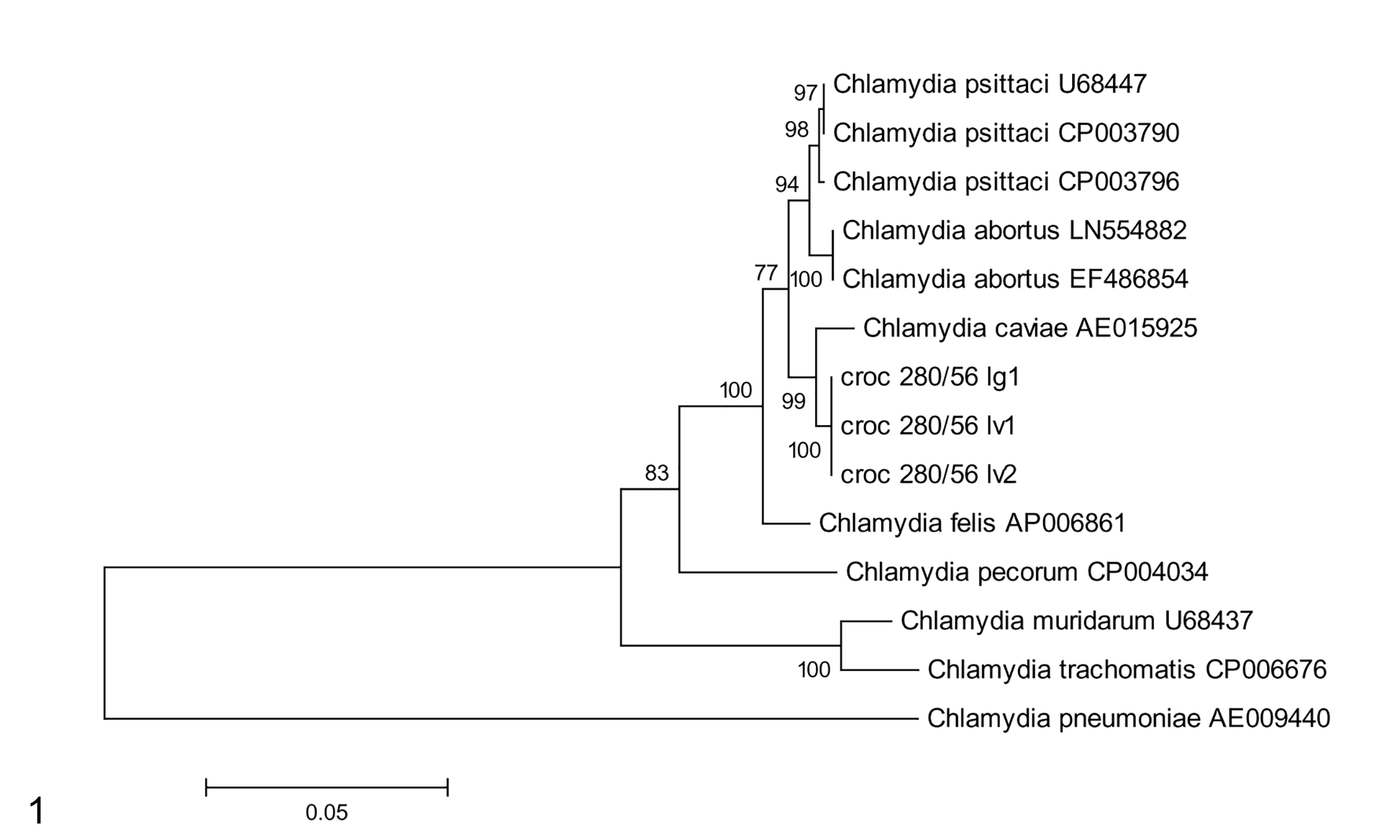
Phylogenetic analysis of 16S-interspacer-23 S sequences comparing the current cases in Siamese crocodiles (croc 280/56) with other Chlamydiaceae gene sequences. The numbers represent the percentage measure of support for each node; high numbers indicate strong evidence that the corresponding gene sequences cluster to the exclusion of other sequences. Scale bar indicates nucleotide substitutions per site. The phylogeny was reconstructed using the maximum likelihood statistic method and the bootstrap test for 1000 replications using MEGA version 5.1.
Additional evaluation of the kidney, liver, and splenic samples found that they were PCR-negative for West Nile virus, crocodile poxvirus, crocodile adenovirus, and endogenous retroviridae.
Macroscopic findings in the 3 necropsied animals included marked erosion and ulceration of the scaled skin around the head, fibrinous pleuritis, and necrotizing tracheitis and pharyngitis. The animals also had evidence of mild necrotizing stomatitis, and 1 animal presented with severe fibrinous hepatitis with adhesion of abdominal viscera to the hepatic capsule, while another animal was observed to have hydropericardium and coelomic ascites.
Histopathologic evaluation of the selected tissues from 2 animals revealed severe necrotizing lymphohistiocytic and heterophilic hepatitis that was predominantly concentrated on the portal areas but frequently encompassed the entire lobule. Additionally, there was frequent hepatic thrombosis and frequent intrahistiocytic intracytoplasmic amphophilic to basophilic granular material interpreted to be bacteria in which individual organisms measured less than 1 μm (Fig. 2). Additionally, there was severe diffuse histiocytic, heterophilic, and lymphocytic splenitis with intrahistiocytic, intracytoplasmic pigment interpreted as hemosiderin as well as erythrophagocytosis, thrombosis, and frequent intrahistiocytic intracytoplasmic bacteria. Both animals had a marked histiocytic interstitial pneumonia with thrombosis, hemosiderosis, and intracytoplasmic bacteria within the histiocytes as well as a multifocal to coalescing necrotizing lymphohistiocytic myocarditis. Vasculitis was observed in multiple tissues, but in both the spleen and the liver, the extensive and coalescing necrosis often masked the specific vascular mural injury. There was a lesser degree of vasculitis and thrombosis in the kidneys, thymus, stomach (Fig. 3), and trachea. Additional findings included necrotizing tonsillitis and multifocal enteritis in both animals.
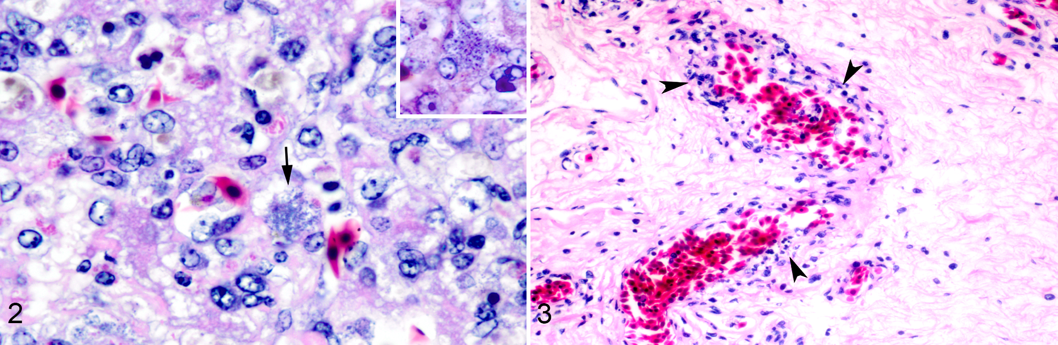
Chlamydiosis, Siamese crocodile.
Transmission electron microscopy of sections of the liver demonstrated several 300- to 400-nm diameter circular organisms comprised either Chlamydia sp. elementary bodies (EB), reticulate bodies (RB), and intermediate bodies (IB). The structures interpreted as EB had an electron-dense nucleoid, which was often peripheralized and surrounded by an electron-lucent halo and further bounded by electron dense cytoplasm (Fig. 4). Additionally, several RB were observed undergoing binary fission with a characteristic hourglass shape (Fig. 4). Finally, admixed within aggregates of the EB and RB were occasional IB, which were comprised of a central intensely electron-dense core, bounded by a thin clear halo and further bounded by tightly pack peripheral cytoplasmic granules and a thin cell wall (Fig. 5). The cellular material was often fragmented and nondiagnostic, although there appeared to be swollen membrane-bound structures that were interpreted to be ghost mitochondria. No evidence of the cultured aeromonad bacterium was observed ultrastructurally.
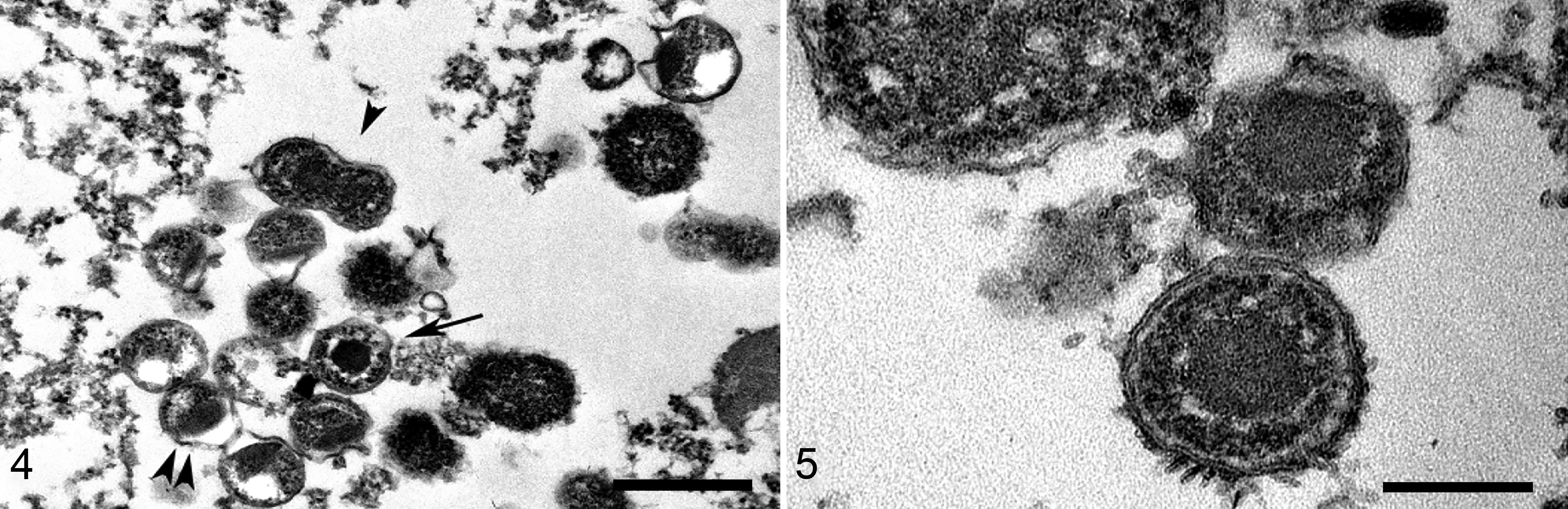
Chlamydiosis, liver, Siamese crocodile.
While chlamydial infections in hatchling and juvenile Indo-Pacific crocodiles (Crocodylus porosus) from Australia and Papau New Guinea 5 –7 and Nile crocodiles (Crocodylus niloticus) from South Africa and Zimbabwe have been described, 4 the reported lesions were typically associated with the conjunctiva and upper respiratory tract, with death occurring secondary to advanced respiratory disease involving occlusion of the trachea by fibrinous exudate. The fact that the pharynx appears to be directly targeted is significant in that both the particular anatomy of the crocodile pharynx and the inability of crocodiles to cough make them particularly susceptible to obstruction. 6 Similar to the findings in this epizootic in Thailand, the outbreak of Chlamydia sp. in hatchling Nile crocodiles in Zimbabwe presented with a severe necrotizing lymphoplasmacytic hepatitis without evidence of conjunctivitis. 4 A large study conducted in both farmed and wild estuarine crocodiles in northern Australia employed molecular techniques to attempt to identify the causative species of fatal “eye and throat” disease. Their findings suggest that there are unique crocodile strains of Chlamydia and that clinically normal animals may act as carriers. Furthermore, the authors suggested that the crocodile Chlamydia may act either as an opportunist or in concert with other pathogens. 7
Crocodile farming in central Thailand is an important industry and one that is affected by a significant amount of unexpected mortality. Other groups who have conducted necropsies of young farmed crocodiles with unexplained sudden death have noted a disease presentation more consistent with the descriptions of conjunctivitis-pharyngitis in South Africa, Papau New Guinea, and Australia. It is important to note that while none of the viral agents associated with crocodile disease were found, A. sobria was cultured from these animals and may have played an important role in the propagation and exacerbation of the disease in these animals.
In birds, C. psittaci is transmitted through inhalation or ingestion of infective particles found in nasal and cloacal discharge, feather dust, and fecal matter. Affected animals develop severe disease comprised primarily of histiocytic inflammation in multiple organ systems. 1 –3,10 The mechanism of transmission in crocodiles has not been established; however, we posit that overcrowding of hatchling animals combined with poor water quality and concurrent opportunistic pathogens all contributed to the perpetuation and severity of disease in crocodiles. The water quality on the farms was extremely poor and combined with overcrowding could certainly induce significant stress and allow for suppression of the immune system and co-infection with both Chlamydia sp. and A. sobria in juvenile animals. As such, it is possible that unidentified pathogens or other environmental factors contributed to the skin disease in these cases as well as potentially playing a role in exacerbating the systemic pathology.
This report is the first histopathological and ultrastructural description of Chlamydia spp. infecting Siamese crocodiles, and the first report of combined Chlamydia spp. and A. sobria associated systemic disease affecting all of the examined viscera in crocodiles.
Footnotes
Disclaimer
E.K. Morris is a major and E.D. Lombardini a lieutenant colonel in the US Army. The opinions or assertions herein are those of the authors and do not necessarily reflect the view of the Department of the Army or the Department of Defense.
Declaration of Conflicting Interests
The author(s) declared no potential conflicts of interest with respect to the research, authorship, and/or publication of this article.
Funding
The author(s) received no financial support for the research, authorship, and/or publication of this article.