
Abstract
Uteri from 50 four-toed hedgehogs (Atelerix albiventris) with clinical signs of uterine disease were histopathologically examined. Sixteen animals (32%) were diagnosed with endometrial hyperplasia, 7 animals (14%) were diagnosed with endometrial polyp, and 27 animals (54%) were diagnosed with endometrial neoplasia. The mean ages of the animals with endometrial hyperplasia, polyp, and neoplasia were 28.7 months, 29.4 months, and 25.2 months, respectively. The neoplasms were classified into 7 endometrial mixed tumors, 12 endometrial stromal nodules, and 8 endometrial stromal sarcomas. However, the endometrial stromal nodules and endometrial stromal sarcomas often developed within or were contiguous with an endometrial polyp or mixed tumor. Interestingly, the stromal tumors and the stromal components of the endometrial polyp and mixed tumor displayed extraendometrial differentiation (eg, into adipocytes, granular cells, smooth muscle cells, and osteoid tissue). The endometrial stromal sarcomas exhibited severe cellular atypia and invaded subendometrial tissue. Immunohistochemical examinations demonstrated that the stromal cells of the hyperplastic lesions as well as the neoplastic lesions were positive for CD10, the progesterone receptor, and Wilms tumor 1. The four-toed hedgehog develops unique uterine neoplasms that are mainly composed of endometrial stromal cells and probably arise from endometrial polyps and/or mixed tumors.
In recent years, the four-toed hedgehog (Atelerix albiventris) has become a popular household pet in Japan. At exotic animal clinics, uterine tumors are commonly encountered in this animal species. Interestingly, the four-toed hedgehog is known to develop mixed uterine tumors and endometrial stromal tumors. 34
Uterine mixed tumors, consisting of both malignant epithelial and mesenchymal elements, have been reported in cats, 36,37,40 rabbits, 20,57 rodents, 25,54 and a sow. 8 However, tumors composed of endometrial stromal cells have only been reported in a chimpanzee, 52 a cat, 46 and rodents. 42 To our knowledge, endometrial stromal tumors have never been immunohistochemically characterized in animals.
The current histological classification categorizes uterine proliferative lesions into epithelial tumors and precursors, mesenchymal tumors, and mixed epithelial and mesenchymal tumors. 29 Epithelial tumors and precursors include endometrial hyperplasia, tumor-like lesions (polyp), and endometrial carcinomas. Endometrial hyperplasia is a significant precancerous lesion in humans. However, it is not considered so in domestic animals because endometrial hyperplasia is very common (especially the cystic type), but endometrial carcinoma is rather rare except in the rabbit. 5,56 Segmental hyperplasia is a localized proliferation of the endometrial glands that has been described in the dog. 47 Endometrial polyp is commonly seen in the dog and the cat and is suggested to arise from cystic endometrial hyperplasia and does not show any preneoplastic changes. 18,32 Mesenchymal tumors include endometrial stromal tumors and smooth muscle tumors. As for neoplastic lesions of the uterus, leiomyoma is the most common in domestic animals. 26,42 Endometrial stromal tumors are further classified into endometrial nodule and endometrial sarcoma (low and high grade), based on the infiltrative growth pattern invading the myometrium in endometrial sarcoma. 14 Mixed epithelial and mesenchymal tumors are further classified based on their benign or malignant nature of epithelial and mesenchymal components (ie, carcinosarcoma, adenosarcoma, and others). However, carcinosarcoma (previously called malignant mixed Müllerian tumor) is considered a subset of endometrial carcinoma based on evidence of the monoclonality of their epithelial and stromal components. 23 Subinvolution of placental site and placental site nodule should be differentiated from the above proliferative lesions of the endometrium.
The uterine endometrium is composed of epithelial cells and stromal cells, which are characterized by cytokeratin and CD10 expression, respectively, in both mouse and human. 21 Stromal cells interact with epithelial cells during development as well as during the pathological and physiological proliferation of the endometrium. 4,16,50 Moreover, experimental studies of mouse and human cells have suggested that endometrial stem cells can differentiate into both epithelial and stromal cells. 17 Wilms tumor (WT) 1, a tumor suppressor protein, plays an essential role in the development of the urogenital system. 27,28 A study of human endometrial stromal cells has shown that the expression of WT1 in endometrial stromal cells is regulated by progesterone via the progesterone receptor (PR). 3
In the present study, we examined the uteri of 50 four-toed hedgehogs with clinical signs of uterine disease. The clinical and histopathological findings of the animals’ endometrial cells, including their immunohistochemical phenotypes, are described.
Materials and Methods
Tissue Samples and Histopathology
Uteri from 50 four-toed hedgehogs that were collected at Miwa Exotic Animal Hospital between 2014 and 2016 were included in this study. The ages of the animals ranged from 5 to 51 months (mean age, 27.0 months). In all cases, ovariohysterectomy was conducted via a laparotomy (a median incision), and the abdominal cavity was explored for other lesions. All surgical procedures were performed under isoflurane-induced general anesthesia. The collected tissues were readily fixed in 10% neutral-buffered formalin and sent to the Laboratory of Veterinary Pathology, University of Tokyo, for pathological examination. The tissues were routinely embedded in paraffin wax and cut into 4-μm-thick serial sections, and deparaffinized sections were stained with hematoxylin and eosin (HE). The resultant slides were reviewed by 2 veterinary pathologists certified by the Japanese College of Veterinary Pathologists (J.K.C. and K.U.). Histopathological diagnoses were made on the consensus of the 2 pathologists based on the following criteria. Thickening of the endometrium without any solid mass was diagnosed as endometrial hyperplasia. Intraluminal mass attached to the endometrium by a thin stalk (less than one-fourth of the endometrial surface) was diagnosed as endometrial polyp. Mural mass of the uterus that did not form a polyp (ie, involved more than one-fourth of the endometrial surface) was diagnosed as endometrial neoplasia. A neoplasm composed of both endometrial epithelial and stromal cells was diagnosed as endometrial mixed tumor. A well-demarcated neoplasm composed solely of stromal cells was diagnosed as endometrial stromal nodule. A nondemarcated neoplasm composed solely of endometrial stromal cells with significant atypia was diagnosed as endometrial stromal sarcoma. If a mass was composed of both hyperplastic and neoplastic lesions, it was diagnosed as neoplastic.
Immunohistochemistry
After deparaffinization and antigen retrieval, the sections were treated with 3% hydrogen peroxide in methanol at room temperature for 5 minutes and then incubated with 8% skimmed milk in Tris-buffered saline at 37°C for 40 minutes to block nonspecific reactions. Antigen retrieval was performed except for anti–smooth muscle actin (SMA) antibody by heating the sections with an autoclave, in pH 6.0 citrate buffer, at 121°C for 10 minutes. Subsequently, the sections were incubated with primary antibodies at 4°C overnight. The following primary antibodies were used in this study: mouse anti-human CD10 antibody (clone 56C6; Invitrogen, Camarillo, CA), mouse anti-human SMA antibody (clone 1A4; Dako, Tokyo, Japan), mouse anti-human PR antibody (clone hPRa2/hPRa3; Thermo Scientific, Fremont, CA), mouse anti-human cytokeratin antibody (clone AE1/AE3; Dako), rabbit anti-human WT1 antibody (C-19; Santa Cruz Biotechnology, Dallas, TX), mouse anti-human estrogen receptor (ER) antibody (clone 6F11; Leica, Newcastle Upon Tyne, UK), mouse anti-rat calretinin antibody (clone 6B8.2; Millipore, Temecula, CA), mouse anti-human inhibin α antibody (clone R1; AbD Serotec, Oxford, UK), rabbit anti-cow S100 antibody (Dako), and rabbit anti-human CD117 antibody (Dako). After being washed in Tris-buffered saline, the sections were then incubated with horseradish peroxidase–labeled polymer (Dako Envision System; Dako). Finally, the reaction products were visualized with 0.05% 3-3′-diaminobenzidine and 0.03% hydrogen peroxide in Tris/HCl buffer, and then the sections were counterstained with Mayer’s hematoxylin. For positive control tissue, formalin-fixed uterus of hedgehog with hyperplastic endometrium was used. For negative controls, primary antibody was replaced by antibody diluent, irrelevant antibody, and buffer. 43 The limitation of this study is that the primary antibodies were raised against human proteins and thus may have bound to unrelated various antigens of hedgehog.
Results
Clinical Findings
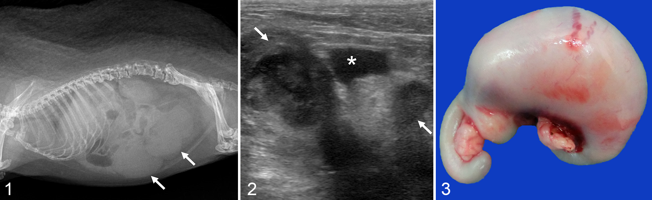
At the time of their clinical presentation, 43 of 50 animals (86%) exhibited hematuria or blood attached to the vulva, indicating hematometria. These signs were observed in 62.5% (10/16) of cases with endometrial hyperplasia, 100.0% (7/7) of cases with endometrial polyp, and 96.2% (26/27) of cases with endometrial neoplasia (Suppl. Table S1). The animals’ other clinical signs included colpoptosis, abdominal distention, and other non-specific signs, such as a reduction in activity, anorexia, and soft feces. Radiography was employed in 42 cases, and enlargement of the uterus was suspected in 8 cases (Fig. 1). Ultrasonography was performed in 40 cases, and enlargement of the uterus or an abdominal mass was suspected in 33 cases (Fig. 2).
Preanesthetic blood tests were conducted in 15 cases. The mean white blood cell count was 12,700/μl (reference value: 11,000 ± 6000/μl), the mean packed cell volume was 28.1% (reference value: 36 ± 7), and the mean total protein level was 5.3 g/dl (reference value: 5.8 ± 0.7 g/dl). 9 Reproductive history was available in 48 cases: 3 animals were parous, and 45 animals were nulliparous (Suppl. Table S1). In the 3 parous animals, deliveries were more than 1 year before clinical presentation. Placental sites were not found grossly or microscopically in any of the cases that were examined.
Ascites was observed during surgery in 4 cases. Marked enlargement of the uterus was grossly observed in 35 cases (Fig. 3). Three of 50 animals died within 14 days of surgery (2 cases of endometrial mixed tumor and 1 case of endometrial stromal sarcoma). The other 47 animals recovered, and their sutures were removed at 10 to 14 days after surgery. Fourteen animals died during the study (3 cases of endometrial hyperplasia, 3 cases of endometrial polyp, 4 cases of endometrial mixed tumor, and 4 cases of endometrial stromal sarcoma), but no necropsy procedures were performed (Suppl. Table S1). Two animals that were diagnosed with endometrial stromal nodules developed suspected recurrent tumors involving the vagina and vulva. One animal developed a mass at 25 days after undergoing ovariohysterectomy.
The mass was surgically resected at 59 postoperative days, and it was diagnosed as an invasive lesion of endometrial stromal sarcoma. The other animal developed a mass at 183 postoperative days; however, no histopathological examination was performed. Two animals that were diagnosed with endometrial stromal sarcomas developed suspected recurrent tumors. One animal developed a suspected recurrent tumor in the cervix. The tumor was surgically resected at 346 postoperative days and was diagnosed as an invasive lesion of endometrial stromal sarcoma. The other animal developed a suspected recurrent tumor in the lower abdomen. The tumor was surgically resected at 364 postoperative days and again at 441 postoperative days, and both were diagnosed as invasive lesion of endometrial stromal sarcoma. Survival rates at 180 days after ovariohysterectomy were 66.6% (6/9) in endometrial hyperplasia, 100.0% (4/4) in endometrial polyp, 60.0% (3/5) in endometrial mixed tumor, 100.0% (3/3) in endometrial stromal nodule, and 42.9% (3/7) in endometrial stromal sarcoma (Suppl. Table S1).
Histopathology and Immunohistochemistry
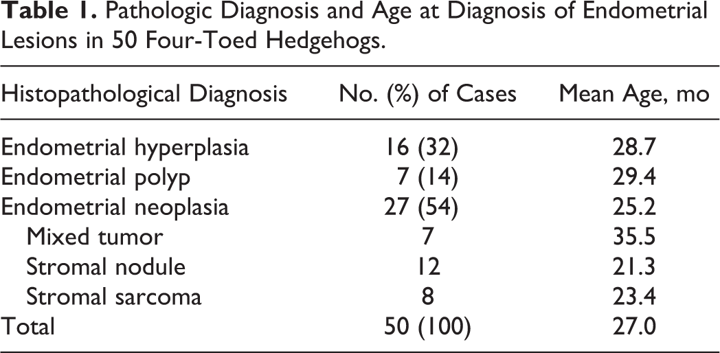
Based on the histopathological findings, 16 animals (32%) were diagnosed with endometrial hyperplasia and 7 animals (14%) were diagnosed with endometrial polyp. Twenty-seven animals (54%) were diagnosed with neoplasia: 7 (14%) endometrial mixed tumor, 12 (24%) endometrial stromal nodule, and 8 (16%) endometrial stromal sarcoma. The mean ages of the animals were follows: endometrial hyperplasia, 28.7 months; endometrial polyp, 29.4 months; endometrial mixed tumor, 35.5 months; endometrial stromal nodule, 21.3 months; and endometrial stromal sarcoma, 23.4 months (Table 1).
Pathologic Diagnosis and Age at Diagnosis of Endometrial Lesions in 50 Four-Toed Hedgehogs.
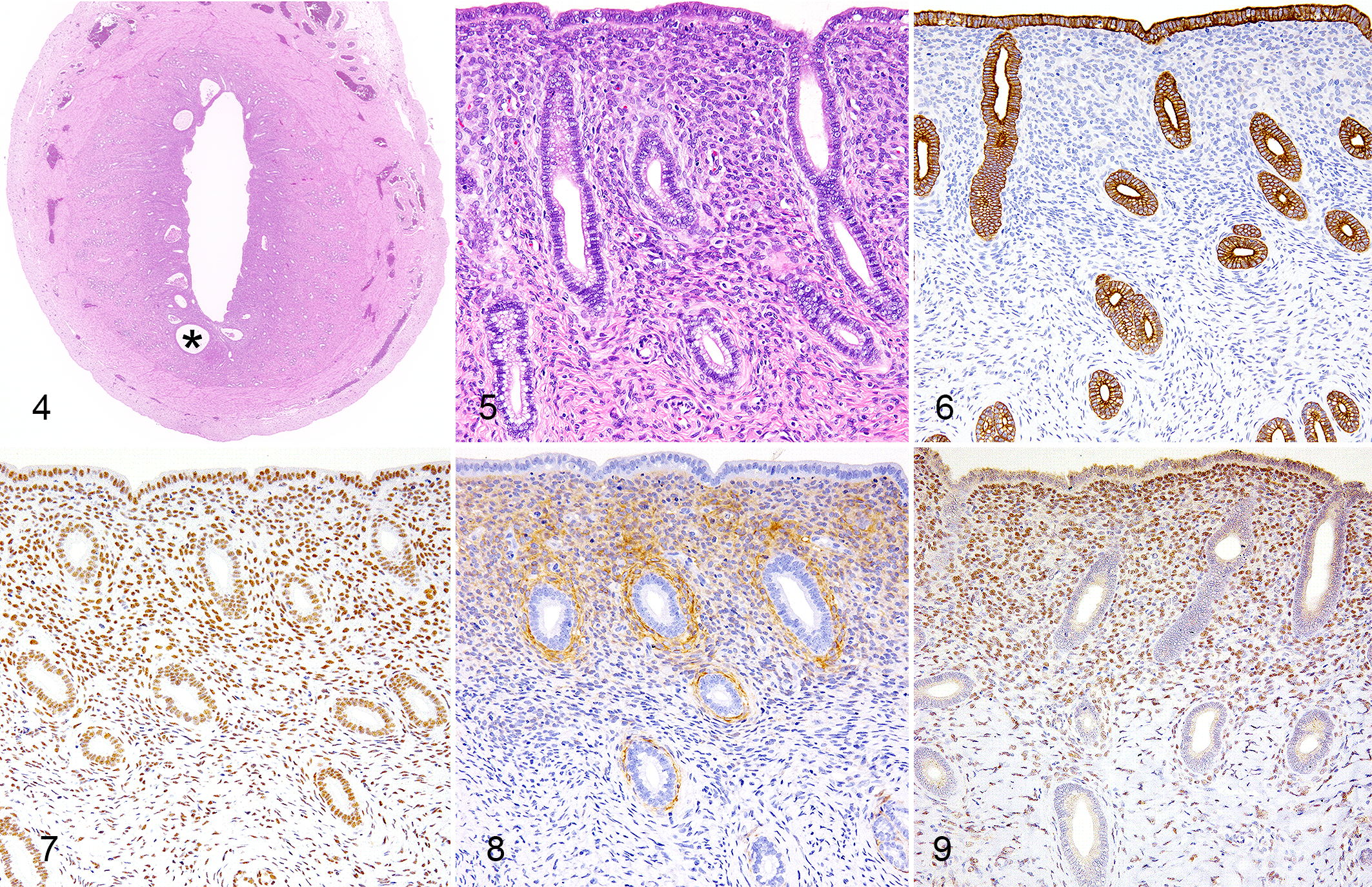
In endometrial hyperplasia, the endometrium was thickened but well organized and did not form any distinct masses (Fig. 4). Hyperplastic endometrium was composed of cuboidal-shaped epithelial cells and spindle-shaped stromal cells (Fig. 5). Dilation of the lumina of the endometrial glands was occasionally observed (Fig. 4). Immunohistochemical examinations demonstrated that the endometrial epithelial cells were positive for cytokeratin and PR but negative for CD10, WT1, and SMA (Figs. 6–9). On the other hand, the endometrial stromal cells were positive for PR, CD10, and WT1 but negative for cytokeratin and SMA (Figs. 6–9). Stromal cells were densely distributed beneath the surface epithelium and around the endometrial glands. The walls of the surrounding arteries as well as the muscular layer of the uterus were positive for SMA (not shown).
Endometrial polyps were composed of both epithelial and stromal cells together with fibrous tissue (Fig. 10). Epithelial cells were arranged in a single layer and often formed dilated glandular structures, and stromal cells surrounded the glandular structure (Fig. 11). Fibrous connective tissue was often edematous with dilated vessels and scant cellular component.
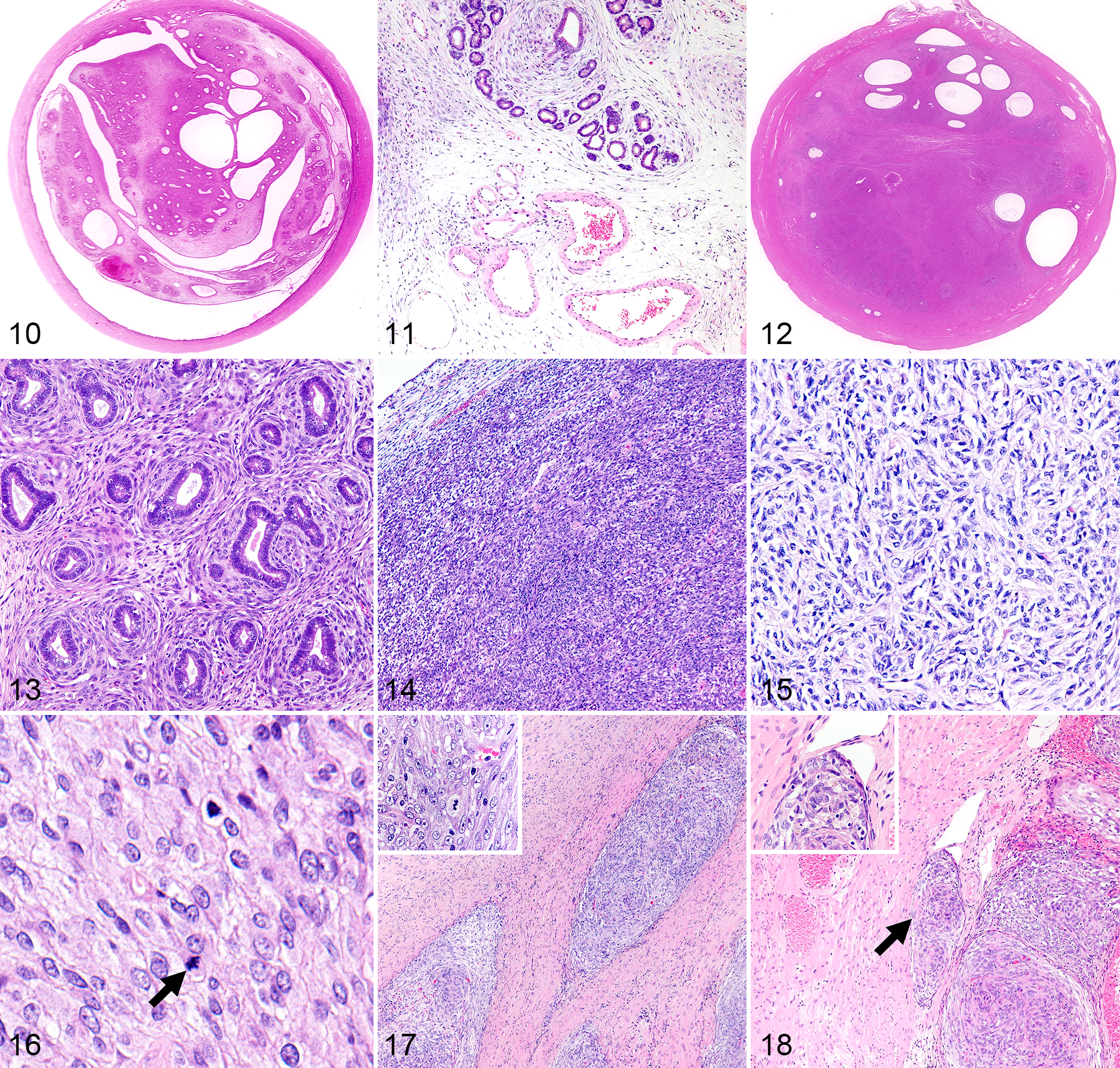
Mixed tumors were composed of both epithelial and stromal cells with higher cellularity compared to endometrial polyps (Fig. 12). Stromal cells were arranged in whorls surrounding the glandular structures (Fig. 13). The 2 components were always found together in the mixed tumors.
Endometrial stromal nodules lacked epithelial components and were solely composed of spindle-shaped stromal cells (Fig. 14). Stromal cells were densely packed in fascicles or a storiform pattern (Fig. 15). These cells were uniform, and their morphology was comparable to that of the stromal cells found in the mixed tumors. Modest cellular and nuclear atypia and occasional mitotic figures were found (Fig. 16).
Endometrial stromal sarcomas were composed of spindle-shaped cells. Necrosis and invasion into the uterine muscular layer and vasculature were often observed (Figs. 17, 18). The neoplastic cells displayed significant pleomorphism and atypia, such as anisocytosis, large nuclei, and prominent nucleoli (Fig. 19).
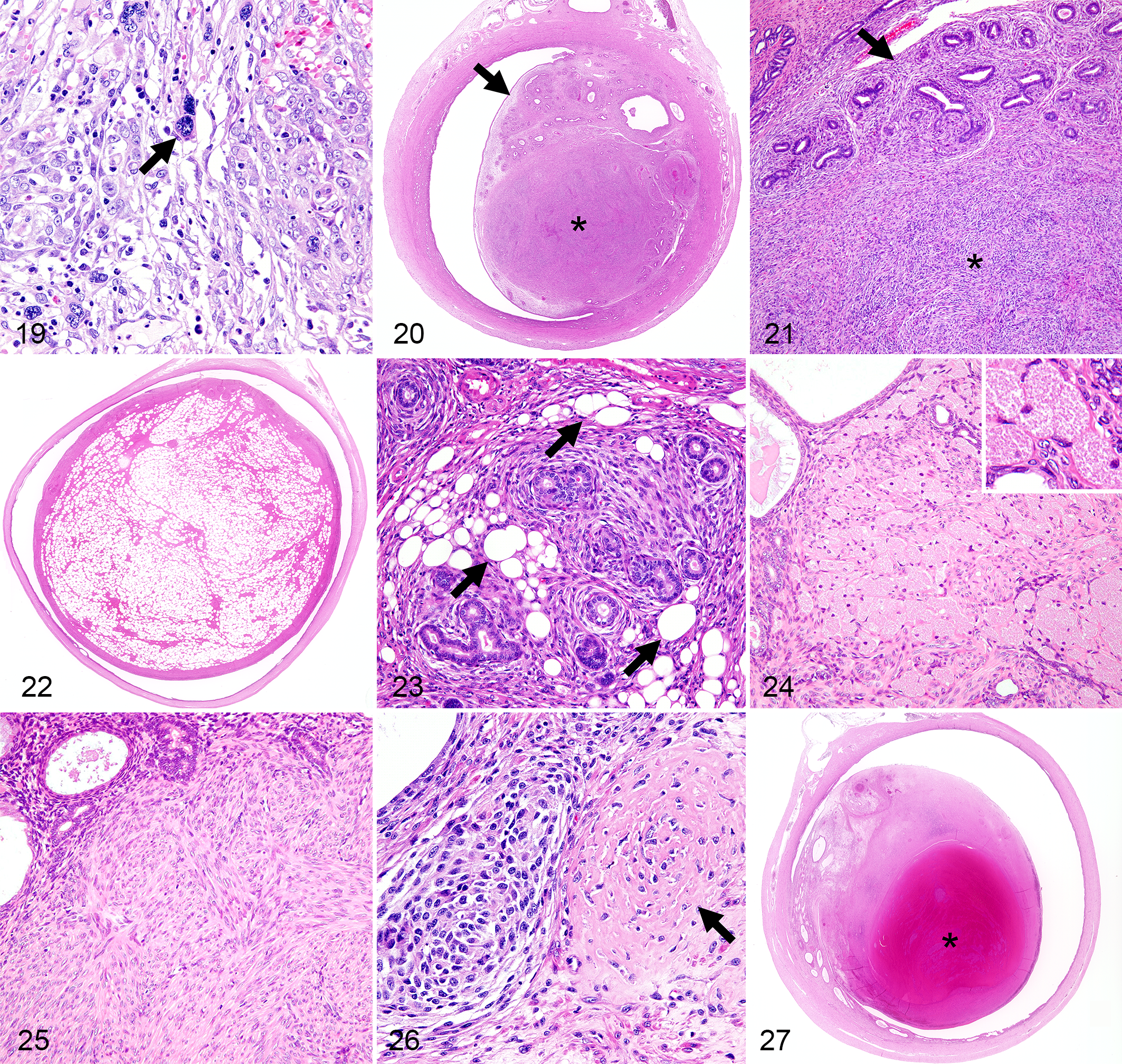
Endometrial stromal nodule and endometrial stromal sarcoma were often developed within or were contiguous with a polyp or a mixed tumor (Suppl. Table S1; Figs. 20, 21). In the endometrial polyps, mixed tumors, and stromal nodules, the tumor cells had differentiated into various types of cells, including adipocytes (Figs. 22, 23), granular cells (Fig. 24), smooth muscle cells (Fig. 25), and osteoid tissue (Fig. 26). The smooth muscle cells were positive for SMA. In 2 cases, more than 80% of the tumor tissue was composed of adipose cells (Fig. 22). Adipocytes, granular cells, smooth muscle cells, and osteoid tissue were observed in 12 cases, 1 case, 4 cases, and 2 cases, respectively. These components were well differentiated and did not exhibit any malignant findings, such as invasiveness, mitosis, or nuclear atypia. Hemorrhage was often observed in endometrial polyps and neoplastic lesions (Fig. 27).
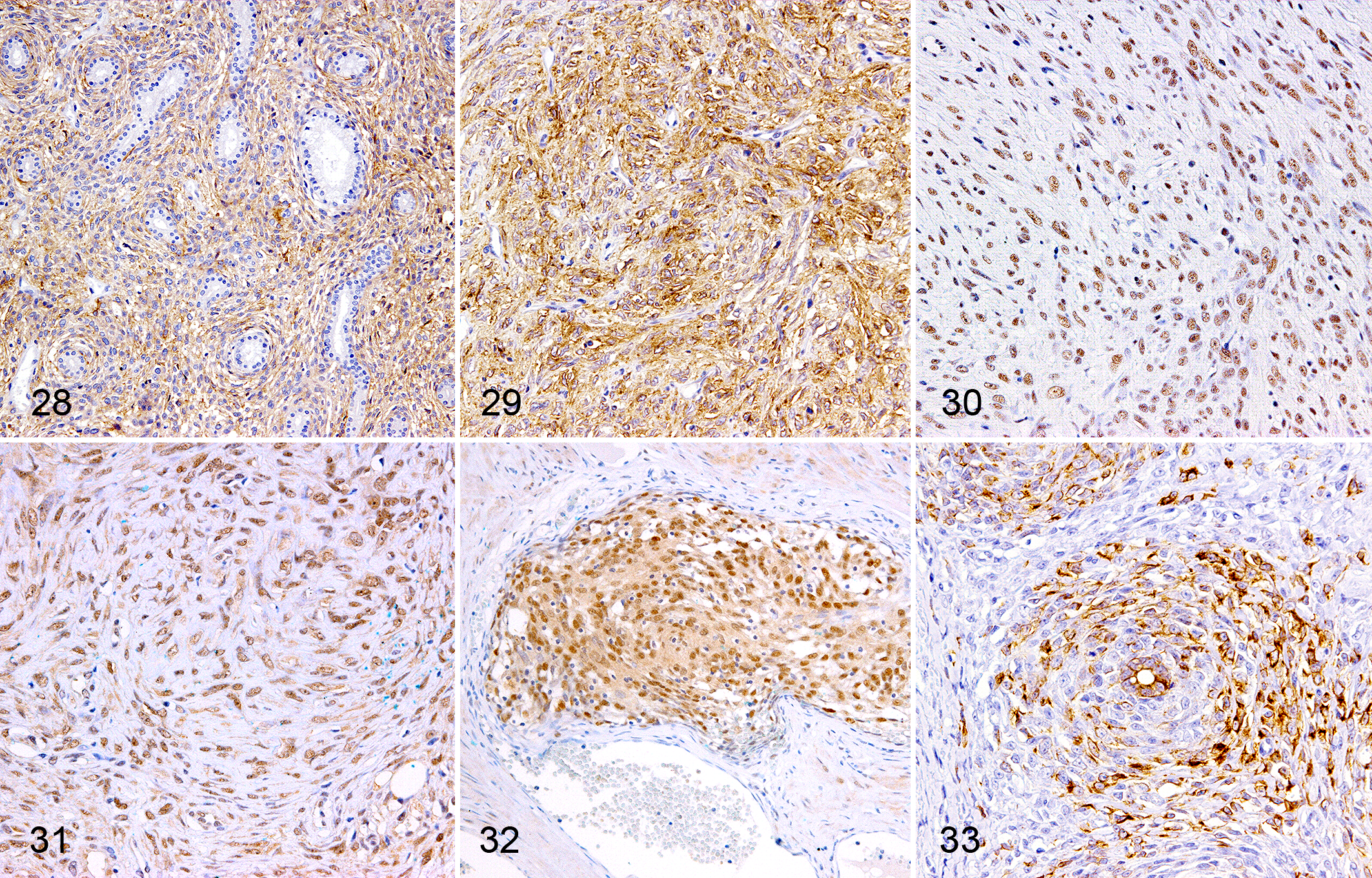
The spindle cells of the endometrial polyps, endometrial mixed tumors, and endometrial stromal nodules were positive for CD10 (Figs. 28, 29), PR (Fig. 30), and WT1 (Fig. 31), and they retained the same immunophenotype as the endometrial stromal cells seen in endometrial hyperplasia. In the endometrial stromal sarcomas, the tumor cells were positive for PR and WT1 (Fig. 32), whereas the degree of CD10 positivity varied. Interestingly, the spindle-shaped cells in the endometrial mixed tumors, endometrial stromal nodules, and endometrial stromal sarcomas were occasionally positive for cytokeratin (Fig. 33), which differed from the results obtained for the stromal cells observed in endometrial hyperplasia (Fig. 6). Antibodies for ER, calretinin, and inhibin alpha did not show cross-reactivity to uterine tissue of this animal species. Inconsistent staining results were obtained with anti-S100 and anti-CD117 antibodies, possibly due to low cross-reactivity.
Discussion
In the present study, proliferative lesions of the endometrium in four-toed hedgehogs with clinical signs were examined. In regard to the design of this study, all the cases that underwent ovariohysterectomy during the study period were included. The limitation and potential bias of this study is that we did not examine the following cases: cases with latent (clinically undetectable) uterine diseases, cases where the clinician did not recommend ovariohysterectomy due to the condition of the animal, or cases where the owner refused ovariohysterectomy.
Hematuria or blood attached to the vulva is indicative of hemorrhage in the urogenital tract. Almost all the cases (97.0%) with endometrial neoplasia showed these signs, indicating hemorrhage in the neoplastic tissue or in the surrounding endometrium due to compression or invasion by the neoplastic tissue. Larger proliferative lesions are more likely to suffer ischemic change and hemorrhage; therefore, smaller lesions that did not develop such clinical signs were not likely to be included in this study. It is suggested that further clinical examinations are necessary for detecting endometrial lesions in the hedgehog. In the present study, ultrasonography was more effective than radiography at detecting uterine lesions. However, since the age, signs, and clinical findings of the patients were similar, it was difficult to distinguish between endometrial hyperplasia and neoplasia without performing histopathological examinations. Moreover, no specific results were obtained by general blood tests. The clinical outcomes of surgical treatment were generally good, although 8 animals (16%) were diagnosed with endometrial stromal sarcoma. Therefore, veterinary clinicians might consider performing surgical resection to histopathologically evaluate and treat uterine disease in four-toed hedgehogs.
Hyperplastic lesions of the endometrium are often seen in domestic animals, although neoplastic lesions are comparably rare. Cystic endometrial hyperplasia is the most common type of hyperplastic lesion in domestic animals but not in the mare. 47 In the dog and cat, progesterone plays the major role in induction of the lesion, while hyperestrogenism due to ovarian cysts or granulosa cell tumors is involved in the cow. In the dog, it may progress into cystic endometrial hyperplasia-pyometra syndrome in association with bacterial infection. Pseudoplacentational endometrial hyperplasia has been demonstrated in the dog. 48 This lesion is characterized by segmental hyperplasia of the endometrium and often seen with pseudopregnancy. Adenomyosis is not common in animals but seen in the dog with cystic endometrial hyperplasia. In this condition, ectopic endometrial tissues appear in the myometrial layers. Hyperplastic endometrium of the hedgehog showed diffuse thickening of the endometrium with mild cystic dilation of the glands. Ectopic endometrium was not observed.
Endometrial polyp has been described not only in humans but also in chimpanzees, 10 dogs, 18,29 cats, 18,29 , horses, 22 elephants, 2 African wild dog, 12 rock hyrax, 23 mice, 45 guinea pigs, 30,55 fat sand rats, 15 and hedgehogs. 41 Clinical and pathological features of endometrial polyp have been well studied in the dog and cat. 18,32 In the study of the fat sand rat, 3 of 19 cases with spontaneous uterine masses were diagnosed as endometrial polyp. In a study of the guinea pig, 2 of 37 cases with spontaneous uterine lesions were diagnosed as endometrial polyp. 55 In another study of the guinea pig, 8 of 64 masses in the uterus or cervix were diagnosed as polyp. In the present study, 7 of 34 cases that developed a mass lesion in the uterus were diagnosed with endometrial polyp (Suppl. Table S1). Endometrial polyp is defined by nodular protrusion of the endometrium consisting of glands and fibrous stroma. Although malignant tumors may arise in endometrial polyps of humans, it is not likely so in most animal species. Histopathological findings of endometrial polyps in hedgehogs were comparable to that of other species, but the abundant stromal cells are characteristic in the endometrial polyp of the hedgehog.
In endometrial mixed tumors of the hedgehogs, the stromal component was hypercellular and fibrous connective tissue was sparse compared to endometrial polyp. In contrast to human endometrial carcinosarcomas, the epithelial components of the uterine tumors that arose in four-toed hedgehogs were well differentiated and did not show any signs of malignancy, such as marked atypia or invasion. The good clinical outcome and the absence of malignancy on histopathological examination (cellular atypia and invasion) suggest that both epithelial and mesenchymal components of endometrial mixed tumor in the hedgehog are benign in nature. However, taking into consideration the high occurrence of endometrial stromal tumors in this animal species, the stromal cells in endometrial polyp and endometrial mixed tumor of the hedgehog may be regarded as preneoplastic and/or premalignant. The present study shows that uterine tumors commonly arise in hedgehogs. However, no examples of adenocarcinoma or leiomyoma were found, even though these are the most common uterine tumors in humans and domestic animals. Moreover, the uterine tumors detected in hedgehogs exhibited distinctive histopathological features. Unlike other animal species, proliferation of the stromal component was often observed in the uterine tumors that developed in the four-toed hedgehogs, which is also seen in human adenosarcoma and stromal tumors (ie, stromal nodule and stromal sarcoma). Moreover, in hedgehogs, foci of stromal cells extended continuously from endometrial polyps and mixed tumors. Therefore, the stromal cells in the endometrial polyps and mixed tumors might have lost their connection with the epithelial component and developed into components that consisted solely of stromal cells (ie, a stromal tumor within a polyp or a mixed tumor). According to the human tumor classification, endometrial stromal nodule and endometrial stromal sarcoma in the hedgehog were classified based on an infiltrative growth pattern invading the myometrium in endometrial sarcoma. 14,29 During the study, 2 animals with endometrial stromal nodule and 2 with an endometrial stromal sarcoma developed suspected recurrent lesions after ovariohysterectomy, suggesting potential malignancy of these tumors.
In humans, CD10 is expressed in endometrial stromal and decidual cells and has been used as a marker of uterine stromal cell tumors. 13,24,33,38,53 The stromal components of carcinosarcoma and adenosarcoma are also positive for CD10. 35 In animals, CD10 expression has been detected in the endometrial stromal and decidual cells of monkeys. 6 However, the uterine stromal cells of cats and rabbits are negative for CD10 according to tests performed using the same monoclonal antibody. 11,46
PR and WT1 are expressed in both normal and neoplastic endometrial stromal cells. 31,44,51 Progesterone upregulates WT1 expression via the PR during decidualization, while WT1 downregulates the expression of the androgen receptor in endometrial stromal cells. 3,19 The current study revealed that the endometrial stromal cells in the nonneoplastic uteri of hedgehogs were positive for PR, CD10, and WT1. These markers were also consistently positive in the stromal cell components of endometrial mixed tumors and endometrial stromal tumors. However, the degree of CD10 expression in endometrial stromal sarcomas varied, as it does in endometrial stromal sarcoma cells in humans. 7 Therefore, we presume that CD10 expression was lost during the dedifferentiation of the anaplastic tumor cells.
In humans, endometrial nodule, endometrial stromal sarcoma, and carcinosarcoma exhibit differentiation into various types of extraendometrial tissue, such as smooth muscle, striated muscle, adipose tissue, and bone. 29,33,39 In vitro studies have shown that stromal cells derived from the human endometrium undergo adipogenic, osteogenic, myogenic, and chondrogenic differentiation. 49 In the present study, endometrial polyps, mixed tumors, and stromal nodules demonstrated extraendometrial differentiation. Adipocytes were the most common type of differentiated cells (12 cases), and adipose tissue was the major tumor component in 2 of these cases. In 4 cases, smooth muscle tissue developed to various degrees, and the smooth muscle cells were positive for SMA and WT1 but negative for CD10. In humans, the overlapping WT1 immunophenotype of endometrial stromal and leiomyomatous tumors is considered to reflect the same origin of both cell types from a common progenitor within the uterus during development. 51 Interestingly, the tumor cells in almost half of human endometrial stromal sarcoma cases are positive for cytokeratin. 1 In the present hedgehog study, cytokeratin expression was occasionally observed in the neoplastic endometrial stromal cells but not in the hyperplastic endometrial stromal cells.
The current study provides information about the immunohistochemical profiles of endometrial proliferative lesions in four-toed hedgehogs with hematuria and expands on the findings of the previous study. 34 This species develops unique uterine tumors composed of endometrial stromal cells.
Supplemental Material
Supplemental Material, DS1_VET_10.1177_0300985818758467 - Proliferative Lesions of the Endometrium of 50 Four-Toed Hedgehogs (Atelerix albiventris)
Supplemental Material, DS1_VET_10.1177_0300985818758467 for Proliferative Lesions of the Endometrium of 50 Four-Toed Hedgehogs (Atelerix albiventris) by James K. Chambers, Takanori Shiga, Haruka Takimoto, Atsushi Dohata, Yasutsugu Miwa, Hiroyuki Nakayama, and Kazuyuki Uchida in Veterinary Pathology
Footnotes
Acknowledgements
We thank Ms. S. Kato of the Laboratory of Veterinary Pathology, the University of Tokyo, for her technical assistance.
Declaration of Conflicting Interests
The author(s) declared no potential conflicts of interest with respect to the research, authorship, and/or publication of this article.
Funding
The author(s) received no financial support for the research, authorship, and/or publication of this article.
Supplementary material for this article is available online.
References
Supplementary Material
Please find the following supplemental material available below.
For Open Access articles published under a Creative Commons License, all supplemental material carries the same license as the article it is associated with.
For non-Open Access articles published, all supplemental material carries a non-exclusive license, and permission requests for re-use of supplemental material or any part of supplemental material shall be sent directly to the copyright owner as specified in the copyright notice associated with the article.